. 2001 May 22;98(11):6336-41.
doi: 10.1073/pnas.101133498.

A rescue factor abolishing neuronal cell death by a wide spectrum of familial Alzheimer's disease genes and Abeta

Affiliations

A rescue factor abolishing neuronal cell death by a wide spectrum of familial Alzheimer's disease genes and Abeta

Y Hashimoto et al. Proc Natl Acad Sci U S A. .

Erratum in

  • Proc Natl Acad Sci U S A 2001 Oct 23;98(22):12854

Abstract

Through functional expression screening, we identified a gene, designated Humanin (HN) cDNA, which encodes a short polypeptide and abolishes death of neuronal cells caused by multiple different types of familial Alzheimer's disease genes and by Abeta amyloid, without effect on death by Q79 or superoxide dismutase-1 mutants. Transfected HN cDNA was transcribed to the corresponding polypeptide and then was secreted into the cultured medium. The rescue action clearly depended on the primary structure of HN. This polypeptide would serve as a molecular clue for the development of new therapeutics for Alzheimer's disease _targeting neuroprotection.


Figures

Figure 1
Figure 1
Effects of DT clones on V642I-APP-induced death of neuronal cells. The region encoding the activity suppressing F11/EcR cell death by V642I-APP. The cDNA fragments are aligned within the longest clone [from −934 to 600; No. 1 base corresponds to the first base of the HN ORF (shaded area), one base before which is numbered −1], and their activities against V642I-APP-induced death of F11/EcR cells are indicated. F11/EcR cells were transfected with pIND-V642I-APP (1 μg) with 1 μg of either pEF-BOS or each cDNA fragment-coding pEF-BOS, followed by 72 h treatment with EcD, and then cell mortality was measured. When there was a statistically significant difference (P < 0.01) in cell mortality between pEF-BOS-transfected cells and each cDNA fragment-transfected cells, the protective activity was assessed to be present in the cDNA fragment and was indicated as Y; N indicates the absence of significant activity. All of the DT cDNAs with significant activity, shown as Y, caused almost complete suppression of V642I-APP-induced F11/EcR cell death. Nonprotective HN cDNAs such as DT171 were selected for the following reason: in death-trap screening, one way to augment the recovery of the plasmids from survived cells is to culture insult-treated cells for a short period and to harvest the cells at the time point when 70–80% of total cells are dead. By repeating the recovery and the retransfection, the antagonizing clones are concentrated. As in the text, we repeated this selection for a total of four rounds, picked out the final clones, and classified them into crosshybridizable groups by using dot blot hybridization with randomly selected clones. Therefore, some of the obtained clones were false positive, and each crosshybridizable group included inactive fragments, more or less.
Figure 2
Figure 2
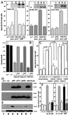
Effect of pHN and secretion of the transcribed HN into cultured media. (A and B) F11 cells were transfected with pcDNA or either V642I-APP, M146L-PS1, or N141I-PS2 cDNA with pFLAG or pHN for 72 h. In A and B, cell mortality and viability were measured, respectively. Negative controls without transfection (no T) or with empty plasmid transfection (vec) were also examined. (A Insets) Expression of cognate FAD proteins by transfection with FAD genes. Arrows indicate the cognate holoproteins (1, no T; 2, vec; 3, FAD gene; 4, FAD gene + pHN). (C) F11 cells were transfected with pcDNA or V642I-APP cDNA in the absence of serum for 3 h, incubated with HF-18% for 2 h, and cultured with CM/pHN, other transfected CM, or fresh media (fresh HF-10%) for 67 h. Cell mortality was measured 72 h after transfection. (D) F11 cells were transfected with pHN, pS7A (pHN mutant with S7A), or pHNR for 72 h, and the cell lysates (Lower) and media (Upper) were analyzed by M2 antibody. (E) F11 cells were transfected with pHN for 24 h and treated with or without secretagogues or an inhibitor for 6 h. CM and cell lysates were submitted to immunoblot analysis with M2 antibody [1, no transfection; 2, pHN transfection alone; 3, pHN + 48 mM KCl; 4, pHN + 1 μM forskolin; 5, pHN + 0.1 mM 3-isobutyl-1-methylxanthine (IBMX); 6, pHN + 1 μM forskolin + 0.1 mM IBMX; 7, pHN + 20 ng/ml (+)Brefeldin A]. (F) F11 cells were transfected with pcDNA or V642I-APP cDNA with cotransfection of pFLAG or pHN or with treatment of 10 μM HN peptides. After 72-h culture, cell mortality was measured. *, significant suppression (P < 0.01); **, no significant suppression vs. V642I-APP-induced cell death.
Figure 3
Figure 3
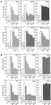
Effect of HN polypeptides on neuronal cell death by FAD genes. (A) F11 cells were transfected with V642I-APP cDNA and were treated with various concentrations of sHN, sHNG, sHNA, the dimer form of sHN through C8 (C8-C8), and sHN whose C-terminal KRRA was replaced with AAAA (KRRA to AAAA). Seventy-two hours after transfection, cell mortality was measured by Trypan blue exclusion assay. (B) F11 cells were transfected with M146L-PS1 or N141I-PS2 cDNA, and were treated with various concentrations of sHN, sHNG (S14G), or sHNA (C8A). Seventy-two hours after transfection, cell mortality was similarly measured.
Figure 4
Figure 4
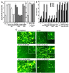
Effect of HN on Aβ-induced cell death in primary neurons. (A and B) Primary cortical neurons were treated with 25 μM Aβ1–43 in the presence or absence of sHN or its derivatives. In A, cell mortality was measured 72 h after Aβ treatment with or without HN peptides. Neurons were similarly treated, as a positive control, with 20 μM etoposide in the presence or absence of 10 μM HN peptides for 72 h. In B, the LDH activity in the culture media was monitored by sampling 6 μl of the media culturing neurons treated with 25 μM Aβ1–43 in the presence or absence of HN peptides at 24, 48, or 72 h after the onset of Aβ treatment. The LDH release was also measured in neurons treated with 20 μM etoposide in the presence or absence of HN peptides at 24, 48, or 72 h after the onset of etoposide treatment. These experiments were performed independently for both assays. (C) Fluorescence microscopic views by Calcein-AM staining for neuronal viability. Seventy-two hours after treatment with 25 μM Aβ1–43, in the presence or absence of HN peptides, neurons were stained with Calcein-AM. Insets indicate magnified views of similarly treated independent cultures. Representative views are indicated.
Figure 5
Figure 5
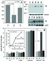
Analysis of the action of HN. (A) F11 cells were incubated with radiolabeled sHNG in the presence or absence (1) of 2 μM unlabeled sHNG (2) or sHNA (3) for 2 h at 4°C and then treated with 200 μM BS3 for 20 min at 4°C. After adding Tris⋅HCl buffer and washing cells with PBS four times, cells were resuspended in PBS and subjected to radiocounting. The total input of radioactivity was 590,990 cpm/sample. (B) F11 cells were transfected with pcDNA or V642I-APP cDNA and incubated with or without 10 μM sHN in the presence or absence of 10 nM wortmannin (W), 100 μM genistein (G), or 50 μM PD98059 (PD). Seventy-two hours after the onset of transfection, cell mortality was similarly measured. (Inset) Jurkat cells were incubated with or without 100 ng/ml CH-11 (anti-Fas; MBL) in the presence or absence of 10 μM sHN (anti-Fas + sHN) for various periods, and cell mortality was similarly measured. (C) F11 cells were transfected with CN-procaspase-3 cDNA with pcDNA or V642I-APP cDNA by lipofection [0.2 μg CN-procaspase-3 cDNA, 0.8 μg V642I-APP cDNA, LipofectAMINE 2 μl, PLUS Reagent 4 μl (Upper); 0.4 μg CN-procaspase-3 cDNA, 0.6 μg V642I-APP cDNA, LipofectAMINE 2 μl, PLUS Reagent 4 μl (Lower)] in the presence or absence of 10 μM sHN or 100 μM Ac-DEVD-CHO [no transfection (lanes 1, 6); pcDNA transfection (lanes 2, 7); CN-procaspase-3 transfection (lanes 3, 8); CN-procaspase-3 + V642I-APP (lanes 4, 9); CN-procaspase-3 + V642I-APP + sHN (lanes 5, 10); CN-procaspase-3 + V642I-APP + Ac-DEVD-CHO (lane 11)]. Forty-eight hours after onset of transfection, cell lysates were submitted to immunoblot analysis with anti-caspase-3, anti-APP, or anti-tubulin antibody. Arrows, procaspase-3.

Similar articles

Cited by

References

    1. Shastry B S, Giblin F J. Brain Res Bull. 1999;48:121–127. - PubMed
    1. Yamatsuji T, Okamoto T, Takeda S, Fukumoto H, Iwatsubo T, Suzuki N, Asami-Odaka A, Ireland S, Kinane T B, Nishimoto I. Science. 1996;272:1349–1352. - PubMed
    1. Zhao B, Chrest F J, Horton W E, Jr, Sisodia S S, Kusiak J W. J Neurosci Res. 1997;47:253–263. - PubMed
    1. Luo J J, Wallace W, Riccioni T, Ingram D K, Roth G S, Kusiak J W. J Neurosci Res. 1999;55:629–642. - PubMed
    1. Wolozin B, Iwasaki K, Vito P, Ganjei J K, Lacana E, Sunderland T, Zhao B, Kusiak J W, Wasco W, D'Adamio L. Science. 1996;274:1710–1713. - PubMed

Publication types

Associated data

  NODES
Note 1
twitter 2