. 2002 Jun 15;30(12):e57.
doi: 10.1093/nar/gnf056.

Relative quantification of 40 nucleic acid sequences by multiplex ligation-dependent probe amplification

Affiliations

Relative quantification of 40 nucleic acid sequences by multiplex ligation-dependent probe amplification

Jan P Schouten et al. Nucleic Acids Res. .

Abstract

We describe a new method for relative quantification of 40 different DNA sequences in an easy to perform reaction requiring only 20 ng of human DNA. Applications shown of this multiplex ligation-dependent probe amplification (MLPA) technique include the detection of exon deletions and duplications in the human BRCA1, MSH2 and MLH1 genes, detection of trisomies such as Down's syndrome, characterisation of chromosomal aberrations in cell lines and tumour samples and SNP/mutation detection. Relative quantification of mRNAs by MLPA will be described elsewhere. In MLPA, not sample nucleic acids but probes added to the samples are amplified and quantified. Amplification of probes by PCR depends on the presence of probe _target sequences in the sample. Each probe consists of two oligonucleotides, one synthetic and one M13 derived, that hybridise to adjacent sites of the _target sequence. Such hybridised probe oligonucleotides are ligated, permitting subsequent amplification. All ligated probes have identical end sequences, permitting simultaneous PCR amplification using only one primer pair. Each probe gives rise to an amplification product of unique size between 130 and 480 bp. Probe _target sequences are small (50-70 nt). The prerequisite of a ligation reaction provides the opportunity to discriminate single nucleotide differences.


Figures
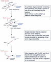
Figure 1
Figure 1
Preparation of the long M13-derived MLPA probe oligonucleotides. The basic MLPA vector M214 was derived from M13mp18 (24) by destroying the single BsmI site and replacement of the polylinker site by a synthetic oligonucleotide containing from 5′ to 3′, a new BsmI site, an SphI and XbaI site, a sequence complementary to one of the two SALSA PCR primer sequences and an EcoRV site. A collection of 118 different SALSA vectors was prepared by insertion of stuffer fragments in the SphI and XbaI sites. Each stuffer fragment has a different sequence and length (19–370 bp, 3 bp increments). Stuffer fragments up to 55 nt were synthetic; longer fragments were made by PCR amplification of T7 phage sequences. For each MLPA probe, a synthetic 30–43 nt long oligonucleotide containing the hybridising sequence is cloned in the SphI and BsmI sites of one of the SALSA vectors. Single stranded DNA is prepared from the clone obtained and is digested by BsmI and EcoRV as indicated in the figure.
Figure 2
Figure 2
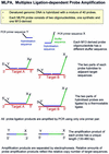
Outline of the MLPA reaction.
Figure 3
Figure 3
Sensitivity of MLPA analysis to mismatches in the short probe oligonucleotide. MLPA reactions were performed on 100 ng samples of human DNA. Amplification products were separated on a denaturing acrylamide gel (LICOR). Length (bp) as well as gene HUGO name is indicated for each probe amplification product. The probe mix contained 33 complete probes. For seven other (underlined) probes, only the long M13-derived oligonucleotide was included in each test. The part of the gel shown shows the result of addition of different short probe oligonucleotides for two of these probes to the probe mix: lane 1, Bax-specific short probe oligonucleotide, resulting in a 301 bp extra amplification product. Lane 2, as lane 1 but with a mismatch (T/T) at the fourth nucleotide from the ligation site. Lane 3, as lane 1 but with a mismatch (A/C) at the 3′ nucleotide of the BAX short probe oligonucleotide. Lane 4, F3-specific short probe oligonucleotide, resulting in a 328 bp extra amplification product. Lane 5, as lane 4 but with a mismatch (G/G) at the fourth nucleotide from the ligation site. Lane 6, as lane 4 but with a mismatch (G/G) at the 3′ nucleotide of the F3 short probe oligonucleotide.
Figure 4
Figure 4
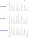
Detection of trisomies by MLPA. Samples containing 100 ng DNA were analysed by MLPA using probe mix P001. Male and female control DNA was obtained from Promega. Blood-derived DNA from a triple X and a female triple 21 individual were provided by the Department of Clinical Genetics, Free University of Amsterdam. Reactions were analysed by capillary electrophoresis (Beckman CEQ2000). In each case the female control DNA is shown in red. Probe mix P001 contains 40 probes. Only part of the resulting electropherogram is shown. Arrows indicate the positions of a 292 bp amplification product of a probe specific for the TFF1 gene on chromosome 21, a 319 bp amplification product of a probe specific for the L1CAM gene on the X chromosome and a 337 bp amplification product of a probe specific for the APP gene on chromosome 21. The other probes were specific for chromosome 3 (283 bp), chromosome 18 (301 and 346 bp), chromosome 13 (310 bp) and chromosome 1 (328 bp). A complete list of genes in this probe mix can be found in Table 1.
Figure 5
Figure 5
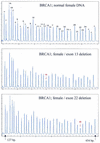
Detection of BRCA1 exon deletions by MLPA. Samples containing ∼100 ng DNA were analysed by MLPA using probe mix P002. Female control DNA was obtained from Promega. Blood-derived DNA from individuals known to contain an exon 13 or an exon 22 deletion were provided by the Department of Clinical Genetics, Free University of Amsterdam. Reactions were analysed by capillary electrophoresis. Probe mix P002 contains 34 probes. Nine probes recognising non-BRCA1 sequences on various chromosomes are indicated by a ‘c’. The exon recognised by the BRCA1-specific probes is indicated by a number. Probes for both alternative exons 1 are indicated as 1A and 1B. Exon 4 is not present in the normal BRCA1 gene transcript. Two probes specific for the first and last parts of exon 11 are included as this exon is very large (3.4 kb). Exon deletions are apparent by an ∼50% reduction in peak area of a specific probe. The exact gene and sequence recognised by each probe of the P002 probe mix can be found on the www.mrc-holland.com website.
Figure 6
Figure 6
Detection of MSH2 exon deletions by MLPA. Samples containing ∼100 ng DNA were analysed by MLPA using probe mix P003. Female control DNA was obtained from Promega. Blood-derived DNA from an individual known to contain a deletion of exons 1–6 of the MSH2 gene was provided by the Department of Clinical Genetics, Free University of Amsterdam. Reactions were analysed by capillary electrophoresis (Beckman CEQ2000). Probe mix P003 contains 42 probes. Probes are present for each of the 19 MLH1 and each of the 16 MSH2 exons. Seven probes recognise other sequences on various chromosomes. Only part of the electropherogram is shown. The exon recognised by the MSH2-specific probes is indicated by a number. Control probes are indicated by a ‘c’. The remaining probes are specific for MLH1 exons 1–9. Exon deletions are apparent by an ∼50% reduction in peak area of a specific probe. The exact gene and sequence recognised by each probe of the P003 probe mix can be found at the www.mrc-holland.com website.
Figure 7
Figure 7
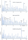
Detection of gains and losses of human chromosomal sequences by MLPA. Samples containing ∼100 ng DNA were analysed by MLPA using probe mix P006. Male control DNA was obtained from Promega. DNA isolated from a frozen DLBCL lymphoma was provided by the Department of Pathology, Amsterdam Medical Center. DNA isolated from the SkBr3 cell line was provided by the Henry Ford Hospital in Detroit. Reactions were analysed by capillary electrophoresis (Beckman CEQ2000). Probe mix P006 contains 41 probes. Probes specific for the chromosome 18 genes PMAIP1, BCL2 and DCC, as well as the CDKN2B gene on chromosome 9 and the ERBB2 gene on chromosome 17 are indicated. A probe recognising the SRY gene on the Y chromosome is indicated by Y. Four probes recognising the KIAA0170, BAK1, TNF and IER3 genes, all in the HLA region of chromosome 6, are indicated by 6p. The exact gene, sequence and chromosomal location recognised by each probe of the P006 probe mix can be found at the www.mrc-holland.com website.
Figure 8
Figure 8
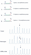
MLPA analysis of SNPs/mutations resulting in amplification products for both alleles with different lengths. (Top) Schematic drawing. _target sequences A and B differ in 1 nt. The short synthetic probe oligonucleotides S1 and S2 differ also in 1 nt, each being specific for one SNP sequence. In addition, S1 and S2 differ in length due to the presence of a 3 nt stuffer sequence between the hybridising sequence and the PCR primer sequence of oligonucleotide S2. Both oligonucleotides can anneal to _target sequences A and B and will compete with each other for binding. A mismatch adjacent to the ligation site, prevents oligonucleotide ligation to the common M13-derived probe oligonucleotide M when S1 is bound to B, or when S2 is bound to _target sequence A. (Bottom) Capillary electrophoresis profile. Detection of the CFTR ΔF508 mutation by MLPA. DNA samples (100 ng) of a control individual, a heterozygote and a homozygote carrier of the CFTR ΔF508 mutation (3 nt deletion) were tested by MLPA. The probe mix contained 40 probes including a probe for the wild type CFTR sequence and one specific for this mutation. These two probes have the M13-derived probe oligonucleotide in common. The short synthetic probe oligonucleotides differ at the site of the mutation, which is located at the 3′ end of the oligonucleotide as well as in length. Only part of the capillary electrophoresis profile is shown. Arrows indicate wild type (202 bp) and mutation specific probe (205 bp) amplification products.

Similar articles

Cited by

References

    1. Norman A.M., Thomas,N.S.T., Kingston,H.M. and Harper,P.S. (1990) Becker muscular dystrophy: correlation of deletion type with clinical severity. J. Med. Genet., 27, 236–239. - PMC - PubMed
    1. Petrij-Bosch A., Peelen,T., van Vliet,M., van Eijk,R., Olmer,R., Drusedau,M., Hogervorst,F.B., Hageman,S., Arts,P.J., Ligtenberg,M.J. et al. (1997) BRCAI genomic deletions are major founder mutations in Dutch breast cancer patients. Nature Genet., 17, 341–345. - PubMed
    1. Wijnen J., van der Klift,H., Vasen,H., Khan,P.M., Menko,F., Tops,C., Meijers Heijboer,H., Lindhout,D., Moller,P. and Fodde,R. (1998) MSH2 genomic deletions are a frequent cause of HNPCC. Nature Genet., 20, 326–328. - PubMed
    1. Kauraniemi P., Barlund,M., Monni,O. and Kallioniemi,A. (2001) New amplified and highly expressed genes discovered in the ERBB2 amplicon in breast cancer by cDNA microarrays. Cancer Res., 61, 8235–8240. - PubMed
    1. Leyland-Jones B. and Smith,I. (2001) Role of herceptin in primary breast cancer. Oncology, 61 (Suppl. 2), 83–91. - PubMed

Publication types

MeSH terms

  NODES
twitter 2