Comparative Study
. 2004 Mar 15;199(6):785-95.
doi: 10.1084/jem.20031109. Epub 2004 Mar 8.

Btk is required for an efficient response to erythropoietin and for SCF-controlled protection against TRAIL in erythroid progenitors

Affiliations
Comparative Study

Btk is required for an efficient response to erythropoietin and for SCF-controlled protection against TRAIL in erythroid progenitors

Uwe Schmidt et al. J Exp Med. .

Erratum in

  • J Exp Med. 2004 Apr 5;199(7):following 1031

Abstract

Regulation of survival, expansion, and differentiation of erythroid progenitors requires the well-controlled activity of signaling pathways induced by erythropoietin (Epo) and stem cell factor (SCF). In addition to qualitative regulation of signaling pathways, quantitative control may be essential to control appropriate cell numbers in peripheral blood. We demonstrate that Bruton's tyrosine kinase (Btk) is able to associate with the Epo receptor (EpoR) and Jak2, and is a substrate of Jak2. Deficiency of Btk results in reduced and delayed phosphorylation of the EpoR, Jak2, and downstream signaling molecules such as Stat5 and PLCgamma1 as well as in decreased responsiveness to Epo. As a result, expansion of erythroid progenitors lacking Btk is impaired at limiting concentrations of Epo and SCF. In addition, we show that SCF induces Btk to interact with TNF-related apoptosis-inducing ligand (TRAIL)-receptor 1 and that lack of Btk results in increased sensitivity to TRAIL-induced apoptosis. Together, our results indicate that Btk is a novel, quantitative regulator of Epo/SCF-dependent expansion and survival in erythropoiesis.


Figures
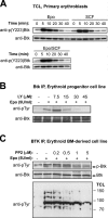
Figure 1.
Figure 1.
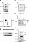
Epo and SCF induce Btk tyrosine phosphorylation. (A) Primary, fetal liver–derived progenitors were factor deprived and restimulated with Epo, SCF, or Epo/SCF as shown. Stimulation time is indicated in minutes. Btk phosphorylation was analyzed using an anti-Y223 phospho-specific antibody. Total Btk was stained to confirm equal loading. (B and C) wt erythroid progenitor cells (2B6) were factor deprived in the absence or presence of various concentrations of the PI3 kinase inhibitor LY294002 (B, LY) or the Src kinase inhibitor PP2 (C) as shown and restimulated with Epo for 10 min. Btk immunoprecipitates were analyzed for phosphorylation using antiphosphotyrosine antibodies (PY99; p-Btk is the bottom band) and restained for total Btk (B) or for phosphorylated proteins in total cell lysates (TCLs) as a control (C). Size markers are indicated in kilodaltons.
Figure 2.
Figure 2.
Btk coimmunoprecipitates with and is a substrate of Jak2. (A–D and F) COS cells were transfected with pSG5-based expression constructs encoding the indicated proteins. Expression of all proteins was verified in Western blots using specific antibodies (not depicted). After 48 h, cells were stimulated with 5 U/ml Epo (B) or 500 ng/ml SCF (C, lane 4; D and E, lane 4) for 10 min, harvested, and lysed, and Btk (A and C), EpoR (B), Jak2 (D), and c-Kit (F) were immunoprecipitated. (top panels) Immunoblots were stained with antiphosphotyrosine antibodies (PY99). The blots were restained with antibodies recognizing the immunoprecipitated proteins to check for equal loading. Arrows indicate the position of the immunoprecipitated and coimmunoprecipitating proteins. (E) wt erythroid progenitors (2B6) were factor deprived in the absence or presence of the Src kinase family inhibitor PP2 as indicated and stimulated with Epo for 10 min. Epo-induced Jak2 phosphorylation was assayed on immunoblots using PY99 (top). (bottom) Blots were restained with anti-Jak2 to confirm equal loading. (F) c-Kit immunoprecipitates were stained with antiphosphotyrosine antibodies (PY99), and blots were restained with anti–c-Kit and anti-Btk antibodies, whereas total cell lysates (TCLs) were stained with anti–c-Kit and anti-Btk to check expression levels.
Figure 3.
Figure 3.
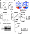
Btk erythroid progenitors show enhanced differentiation at the expense of renewal. (A) Btk primary fetal liver progenitors were seeded in medium containing high (2 U/ml Epo and 100 ng/ml SCF) or low (0.5 U/ml Epo and 25 ng/ml SCF) concentrations of SCF and Epo, while maintaining normal concentrations of Dex, and cumulative cell numbers were determined. The different symbols represent two independent experiments using wt and Btk erythroid progenitors as indicated. (B) Histological analysis of wt and Btk cells described in A at day 6 (A, arrow). The percentages of erythroid cells at different stages of maturity (as indicated) were scored for wt and Btk cultures and plotted in a pie chart. Percentage of immature cells: wt, 47%, and Btk, 22%. Partially mature cells: wt, 21%, and Btk, 28%. Mature cells: wt, 15%, and Btk, 37%. Dead cells: wt, 17%, and Btk, 12%. (C) Hemoglobin content (Hb/cell volume in arbitrary units) of cells as described in A was measured at days 4 and 6. (D) Btk bone marrow cell lines ectopically Btk-reexpressing (BtkRE) and Btk cell lines (Btk) were treated as described in A. Open symbols represent BtkRE, and closed symbols represent Btk cells cultured on low, high, or no-growth factors (NF; see A). (E) Primary fetal liver progenitors were seeded in differentiation medium. Expression of Btk and the EpoR during differentiation was checked by Western blotting using Btk and EpoR antibodies. Equal loading was examined using an Actin antibody. (F) Cells were seeded in differentiation conditions at 10, 0.1, and 0.01 Epo U/ml. Cumulative cell numbers were determined. Symbols indicate the different growth conditions as well as wt and Btk cultures.
Figure 4.
Figure 4.
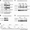
Btk cells are perturbed in Epo-induced signaling. (A–D) wt (Btkwt), Btk (Btk), Btk-reexpressing (BtkRE) bone marrow–derived erythroid cell lines (A, C, and D), and primary wt and Btk5 fetal liver progenitors (B) were factor deprived and restimulated with 5 U/ml Epo or SCF as indicated for 10 min. Cells were lysed, and the immunoprecipitated proteins as well as total cell lysates (TCLs) were analyzed by Western blotting. (top) Blots were stained with the antiphosphotyrosine antibody PY99, except for the TCLs (B), which were stained in B with anti–phospho-Tyr694/699 Stat5a/b. (bottom) The same blots were restained with antibodies recognizing the immunoprecipitated protein as indicated or with anti–total Stat5a/b for the TCLs. (E) Flow cytometric analysis of cell surface expression of c-Kit, TER119, Integrinα4 by specific antibodies, and EpoR by biotinylated Epo in wt (thick black line) and Btk (thin black line) erythroid progenitors. Expression levels are also shown as histograms depicting control stainings (dotted line) and addition of excess Epo (gray line) for EpoR detection. Mean fluorescence values varied <10% for each staining in three independent experiments, indicating that wt and Btk erythroid progenitors express similar levels of the respective cell surface markers.
Figure 5.
Figure 5.
Btk erythroid progenitors have a delayed Epo-induced Jak2 and Stat5 phosphorylation. (A–E) wt (Btkwt), Btk (Btk), and Btk-reexpressing (BtkRE) erythroid cell lines (A–C) or primary fetal liver progenitors (D) were factor deprived and restimulated with 5 U/ml Epo for the indicated times. Cells were lysed, and Jak2 (B) or the EpoR (C) was immunoprecipitated. The immunoprecipitations as well as total cell lysates (TCLs) were analyzed by Western blotting. (A and D) Blots were stained with anti–phospho-Tyr694/699 Stat5a/b (top) and restained with anti–total Stat5a/b to confirm equal loading (bottom). (B and C, top) Blots were stained with antiphosphotyrosine PY99. (bottom) The same blots were restained with anti-Jak2 or anti-EpoR to confirm equal loading. PY-staining of these proteins in Btk cells was much lower than in wt cells. Therefore, Btk panels were exposed much longer than wt panels to enable proper comparison of kinetics of deactivation. (E) Erythroid progenitors were restimulated with increasing concentrations of Epo for 10 min as indicated. (top) Blots stained with anti–phospho-Tyr694/699 Stat5a/b; (bottom) the same blots were restained with anti–total Stat5a/b.
Figure 6.
Figure 6.
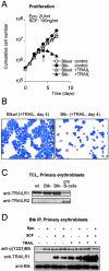
Btk erythroid progenitors are no longer protected by SCF from TRAIL-induced apoptosis. (A) wt and Btk fetal liver cells were seeded in medium containing Epo, SCF, and Dex in the presence and absence of 20 ng/ml TRAIL, and cumulative cell numbers were determined. (B) At day 4, cytospins were prepared from cultures of wt and Btk cells in the presence of TRAIL (A, arrow) and stained with neutral benzidine plus histological dyes. (C) Western blots from cell lysates of wt and Btk primary fetal liver erythroid progenitors and primary pro–B cells were performed using anti-TRAILR1 and anti-TRAILR2 antibodies as indicated. (D) wt primary fetal liver progenitors were factor deprived and restimulated (for 10 min) with 5 U/ml Epo, 500 ng/ml SCF, and 200 ng/ml TRAIL or combinations of factors as indicated. Btk was immunoprecipitated from cell lysates and analyzed by Western blotting. Btk tyrosine phosphorylation was analyzed using an anti–phospho-Tyr223 Btk antibody (top), whereas coimmunoprecipitation of TRAILR1 was checked with anti-TRAILR1 (middle). Equal loading was confirmed by restaining the same blot with total anti-Btk (bottom).

Similar articles

Cited by

References

    1. Wu, H., X. Liu, R. Jaenisch, and H.F. Lodish. 1995. Generation of committed erythroid BFU-E and CFU-E progenitors does not require erythropoietin or the erythropoietin receptor. Cell. 83:59–67. - PubMed
    1. Flanagan, J.G., D.C. Chan, and P. Leder. 1991. Transmembrane form of the kit ligand growth factor is determined by alternative splicing and is missing in the Sld mutant. Cell. 64:1025–1035. - PubMed
    1. Shetty, V., S. Hussaini, L. Broady-Robinson, K. Allampallam, S. Mundle, R. Borok, E. Broderick, L. Mazzoran, F. Zorat, and A. Raza. 2000. Intramedullary apoptosis of hematopoietic cells in myelodysplastic syndrome patients can be massive: apoptotic cells recovered from high-density fraction of bone marrow aspirates. Blood. 96:1388–1392. - PubMed
    1. De Maria, R., A. Zeuner, A. Eramo, C. Domenichelli, D. Bonci, F. Grignani, S.M. Srinivasula, E.S. Alnemri, U. Testa, and C. Peschle. 1999. Negative regulation of erythropoiesis by caspase-mediated cleavage of GATA-1. Nature. 401:489–493. - PubMed
    1. Dai, C.H., J.O. Price, T. Brunner, and S.B. Krantz. 1998. Fas ligand is present in human erythroid colony-forming cells and interacts with Fas induced by interferon gamma to produce erythroid cell apoptosis. Blood. 91:1235–1242. - PubMed

Publication types

MeSH terms

  NODES
twitter 2