. 2006 Dec 1;400(2):315-25.
doi: 10.1042/BJ20060259.

Gangliosides play an important role in the organization of CD82-enriched microdomains

Affiliations

Gangliosides play an important role in the organization of CD82-enriched microdomains

Elena Odintsova et al. Biochem J. .

Abstract

Four-transmembrane-domain proteins of the tetraspanin superfamily are the organizers of specific microdomains at the membrane [TERMs (tetraspanin-enriched microdomains)] that incorporate various transmembrane receptors and modulate their activities. The structural aspects of the organization of TERM are poorly understood. In the present study, we investigated the role of gangliosides in the assembly and stability of TERM. We demonstrated that inhibition of the glycosphingolipid biosynthetic pathway with specific inhibitors of glucosylceramide synthase [NB-DGJ (N-butyldeoxygalactonojirimycin) and PPMP (D-threo-1-phenyl-2-hexadecanoylamino-3-morpholino-1-propanol.HCl)] resulted in specific weakening of the interactions involving tetraspanin CD82. Furthermore, ectopic expression of the plasma-membrane-bound sialidase Neu3 in mammary epithelial cells also affected stability of the complexes containing CD82: its association with tetraspanin CD151 was decreased, but the association with EGFR [EGF (epidermal growth factor) receptor] was enhanced. The destabilization of the CD82-containing complexes upon ganglioside depletion correlated with the re-distribution of the proteins within plasma membrane. Importantly, depletion of gangliosides affected EGF-induced signalling only in the presence of CD82. Taken together, our results provide strong evidence that gangliosides play an important role in supporting the integrity of CD82-enriched microdomains. Furthermore, these results demonstrate that the association between different tetraspanins in TERM is controlled by distinct mechanisms and identify Neu3 as a first physiological regulator of the integrity of these microdomains.


Figures
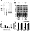
Figure 1
Figure 1. Effect of ganglioside depletion on the interactions in TERMs
HB2/CD82 cells were incubated with 2 mM NB-DGJ for 4–6 days or 20 μM PPMP for 7 days; control cells were grown in parallel. (A) Lipids were extracted, purified, chromotographed on HPTLC (high-performance TLC) plates and revealed with resorcinol staining. The positions of co-chromotographed ganglioside standards are indicated. (B) Surface expression of gangliosides was analysed by flow cytometry using the Coulter Epics program. Results are presented as mean fluorescence intensity (MnX) and represent one of three experiments. The mAbs used were: mAb 1, negative control, 187.1; mAb 2, anti-α3 integrin subunit, A3-IIF5; mAb 3, anti-CD82, M104; mAb 4, anti-GD1a, GD1a-1; mAb 5, anti-GM1, FITC-conjugated CT. (C) Cells were lysed in 1% Brij98 and the complexes were immunoprecipitated using specific mAbs: anti-CD82, γC11; anti-CD9, TS9. The immunoprecipitated complexes were resolved by SDS/10% PAGE (for reduced samples) or SDS/12% PAGE (for non-reduced samples), transferred to a nitrocellulose membrane and probed with antibodies to α3 integrin subunit (polyclonal sera), anti-EGFR (mAb Ab-15), anti-CD82 (mAb TS82b), anti-CD9 (mAb C9BB), or anti-CD151 (polyclonal sera). Results shown represent one of three independent experiments. WB, Western blot. (D) Quantification of the results of three immunoprecipitation (IP) experiments (treatment with NB-DGJ). Means of band intensities relative to that in the control samples (i.e. non-treated cells) are shown. Error bars indicate standard deviations. (a) IP with anti-CD82 mAb; (b) IP with anti-CD9 mAb.
Figure 2
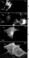
Figure 2. Immunofluorescence staining of HB2/CD82 cells transiently transfected with Neu3 sialidase
Cells grown on glass coverslips were transfected with sialidase Neu3 cDNA. (A, B) After 48 h the cells were fixed with paraformaldehyde and permeabilized with 0.4% saponin. Double staining immunofluorescence was carried out with the anti-HA (F-7) and anti-GD1a (GD1a–1) mAbs. Isotype-specific secondary antibodies conjugated to Alexa Fluor® 488 or Alexa Fluor® 594 fluorochromes were used for detection of the antigens. (A) Staining with the anti-HA antibody. (B) Staining of the same field with the anti-GD1a antibody. Arrows indicate the cell expressing Neu3 sialidase. (C, D) After 48 h cells were incubated with FITC-conjugated CT for 1 h at 4 °C. Cells were washed, fixed with 2% (w/v) paraformaldehyde for 10 min and permeabilized with 0.4% saponin. Subsequently, cells were stained with the anti-HA mAb (F-7). Alexa Fluor® 594-conjugated goat anti-mouse antibody was used to visualize cells expressing HA-tagged Neu3 (indicated with arrows). (C) Staining with the anti-HA mAb. (D) Staining of the same field with FITC-conjugated CT. Note that staining with the anti-HA mAb is more dispersed in the presence of CT, most likely due to the pre-incubation of cells with CT prior to fixation.
Figure 3
Figure 3. Expression of Neu3 sialidase affects the interactions involving CD82 but does not affect glycosylation pattern of the protein
(A and B) HB2/CD82 cells were transiently transfected with the HA-tagged form of sialidase Neu3 or with the control DNA. After 48 h cells were lysed in 1% Brij98 and analysed for antigen expression. Data of one of the two separate experiments are shown. Complexes were immunoprecipitated using specific mAbs: anti-HA-tag, F-7 (lanes 2 and 6); anti-CD82, γC11 (lanes 1 and 5); anti-CD9, TS9 (lanes 3 and 7). The immunoprecipitated complexes were divided into three aliquots (20–40 μl) and resolved by SDS/12% PAGE (non-reduced samples) for probing with the anti-HA-tag polyclonal antibody (Y11), anti-CD82 (mAb TS82b), anti-CD9 (mAb C9BB) or anti-CD151 (polyclonal sera), or in SDS/10% PAGE (reduced samples) for probing with the anti-EGFR (mAb Ab-15). Two bands at the top panel (A, lane 10) developed with the anti-HA antibody most likely represent the monomer and dimer of Neu3 sialidase. Lys1 (lane 9 in A, and lane 6 in B): lysate derived from the control cells; Lys2 (lane 10 in A, and lane 7 in B): lysate derived from the cells transfected with Neu3 sialidase. (C) HB2 cells were transiently transfected with the plasmid encoding CD82 alone or in combination with the plasmid encoding HA-tagged form of sialidase Neu3. After 48 h cells were lysed in 1% Brij98 and the proteins containing sialic acid moiety were precipitated using the immobilized SNA lectin. Proteins were resolved by SDS/12% PAGE, transferred to the nitrocellulose membrane and probed with the anti-CD82 mAb (TS82b). Top panel: lanes 1 and 2, lectin IP; lanes 3 and 4, lysates from the appropriate sample. Lower panel shows results of Western blotting with the anti-HA mAb (lanes 5 and 6). Molecular masses are given in kDa. WB, Western blot.
Figure 4
Figure 4. Expression of Neu3 sialidase does not affect interactions of tetraspanins in the absence of CD82
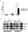
HB2 cells were transfected with the plasmid encoding the HA-tagged form of sialidase Neu3 or with the control DNA. Positive clones were tested by flow cytometry for decrease in GD1a surface expression and by Western blotting for expression of HA-tagged sialidase. Experiments were repeated three times. (A) Surface expression level of GD1a in cells expressing Neu3 was analysed by flow cytometry. The mAbs used were: negative control, 187.1; anti-CD82, M104; anti-GD1a, IgG1. Results are presented as means of fluorescence intensity (MnX). (B) Cells were lysed in 1% Brij98 and the complexes were immunoprecipitated using specific mAbs: anti-CD151, 5C11 (lanes 1 and 6); anti-CD81, M38 (lanes 2 and 7); anti-CD9, TS9 (lanes 3 and 8); negative control, 187.1 (lanes 4 and 9). The immunoprecipitated complexes were resolved by SDS/10% PAGE (for reduced samples) or SDS/12% PAGE (for non-reduced samples), transferred to a nitrocellulose membrane and probed with the antibodies to α3 integrin subunit (polyclonal sera), or anti-CD151 (polyclonal sera), or anti-CD9 (mAb C9BB). WB, Western blot.
Figure 5
Figure 5. Ganglioside GM3 has no effect on the interactions of CD82
GM95/CD82+CD151 and GM95/CGlcT-ER/CD82+CD151 cells were lysed in 1% Brij98 and the complexes were immunoprecipitated using specific mAbs: anti-CD82, γC11 (lanes 1 and 4); anti-CD151, 5C11 (lanes 2 and 5); and negative control, 187.1 (lanes 3 and 6). The immunoprecipitated complexes were resolved by SDS/12% PAGE, transferred to a nitrocellulose membrane and probed with the anti-CD82 (mAb TS82b) or anti-CD151 (polyclonal sera). Upper bands in CD151 immunoprecipitates revealed by the anti-CD82 mAb are non-specific. Lysate 1 (lane 7) corresponds to the lysate derived from GM95/CD82+CD151 cells; lysate 2 (lane 8) corresponds to the lysate derived from GM95/CGlcT-ER/CD82+CD151 cells. WB, Western blot.
Figure 6
Figure 6. Exogenous administration of gangliosides does not affect stability of CD82 complexes
Ganglioside-deficient GM95/CD82+CD151 cells were incubated with 50 μg/ml GD1a or GM3 or 25 μl/ml DMSO in serum-free medium for 24 h at 37 °C. After the incubation, cells were subjected to immunoprecipitation (IP) analyses. The complexes were immunoprecipitated using specific mAbs: anti-CD82, γC11 (lanes 1, 4 and 7); anti-CD151, 5C11 (lanes 2, 5 and 8); and negative control, 187.1 (lanes 3, 6 and 9). Proteins were resolved by SDS/12% PAGE, transferred to a nitrocellulose membrane and probed with the antibodies to CD82 (mAb TS82b) or CD151 (polyclonal sera). WB, Western blot.
Figure 7
Figure 7. Inhibition of ganglioside biosynthesis has a specific effect on the EGFR activity in CD82-expressing cells
Cells (HB2/ZEO, A, or HB2/CD82, B,) were incubated with 20 μM PPMP or 0.1% ethanol (control) for 7 days and then serum-starved for 6 h and stimulated with EGF (100 ηg/ml) for 15 or 60 min. Lysates were prepared in 1×Laemmli buffer. Equal amounts of proteins were resolved by SDS/10% PAGE, transferred to a nitrocellulose membrane and probed with the anti-phosphotyrosine mAb (4G10, Upstate Biotechnology), or phosphospecific anti-EGFR polyclonal antibody (Tyr1068), or anti-EGFR mAb (Ab-15). Data presented are results of one of three independent experiments. Quantification of three independent experiments was performed. Means of ratios of the densities of phosphorylated EGFR to the densities of total EGFR are shown. Error bars indicate standard deviations.
Figure 8
Figure 8. Effect of inhibition of ganglioside biosynthesis on the compartmentalization of membrane proteins
The lysates of HB2/CD82 cells treated with 2 mM NB-DGJ for 7 days (A) or 20 mM MβCD for 1 h (B) and control cells were prepared under the detergent-free conditions as described in the Materials and methods section. Lysates were fractionated by ultracentrifugation in continuous sucrose density gradient (5–45%). Fractions were collected from the top of the gradient. Equal volumes of each fraction were resolved by SDS/10% PAGE or SDS/12% PAGE. Distribution of proteins in the gradient fractions was assessed by Western blotting using specific antibodies: anti-CD82 (mAb TS82b), anti-EGFR (mAb Ab-15), anti-α3 integrin subunit (polyclonal sera), anti-CD151 (polyclonal sera) and anti-CD9 (mAb C9BB). Results of one of two separate experiments are shown. c, Control cells; i, inhibitor-treated cells. Note that distributions of proteins in the gradients obtained from the control cells in the experiments shown in (A) and (B) are slightly different. This may be due to the differences in the culturing conditions of cells prior to lysis. Cholesterol depletion was carried out under serum-free conditions and, consequently, the control cells were kept under serum-free conditions (B). In the experiments depicted in (A), cells were grown in the serum-containing medium. P, pellet. (C) Light fractions (2–5) of sucrose density gradients obtained from the control untreated cells and cells depleted of gangliosides were pooled together, and the CD82-containing complexes were immunoprecipitated using mAb γC11. Immunoprecipitated material was resolved by SDS/12% PAGE, transferred to a nitrocellulose membrane and probed with the anti-CD82 (TS82b) mAb, anti-CD9 (C9BB) mAb and anti-CD151 polyclonal antibody. WB, Western blot.
Figure 9
Figure 9. Effect of ganglioside depletion on the surface distribution of CD82
Cells incubated with NB-DGJ (7 days, 2 mM) and control cells were plated on to coverslips. After 24 h the cells were fixed with 2% paraformaldehyde and stained with primary anti-CD82 (IA4) mAb. Isotype-specific secondary antibodies conjugated to Alexa Fluor® 488 fluorochrome were used for the detection of the antigen. (A) Control cells. (B) Cells depleted of gangliosides. Arrows point to the peripheral area of the cells.

Similar articles

Cited by

References

    1. Brown D. A., Jacobson K. Microdomains, lipid rafts and caveolae (San Feliu de Guixols, Spain, 19–24 May 2001) Traffic. 2001;2:668–672. - PubMed
    1. Rajendran L., Simons K. Lipid rafts and membrane dynamics. J. Cell Sci. 2005;118:1099–1102. - PubMed
    1. Razani B., Woodman S. E., Lisanti M. P. Caveolae: from cell biology to animal physiology. Pharmacol. Rev. 2002;54:431–467. - PubMed
    1. Hakomori S. Glycosylation defining cancer malignancy: new wine in an old bottle. Proc. Natl. Acad. Sci. U.S.A. 2002;99:10231–10233. - PMC - PubMed
    1. Simons K., Ikonen E. Functional rafts in cell membranes. Nature. 1997;387:569–572. - PubMed

Publication types

  NODES
admin 3
Association 3
Note 2
twitter 2