. 2008 Dec 1;68(23):9712-22.
doi: 10.1158/0008-5472.CAN-08-1952.

Bidirectional crosstalk between leptin and insulin-like growth factor-I signaling promotes invasion and migration of breast cancer cells via transactivation of epidermal growth factor receptor

Affiliations

Bidirectional crosstalk between leptin and insulin-like growth factor-I signaling promotes invasion and migration of breast cancer cells via transactivation of epidermal growth factor receptor

Neeraj K Saxena et al. Cancer Res. .

Abstract

Obesity is an independent risk factor for breast cancer, and obese breast cancer patients exhibit a higher risk for larger tumor burden and increased metastasis. Obesity, as associated with metabolic syndrome, results in an increase in circulating insulin-like growth factor (IGF), which acts as a mitogen. In addition, higher plasma level of adipocytokine leptin is associated with obesity. In the present study, we show that cotreatment with leptin and IGF-I significantly increases proliferation as well as invasion and migration of breast cancer cells. We found a novel bidirectional crosstalk between leptin and IGF-I signaling; IGF-I induced phosphorylation of leptin receptor (Ob-Rb) and leptin induced phosphorylation of IGF-I receptor (IGF-IR), whereas cotreatment induced synergistic phosphorylation and association of Ob-Rb and IGF-IR along with activation of downstream effectors, Akt and extracellular signal-regulated kinase. Leptin increased phosphorylation of IGF signaling molecules insulin-receptor substrate (IRS)-1 and IRS-2. Interestingly, we found that leptin and IGF-I cotreatment synergistically transactivated epidermal growth factor receptor (EGFR), depending on the proteolytic release of EGFR ligands, as the broad-spectrum matrix metalloproteinase inhibitor GM6001 could inhibit this effect. Using clinically relevant EGFR inhibitors, erlotinib and lapatinib, we found that inhibition of EGFR activation effectively inhibited leptin- and IGF-I-induced invasion and migration of breast cancer cells. Taken together, these data suggest a novel bidirectional crosstalk between leptin and IGF-I signaling that transactivates EGFR and promotes the metastatic properties as well as invasion and migration of breast cancer cells. Our findings indicate the possibility of using EGFR inhibitors erlotinib and lapatinib to counter the procancerous effects of leptin and IGF-I in breast cancers.


Conflict of interest statement

Disclosure of Potential Conflicts of Interest

No potential conflicts of interest were disclosed.

Figures
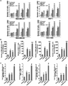
Figure 1
Figure 1
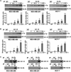
Cotreatment with leptin and IGF-I increases proliferation as well as migration and invasion of breast cancer cells. A, leptin and IGF-I increase proliferation of cells in an anchorage-dependent proliferation assay. MDA-MB-231, MCF-7, MDA-MB-468, and HCC-1806 breast cancer cells were serum starved for 24 h, followed by treatment with 100 ng/mL leptin, 100 ng/mL IGF-I alone, and the combination for 12, 24, 48, and 72 h. XTT assays were then done as described in Materials and Methods. Both leptin and IGF-I increased proliferation of breast cancer cells in a time-dependent manner and combined treatment resulted in further increase in proliferation. Columns, mean of three independent experiments done in triplicates; bars, SE. *, P < 0.001, compared with untreated control cells; #, P < 0.005, compared with untreated control cells; **, P < 0.001, compared with cells treated with leptin alone. B, leptin and IGF-I increase invasion of breast cancer cells. MDA-MB-231, MCF-7, MDA-MB-468, and HCC-1806 breast cancer cells were cultured in Matrigel invasion chambers and serum starved for 24 h, followed by treatment with 100 ng/mL leptin, 100 ng/mL IGF-I alone, and the combination for 24 h. The number of cells that invaded through the Matrigel was counted in five different regions. The slides were blinded to remove counting bias. Columns, mean of three independent experiments done in triplicates; bars, SE. *, P < 0.005, compared with untreated control cells; **, P < 0.005, compared with untreated control cells; ***, P < 0.001, compared with cells treated with leptin alone. C, leptin and IGF-I up-regulate migration of breast cancer cells. MDA-MB-231, MCF-7, MDA-MB-468, and HCC-1806 breast cancer cells were grown to confluence on fibronectin-coated surface, serum starved for 24 h, and scratched with a pipette tip. The plates were photographed immediately following scratching. Culture media were replaced with media containing 100 ng/mL leptin, 100 ng/mL IGF-I alone, and the combination for 24 h. The plates were photographed at the identical location of the initial image at 24 h posttreatment. The fold change in migration was calculated. Columns, mean of three independent experiments done in triplicates; bars, SE. *, P < 0.005, compared with untreated control cells; **, P < 0.005, compared with untreated control cells; #, P < 0.001, compared with cells treated with leptin alone.
Figure 2
Figure 2
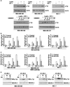
Evidence of crosstalk between leptin receptor (Ob-Rb) and IGF-IR. Leptin and IGF-I act synergistically to increase phosphorylation of Ob-Rb (A) and IGF-IR (B) in MDA-MB-468, MDA-MB-231, and MCF-7 breast cancer cells. Cells were serum starved for 24 h, followed by treatment with 100 ng/mL leptin (L), 100 ng/mL IGF-I alone (IGF-I), and the combination (L + IGF-I) for 30 min. C, untreated controls. Total protein was isolated and equal amounts of proteins were subjected to immunoprecipitation with specific antibodies for leptin receptor (A) or IGF-IR (B). Immunoprecipitates were resolved by SDS-PAGE and subjected to immunoblot analysis with a mouse monoclonal antibody against phospho-tyrosine. Immunoprecipitation with IgG was included as control (IgG). Leptin and IGF-I treatment increased phosphorylation of Ob-Rb whereas combined treatment induced further increase in Ob-Rb phosphorylation (A). Increased phosphorylation of IGF-IR was observed in cells treated with leptin and IGF-I alone, whereas combined treatment induced a synergistic increase in IGF-IR phosphorylation (B). Phosphorylated tyrosine bands shown in all cases correspond to the expected size band (Ob-Rb and IGF-IR). The membranes were reblotted with Ob-Rb and IGF-IR antibodies as control. Representative blots of multiple independent experiments. The histogram is the densitometric analysis of the Western blot signals normalized to total Ob-Rb or IGF-IR. #, P < 0.05, compared with untreated controls; *, P < 0.05, compared with untreated controls; **, P < 0.01, compared with leptin-treated cells. C, association of Ob-Rb and IGF-IR occurs in breast cancer cells. MDA-MB-468, MDA-MB-231, and MCF-7 cells were serum starved for 24 h, followed by no treatment (−) or combined treatment with 100 ng/mL leptin (L) and 100 ng/mL IGF-I for 30 min (+). Total protein was isolated and equal amounts of proteins were subjected to immunoprecipitation with specific antibodies for Ob-Rb. The immunoprecipitates were probed with anti–IGF-IR antibody. In a reverse experiment, equal amounts of proteins were immunoprecipitated with anti–IGF-IR antibody and immunoprecipitates were probed with anti–Ob-Rb antibody. Immunoprecipitation with IgG was included as control (IgG). Both Ob-Rb and IGF-IR were coimmunoprecipitated in the presence of IGF-I and leptin, indicating the association of Ob-Rb and IGF-IR.
Figure 3
Figure 3
Combined treatment with leptin and IGF-I leads to a synergistic increase in phosphorylation of both Akt and ERK in breast cancer cells. MDA-MB-468, MDA-MB-231, and MCF-7 cells were serum starved for 24 h, followed by treatment with 100 ng/mL leptin, 100 ng/mL IGF-I alone, and the combination for 30 min. Total protein was isolated from MDA-MB-468, MDA-MB-231, and MCF-7 cells and equal amounts of proteins were subjected to immunoblot analysis with specific antibodies against phospho-Akt (p-Akt) and Akt (A) or phospho-ERK (p-ERK) and ERK (B). Increased phosphorylation of Akt was observed in MDA-MB-468, MDA-MB-231, and MCF-7 cells treated with leptin or IGF-I, whereas combined treatment induced further increase in Akt phosphorylation. Leptin and IGF-I treatment increased phosphorylation of ERK, whereas combined treatment induced a synergistic increase in ERK phosphorylation. The membranes were reblotted with ERK, Akt, and anti-actin antibodies as control. Representative blots of multiple independent experiments. The representative histogram is the densitometric analysis of the Western blot signals showing fold increase in levels of phospho-Akt and phospho-ERK with respect to total Akt and ERK. #, P < 0.01, compared with untreated controls; *, P < 0.05, compared with untreated controls; **, P < 0.01, compared with leptin-treated cells.
Figure 4
Figure 4
Leptin and IGF-I require EGFR activation for their biological effects. A, leptin and IGF-I increase EGFR phosphorylation. MDA-MB-468, MDA-MB-231, and MCF-7 cells were serum starved for 24 h, followed by treatment with 100 ng/mL leptin, 100 ng/mL IGF-I alone, and the combination for 30 min. Total protein was isolated and equal amounts of proteins were subjected to immunoprecipitation with a specific antibody for EGFR. Immunoprecipitates were resolved by SDS-PAGE and subjected to immunoblot analysis with a mouse monoclonal antibody against phospho-tyrosine. Phosphorylated tyrosine bands shown in all cases correspond to the expected size band (EGFR). Leptin and IGF-I treatment induced a little increase in phosphorylation of EGFR, whereas combined treatment with leptin and IGF-I induced a synergistic increase in EGFR phosphorylation. The membranes were reblotted with anti-EGFR antibody as control. Leptin and IGF-I require MMP activation for EGFR transactivation. MDA-MB-468 and MDA-MB-231 cells were serum starved for 24 h, pretreated with 10 to 100 µmol/L of GM6001 or an equal volume of vehicle (DMSO) for 30 min, and stimulated with combined treatment of 100 ng/mL leptin and IGF-I for 30 min. Total cell lysates were isolated and immunoprecipitated with EGFR; immunoprecipitates were immunoblotted with anti-phosphotyrosine antibody. The membranes were reblotted with anti-EGFR antibody as control. Pretreatment with GM6001 inhibited leptin- and IGF-I–induced EGFR phosphorylation, indicating the requirement of MMP activation. B to D, EGFR inhibition inhibits leptin- and IGF-I–induced Akt and ERK activation and inhibits leptin-induced IRS-1 and IRS-2 activation. MDA-MB-468, MDA-MB-231, and MCF-7 cells were serum starved for 24 h, followed by treatment with 100 ng/mL leptin, 100 ng/mL IGF-I alone, and the combination. Cells were pretreated with 250 nmol/L AG1478 (EGFR inhibitor) for 45 min, followed by leptin and/or IGF-I treatment as indicated. Cell lysates were prepared; Akt and ERK were immunoprecipitated with anti-Akt and anti-ERK antibodies and subjected to Akt and ERK activity assay as detailed in Materials and Methods. Pretreatment with AG1478 inhibits Akt and ERK stimulation in response to combined treatment with leptin and IGF-I. Columns, mean of three independent experiments done in triplicates; bars, SE. *, P < 0.001, AG1478-pretreated and leptin + IGF-I–treated cells compared with leptin + IGF-I–treated cells. IRS-1 and IRS-2 were immunoprecipitated with anti–IRS-1 or anti–IRS-2 antibodies and subjected to immunoblot analysis with p-Tyr antibody. Phosphorylated tyrosine bands shown in all cases correspond to the expected size band (IRS-1 and IRS-2). EGFR inhibition blocks leptin-induced activation of IRS-1 and IRS-2 in MDA-MB-468 and MCF-7 cells.
Figure 5
Figure 5
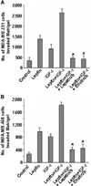
Lapatinib and erlotinib inhibit leptin- and IGF-I–induced increased invasion of breast cancer cells. MDA-MB-231 (A) and MDA-MB-468 (B) cells were cultured in Matrigel invasion chambers and serum starved for 24 h, followed by treatment with 100 ng/mL leptin, 100 ng/mL IGF-I alone, leptin + IGF-I, leptin + IGF-I + 2.5 µmol/L lapatinib, and leptin + IGF-I + 2.5 µmol/L erlotinib for 24 h. The number of cells that invaded through the Matrigel was counted in five different regions. The slides were blinded to remove counting bias. Columns, mean of three independent experiments done in triplicates; bars, SE. *, P < 0.001, compared with leptin + IGF-I–treated cells. #, P < 0.001, compared with leptin + IGF-I–treated cells.
Figure 6
Figure 6
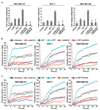
Inhibition of EGFR with lapatinib and erlotinib inhibits leptin- and IGF-I–induced increased migration of breast cancer cells. A, MDA-MB-231, MCF-7, and MDA-MB-468 breast cancer cells were grown to confluence on fibronectin-coated surface, serum starved for 24 h, and scratched with a pipette tip. The plates were photographed immediately following scratching. Culture media were replaced with media containing 100 ng/mL leptin, 100 ng/mL IGF-I alone, leptin + IGF-I, leptin + IGF-I + 2.5 µmol/L lapatinib, and leptin + IGF-I + 2.5 µmol/L erlotinib for 24 h. The plates were photographed at the identical location of the initial image at 24 h posttreatment. The fold change in migration was calculated. Columns, mean of three independent experiments done in triplicates; bars, SE. *, P < 0.005, compared with leptin + IGF-I–treated cells; #, P < 0.001, compared with leptin + IGF-I–treated cells. B and C, MDA-MB-231, MCF-7, and MDA-MB-468 cells were grown to confluence in ECIS plates, serum starved for 16 h, and subjected to an elevated voltage pulse of 40-kHz frequency at 3.5-V amplitude for 30 s to incite a wound. The cells were immediately treated with 100 ng/mL leptin, 100 ng/mL IGF-I alone, leptin + IGF-I, leptin + IGF-I + 2.5 µmol/L lapatinib, and leptin + IGF-I + 2.5 µmol/L erlotinib. The wound was then allowed to be healed by cells surrounding the small active electrode that did not undergo the elevated voltage pulse. Resistance was measured before and after the elevated voltage pulse application as described in Materials and Methods. The measurements were stopped 24 h after the creation of wound. All the experiments were done thrice in triplicates.

Similar articles

Cited by

References

    1. Newell B, Proust K, Dyball R, McManus P. Seeing obesity as a systems problem. N SW Public Health Bull. 2007;18:214–218. - PubMed
    1. Irigaray P, Newby JA, Lacomme S, Belpomme D. Overweight/obesity and cancer genesis: more than a biological link. Biomed Pharmacother. 2007;61:665–678. - PubMed
    1. Calle EE, Rodriguez C, Walker-Thurmond K, Thun MJ. Overweight, obesity, and mortality from cancer in a prospectively studied cohort of U S. adults. N Engl J Med. 2003;348:1625–1638. - PubMed
    1. Berclaz G, Li S, Price KN, et al. Body mass index as a prognostic feature in operable breast cancer: the International Breast Cancer Study Group experience. Ann Oncol. 2004;15:875–884. - PubMed
    1. Grodin JM, Siiteri PK, MacDonald PC. Source of estrogen production in postmenopausal women. J Clin Endocrinol Metab. 1973;36:207–214. - PubMed

Publication types

MeSH terms

  NODES
Association 3
INTERN 1
twitter 2