. 2009 Oct 15;18(20):3926-41.
doi: 10.1093/hmg/ddp335. Epub 2009 Jul 22.

Ahi1, whose human ortholog is mutated in Joubert syndrome, is required for Rab8a localization, ciliogenesis and vesicle trafficking

Affiliations

Ahi1, whose human ortholog is mutated in Joubert syndrome, is required for Rab8a localization, ciliogenesis and vesicle trafficking

Yi-Chun Hsiao et al. Hum Mol Genet. .

Abstract

The primary non-motile cilium, a membrane-ensheathed, microtubule-bundled organelle, extends from virtually all cells and is important for development. Normal functioning of the cilium requires proper axoneme assembly, membrane biogenesis and ciliary protein localization, in tight coordination with the intraflagellar transport system and vesicular trafficking. Disruptions at any level can induce severe alterations in cell function, giving rise to a myriad of human genetic diseases known as ciliopathies. Here we show that the Abelson helper integration site 1 (Ahi1) gene, whose human ortholog is mutated in Joubert syndrome, regulates cilium formation via its interaction with Rab8a, a small GTPase critical for polarized membrane trafficking. We find that the Ahi1 protein localizes to a single centriole, the mother centriole, which becomes the basal body of the primary cilium. In order to determine whether Ahi1 functions in ciliogenesis, loss of function analysis of Ahi1 was performed in cell culture models of ciliogenesis. Knockdown of Ahi1 expression by shRNAi in cells or _targeted deletion of Ahi1 (Ahi1 knockout mouse) leads to impairments in ciliogenesis. In Ahi1-knockdown cells, Rab8a is destabilized and does not properly localize to the basal body. Since Rab8a is implicated in vesicular trafficking, we next examined this process in Ahi1-knockdown cells. Defects in the trafficking of endocytic vesicles from the plasma membrane to the Golgi and back to the plasma membrane were observed in Ahi1-knockdown cells. Overall, our data indicate that the distribution and functioning of Rab8a is regulated by Ahi1, not only affecting cilium formation, but also vesicle transport.


Figures

Figure 1.
Figure 1.
Ahi1 localizes to the mother centriole. (A) IMCD3 cells were immunostained for the centrosomal marker γ-tubulin (green), and for endogenous Ahi1 (red). Arrowheads indicate the co-localization of γ-tubulin and Ahi1 to only one of the paired centrioles. (B) Ahi1-EGFP (green) transfected IMCD3 cells were stained with γ-tubulin (violet) and with ninein (red), a marker for the mother centriole. Arrowheads indicate the co-localization of Ahi1, γ-tubulin and ninein in the mother centriole. (C) Ahi1-EGFP (green) transfected IMCD3 cells were stained with γ-tubulin (violet) and with Odf2 (red), a mother centriole marker. Arrowheads indicate the co-localization of Ahi1, γ-tubulin, and Odf2 only in the mother centriole. DNA is visualized with Hoechst 33258 (blue). Scale bars = 5 µm.
Figure 2.
Figure 2.
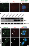
Ahi1 localizes to the basal body of the primary non-motile cilium and its expression is lowered in Ahi1-knockdown IMCD3 cells. (A) Serum-deprived IMCD3 cells have robust cilium formation (one/cell). Ciliated IMCD3 cells were stained with acetylated α-tubulin (green), a ciliary axoneme marker, and for Ahi1 (red). Arrowheads indicate the localization of Ahi1 to the base of a primary cilium. DNA is visualized with Hoechst 33258 (blue). For evaluation of knockdown efficiency of Ahi1, IMCD3 cells stably expressing shRNAi against Ahi1 were established and analysed for Ahi1 protein expression by (B) western blotting and (C) immunostaining. (B) Protein levels of Ahi1 in four independent Ahi1-knockdown IMCD3 stable clones, two control shRNAi scramble stable clones and wild-type cells were analysed by western blotting. Ahi1 (130 kDa, lower band) can be detected in scramble control and wild-type cells, but not in cells expressing a shRNAi against Ahi1. The top band is considered to be a non-specific band (34). (C) Immunostaining for γ-tubulin (green), for Ahi1 (red) and for DNA (Hoechst 33258 (blue)) was performed in wild-type IMCD3 cells and in Ahi1-knockdown cell line 1 (shAhi1-1). Scale bars = 5 µm.
Figure 3.
Figure 3.
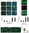
Ahi1 is required for the normal formation of primary non-motile cilium. (A) Control cells (scramble shRNAi), Ahi1-knockdown cell line 1 (shAhi1-1) and Ahi1-knockdown cell line 2 (shAhi1-2) (both shAhi1-1 and shAhi1-2 were RNAi sequence 1) were cultured under serum-deprived conditions, for initiation of robust cilium formation. Cells were immunostained for acetylated α-tubulin (green; marker of the primary cilium) and for Ahi1 (red). Note that the acetylated α-tubulin staining was actually acquired on a Cy5 channel and has been pseudocoloured green (the true FITC channel visualizes the shAhi1 construct that is GFP tagged and is not shown in this image). Higher magnification images of the boxed regions are shown in the right column. The histogram shows the percentage of cells having a primary cilium based on counts of cells with acetylated α-tubulin-positive cilia, for Ahi1-knockdown cells (shAhi1-1, -2 and -3), control-scramble cells and wild-type cells. Ten randomly acquired fields (a minimum of 500 cells were counted for each clone) were analysed. The error bars represent the standard error of the mean. Asterisks indicate significance from scramble and wild-type cells (P < 0.0001) using Chi-square analyses. (B) Ahi1-knockdown cells that had been transiently transfected with RNAi sequence 3 were cultured under serum-deprived conditions, for initiation of robust cilium formation. Cells were immunostained for acetylated α-tubulin (red) with the green channel highlighting cells that had been transfected with the shRNAi construct. The histogram shows the percentage of cells having a primary cilium based on counts of cells with acetylated α-tubulin-positive cilia, for Ahi1-knockdown cells that were transfected with shRNAi constructs against Ahi1 (RNAi sequences 2 and 3), a scramble shRNAi construct or no construct (wild-type cells). Three randomly acquired fields (a minimum of 200 cells were counted for each clone) were analysed. The error bars represent the standard error of the mean. Asterisks indicate significance from scramble and wild-type cells (P < 0.0001) using Chi-square analyses. (C) Mouse embryonic fibroblasts (MEFs) cultured from wild-type (Ahi1+/+) and Ahi1 knockout (Ahi1−/−) mouse embryos. MEFs were immunostained for acetylated α-tubulin (green; marker of the primary cilium). The histogram shows the percentage of Ahi1+/+ and Ahi1−/− MEFs having a primary cilium based on counts of cells with an acetylated α-tubulin-positive cilium. Three randomly acquired fields (a minimum of 100 cells were counted for each field) were analysed for each mouse. The error bars represent the standard error of the mean. Asterisk indicates significance from wild-type (Ahi1+/+) MEFs (P < 0.0001) using Chi-square analysis. Scale bars = 5 µm.
Figure 4.
Figure 4.
Ahi1 is required for the normal formation of primary cilia. (A) Low magnification scanning electron micrographic images of wild-type and Ahi1-knockdown (RNAi sequence 1) IMCD3 cells under conditions optimized for ciliogenesis. Wild-type cells (top) each clearly have an elongated and pronounced primary cilium (black arrowheads) on the cell surface. However, Ahi1-knockdown cells (bottom) have a striking lack of primary cilia. (B) Higher magnification scanning electron micrographs of the primary cilium (black arrowheads) and microvilli (black arrows) in wild-type (left) and Ahi1-knockdown IMCD3 (right) cells. Primary cilia were very difficult to find in Ahi1-knockdown IMCD3 cells; when putatively identified, they tended to be short and stumpy (right). (C) Apical and basal actin filament staining by phalloidin in wild-type, scramble and Ahi1-knockdown cells showing a disorganization and decrease in actin filaments in Ahi1-knockdown cells. Scale bars = 2 µm in (A), 0.3 µm in (B) and 5 µm in (C).
Figure 5.
Figure 5.
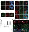
Ahi1 is involved in ciliogenesis through its interactions with the small GTPase, Rab8a. (A, top) Co-immunoprecipitation of Ahi1-EGFP with HA-Rab8a in lysates from HEK293 cells transfected with Ahi1-EGFP and HA-Rab8a, but not in cells transfected with HA-Rab8a alone or Ahi1-EGFP alone. Resin only controls had no labelling (data not shown). (A, bottom) Co-localization of both Ahi1 (green) and Rab8a (red) at the basal body in wild-type cells (denoted by white arrowheads). (B) The protein level of endogenous Rab8a from Ahi1-knockdown (shAhi1-1 and -2) and from scramble shRNAi control cells was analysed by western blotting (top) and graphically displayed (bottom). The level of β-tubulin represents the loading control. The error bars represent the standard error of the mean. (C) Wild-type (top panel) and Ahi1-knockdown IMCD3 cells (lower panel) were stained with antibodies against Rab8a (green) and Arl13b (red; marker for the basal body and the primary cilium). Right panel represents the merged images of Rab8a, Arl13b and DNA (blue) staining. White arrowheads point to the basal body. With this Rab8a antibody, localization is only found at the basal body and does not show any apparent localization to the cilium. Scale bars =5 µm in (A and C).
Figure 6.
Figure 6.
Overexpression of HA-Rab8a in Ahi1-knockdown cells results in an inability to rescue ciliogenesis and an inability for HA-Rab8a to localize to the basal body of the primary cilium. (A) Wild-type IMCD3 cells, cultured under non-cilia forming conditions (top) or under serum-deprived conditions so as to promote cilium formation (bottom), and transfected with an HA-Rab8a expression plasmid were immunostained for Ahi1 (green) and for HA (red). Right panel represents the merged images of Ahi1, HA-Rab8a and DNA (blue) staining. White arrowheads point to the basal body. The bottom panel represents HA-Rab8a localization to the Golgi complex (using the Golgi marker Golph4 (green)). (B) Wild-type, scramble and Ahi1-knockdown cells transfected with HA-Rab8a were immunostained with acetylated α-tubulin (green) for primary cilia and HA for Rab8a (red) distribution. Arrowheads indicate the co-localization of HA-Rab8a and acetylated α-tubulin staining. Right panel represents the merged images of HA-Rab8a, acetylated α-tubulin and DNA (blue) staining. (C) Quantification of cilium formation in HA-Rab8a expressing wild-type, scramble and Ahi1-knockdown cells. Cells were co-stained with HA and Arl13b. Fifty HA-positive cells were counted from each slide and the number of primary cilium was determined. Data were collected from three independent experiments. The error bars represent the standard error of the mean. Asterisks indicate significance from scramble and wild-type cells (P < 0.0001) using Chi-square analyses. (D) Wild-type and scramble cells [grown to sub-confluence (resulting in few cilia); left two panels] and Ahi1-knockdown cells (grown under cilia optimized conditions; right two panels) transfected with HA-Rab8a were stained with antibodies against HA (red, for HA-Rab8a) and Arl13b (green, for the basal body). Arrowheads point to Arl13b labelled basal bodies. The lower panel represents the merged images of HA-Rab8a, Arl13b and DNA (blue) staining. This experiment was designed to capture wild-type and scramble cells at a time when few cells had cilia, so as to easily observe the association of HA-Rab8a in the basal body (before the formation of cilia where HA-Rab8a would enter the cilium making it difficult to differentiate basal body from the primary cilium). Scale bars =5 µm in (A, B and D).
Figure 7.
Figure 7.
Membrane trafficking is altered in Ahi1-knockdown cells. (A) Cholera toxin endocytic assay. Wild-type (top row) and Ahi1-knockdown cells (bottom row) were incubated with Alexa Fluor 546-conjugated Cholera toxin subunit B (CTxB; red) for 30 min after cold incubation. GM130 (green) staining delineates the Golgi. The plot shows the percentages of cells showing co-localization of CTxB and GM130 staining. In each case, >200 cells were scored in three independent tests [asterisk indicates a significant effect (P < 0.0001) using Chi-square analysis in comparing Ahi1-knockdown cells with scramble and wild-type cells]. (B) Transferrin endocytic assay. Wild-type (right) and Ahi1-knockdown (left) cells were incubated with Alexa Fluor 546-conjugated transferrin (Tf; red) for 10, 30 or 60 min after cold incubation. DNA is visualized with Hoechst 33258 (blue). Note that following a 60 min pulse, Tf is found throughout the cell membrane in wild-type cells, but in Ahi1-knockdown cells, Tf has accumulated in the perinuclear region of the cell (and not the membrane). The plot shows the percentages of cells with Tf localized to the perinuclear region of the cell after a 60 min pulse. In each case, >200 cells were scored with the asterisk indicating a significant effect (P < 0.0001) using Chi-square analysis in comparing Ahi1-knockdown cells with scramble and wild-type cells. (C) The Golgi structure and position in Ahi1-knockdown cells. Ahi1-knockdown (shAhi1-1 and -2), control scramble and wild-type IMCD3 cells cultured under serum-deprived conditions were stained with the cis-Golgi compartment marker, GM130 (red), to permit examination of the Golgi structure. Similar results were obtained with the trans-Golgi compartment marker, Golph4 (data not shown). Scale bar=5 µm in (A–C).

Similar articles

Cited by

References

    1. Satir P., Christensen S.T. Overview of structure and function of mammalian cilia. Annu. Rev. Physiol. 2007;69:377–400. - PubMed
    1. Murcia N.S., Richards W.G., Yoder B.K., Mucenski M.L., Dunlap J.R., Woychik R.P. The Oak Ridge Polycystic Kidney (orpk) disease gene is required for left-right axis determination. Development. 2000;127:2347–2355. - PubMed
    1. Caspary T., Larkins C.E., Anderson K.V. The graded response to Sonic Hedgehog depends on cilia architecture. Dev. Cell. 2007;12:767–778. - PubMed
    1. Gerdes J.M., Liu Y., Zaghloul N.A., Leitch C.C., Lawson S.S., Kato M., Beachy P.A., Beales P.L., DeMartino G.N., Fisher S., et al. Disruption of the basal body compromises proteasomal function and perturbs intracellular Wnt response. Nat. Genet. 2007;39:1350–1360. - PubMed
    1. Huangfu D., Liu A., Rakeman A.S., Murcia N.S., Niswander L., Anderson K.V. Hedgehog signalling in the mouse requires intraflagellar transport proteins. Nature. 2003;426:83–87. - PubMed

Publication types

MeSH terms

Substances

  NODES
Association 1
Note 3
twitter 2