. 2010 Oct;120(10):3702-12.
doi: 10.1172/JCI43343. Epub 2010 Sep 13.

Human C3 mutation reveals a mechanism of dense deposit disease pathogenesis and provides insights into complement activation and regulation

Affiliations

Human C3 mutation reveals a mechanism of dense deposit disease pathogenesis and provides insights into complement activation and regulation

Rubén Martínez-Barricarte et al. J Clin Invest. 2010 Oct.

Abstract

Dense deposit disease (DDD) is a severe renal disease characterized by accumulation of electron-dense material in the mesangium and glomerular basement membrane. Previously, DDD has been associated with deficiency of factor H (fH), a plasma regulator of the alternative pathway (AP) of complement activation, and studies in animal models have linked pathogenesis to the massive complement factor 3 (C3) activation caused by this deficiency. Here, we identified a unique DDD pedigree that associates disease with a mutation in the C3 gene. Mutant C(3923ΔDG), which lacks 2 amino acids, could not be cleaved to C3b by the AP C3-convertase and was therefore the predominant circulating C3 protein in the patients. However, upon activation to C3b by proteases, or to C3(H₂O) by spontaneous thioester hydrolysis, C(3923ΔDG) generated an active AP C3-convertase that was regulated normally by decay accelerating factor (DAF) but was resistant to decay by fH. Moreover, activated C(3b923ΔDG) and C3(H₂O)(923ΔDG) were resistant to proteolysis by factor I (fI) in the presence of fH, but were efficiently inactivated in the presence of membrane cofactor protein (MCP). These characteristics cause a fluid phase-restricted AP dysregulation in the patients that continuously activated and consumed C3 produced by the normal C3 allele. These findings expose structural requirements in C3 that are critical for recognition of the substrate C3 by the AP C3-convertase and for the regulatory activities of fH, DAF, and MCP, all of which have implications for therapeutic developments.


Figures

Figure 1
Figure 1. Histology, immunofluorescence, and EM findings.
The first kidney biopsy in GN28 was performed in 1985. Although there was considerable variation in glomerular changes, there was remarkable similarity in the light, immunofluorescence, and ultrastructural findings in the original kidney biopsy and 2 allograft biopsies of GN28 and the kidney biopsies from III-1 and III-2. The characteristic histological lesion consisted of segmental mesangial hypercellularity with thickened, eosinophil-rich segments of basement membrane (A and B, arrows). The affected glomerular segments were PAS positive and reacted to trichrome stain (D, arrow). The affected tufts showed hypercellularity, leukocyte infiltration, and endothelial swelling. The mesangium showed variable expansion, matrix accumulation, and a lobular pattern (C and D, arrows). The main immunofluorescence findings were prominent and diffuse C3 deposits, granular and nodular in some glomerular areas (G and J). Mild deposits of C1q, IgA, and IgM were also associated with these deposits (not shown). All biopsies showed similar ultrastructural alterations consisting of a ribbon-like, osmiophilic deposit present in the GBM (E and I, red arrows). These deposits occasionally showed signs of dissolution with translucent areas (F, red arrows). The mesangial areas showed increased mesangial matrix with electron-dense deposits (H and I, yellow arrows). Original magnification: ×400 (A, B, G, and J); ×500 (C and D); ×2,200 (E and F); ×5,500 (H); ×7,800 (I). Patient number is indicated within each panel.
Figure 2
Figure 2. Mutation in the C3 gene in a multiaffected DDD pedigree.
Pedigree of index case GN28 is illustrated. Individuals are identified by numbers within each generation. Affected individuals are indicated by black symbols. The twin brother III-1 (gray symbol) has not developed ESRD, but shows early signs of disease. II-5 is a sister of GN28 whose death at 13 years of age was attributed to glomerulonephritis. C3 alleles carried by the individuals are shown. The chromatogram corresponding to the DNA sequence surrounding the mutated nucleotides in C3 is shown for GN28 and a control sample. The corresponding amino acid sequences for the WT and mutated alleles are indicated. Amino acid numbering refers to the translation start site (Met +1), and the nucleotide nomenclature refers to nucleotide A in the ATG translation initiation codon, according to Human Genome Variation Society recommendations for description of sequence variants.
Figure 3
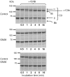
Figure 3. Total C3 from GN28 is only partially cleaved to C3b in the presence of fB and fD.
Coomassie-stained gels correspond to the SDS-PAGE analyses of C3 purified from GN28 and a normal control after incubation with fB and fD. The experiment was repeated twice with identical results. Top: C3 purified from normal individuals was rapidly and completely activated to C3b (determined by cleavage of the C3 α chain) in the presence of fB and fD. This activation correlated with consumption of fB and the appearance of the Bb fragment, indicating formation of the AP C3-convertase. Middle: Same experiment with C3 purified from GN28. Despite formation of the AP C3-convertase (demonstrated by consumption of fB and generation of the Bb fragment), only a small proportion of the C3 was activated to C3b. This suggests that C3 purified from the GN28 plasma contains 2 different C3 forms (WT and mutant protein), and that only C3WT is cleaved to C3b. Bottom: To rule out the presence of inhibitors in the C3 preparation from the GN28 plasma, a mixing experiment (equivalent amounts of control and GN28 C3) showed that addition of fB and fD caused activation of 50% of the C3, likely C3WT.
Figure 4
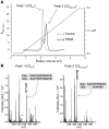
Figure 4. Purification of the mutant allele C3923ΔDG.
(A) C3923ΔDG and C3WT were separated as described in Methods. The elution profiles of this chromatographic separation of C3 prepared from control plasma (dotted line) and from GN28 plasma (solid line) are indicated. (B) The identity of the C3 variants was determined by mass spectrometry, as described in Methods. The minor C3WT peak showed some contamination of the major C3923ΔDG mutant protein (asterisk). Exp, expected; Obs, observed.
Figure 5
Figure 5. Resistance of purified C3923ΔDG to cleavage by the AP C3-convertase.
(A) C3923ΔDG and C3WT were purified to homogeneity and tested for their capacity to be cleaved to C3b in the presence of fB and fD; only the α chain of C3WT was cleaved. Of note, C3923ΔDG consumed fB, illustrating formation of a AP C3-convertase. This experiment was repeated twice. Lanes were run on the same gel but were noncontiguous (white lines). (B) C3WT (1,000 RU) was immobilized via amine coupling to a CM5 Biacore chip. Convertase was formed by flowing fB (2.6 μM) and fD (43 nM) in the presence of Mg2+. At the indicated time (arrows), C3WT (gray line) or C3923ΔDG (black line) was flowed over the surface. Remaining convertase was decayed using sDAF, and deposition of nascent C3b was measured. Resp. diff., response difference.
Figure 6
Figure 6. AP C3-convertase formation by C3923ΔDG and C3WT.
(A) Hydrolyzed C3WT (1,224 RU) or C3923ΔDG (1,067 RU) was thiol-coupled to a CM5 Biacore chip. Convertase formation was analyzed by flowing fB (270 to 17 nM) over the surface in the presence of fD (43 nM). Kinetics were analyzed according to the Langmuir 1:1 binding model. Convertase formation by mutant C3923ΔDG and C3(H2O)WT, measured as KD, was comparable. (B) Hydrolyzed C3WT (1,640 RU) or C3923ΔDG (2,000 RU) was thiol-coupled to a CM5 Biacore chip. fB and fD were flowed over the surfaces to form either C3(H2O)WTBb (black line) or C3(H2O)923ΔDGBb (gray line). C3WT was injected over the surface, where it was cleaved to nascent C3b and deposited on the surface via the thioester group. Remaining convertase after deposition was decayed using sDAF, and bound C3b was measured as change from baseline.
Figure 7
Figure 7. Reduced affinity of fH for hydrolyzed C3923ΔDG impairs both decay of the mutant C3-convertase and fI-mediated inactivation of hydrolyzed C3923ΔDG.
(A) Hydrolyzed C3WT (1,224 RU) or C3923ΔDG (1,067 RU) were thiol-coupled to a CM5 Biacore chip. The affinity for native fH was analyzed by flowing fH (1 μM to 8 nM) over the surface and determined by steady-state analysis. The affinity of fH for C3(H2O)923ΔDG was reduced 2-fold compared with C3WT. Values are mean ± SD of 3 determinations. (B) Convertase was formed on each hydrolyzed C3 surface by flowing fB and fD. After a period of natural decay, fH (0.9 μM) was injected for 60 seconds. Convertase formed by C3(H2O)WT (gray line) was efficiently decayed by fH, whereas the C3(H2O)923ΔDG convertase (black line) was inefficiently decayed. Binding (RU) of fH to the surface in the absence of the convertase was subtracted; curves illustrate decay of Bb. (C) In contrast, mutant convertase was efficiently decayed by DAF (0.4 μM). (D) Hydrolyzed C3WT or C3923ΔDG was thiol-coupled to a CM5 Biacore chip. Initial formation of convertase on each surface was assessed by flowing fB and fD (black line). After complete decay of the convertase, fH (0.33 μM) and fI (0.11 μM) were flowed across the surface for 5 minutes at 5 μl/min. Convertase was then formed again (gray lines) using identical conditions. Enzyme formation by C3(H2O)WT convertase was reduced 50% by fI/fH treatment, whereas enzyme formation from C3(H2O)923ΔDG convertase was hardly affected.
Figure 8
Figure 8. C3b923ΔDG activates C3 cleavage in normal human serum.
The capacity of C3b923ΔDG and C3bWT to activate C3 in normal serum was tested by incubating 180 ng of each C3b with a 1:20 dilution of normal serum at 37°C in AP buffer (5 mM Veronal; 150 mM NaCl; 7 mM MgCl2; 10 mM EGTA, pH 7.4). Samples (5 μl) were taken at the indicated times and loaded into a 10% SDS-PAGE. C3 activation was measured by the appearance of the 43-kDa fragment of C3 α′ chain, as detected by Western blot. The α′ 43 band of iC3b is shown for comparison.
Figure 9
Figure 9. C3b923ΔDG is inactivated by fI and MCP, but not by fH and fI.
Activated C3bWT or C3b923ΔDG was incubated with fI and either fH (A) or sMCP (B). Cleavage of the α′ chain was indicated by generation of the α65 and α43 products. The experiment was repeated twice with identical results.
Figure 10
Figure 10. C3(H2O)923ΔDG is resistant to inactivation by fI in the presence of fH, but not in the presence of sMCP.
Hydrolyzed C3923ΔDG or C3WT were incubated with fI and either fH (A) or sMCP (B). Cleavage of the α′ chain was indicated by generation of the α74 and α43 products. The experiment was repeated twice.

Similar articles

Cited by

References

    1. Amara U, et al. Interaction between the coagulation and complement system. Adv Exp Med Biol. 2008;632:71–79. - PMC - PubMed
    1. Lachmann PJ. The amplification loop of the complement pathways. Adv Immunol. 2009;104:115–149. - PubMed
    1. Walport MJ. Complement- second of two parts. N Engl J Med. 2001;344(15):1140–1144. doi: 10.1056/NEJM200104123441506. - DOI - PubMed
    1. Walport MJ. Complement- first of two parts. N Engl J Med. 2001;344(14):1058–1066. doi: 10.1056/NEJM200104053441406. - DOI - PubMed
    1. Smith RJH, et al. New approaches to the treatment of dense deposit disease. J Am Soc Nephrol. 2007;18(9):2447–2456. doi: 10.1681/ASN.2007030356. - DOI - PMC - PubMed

Publication types

  NODES
Note 1
twitter 2