Critical roles of macrophages in the formation of intracranial aneurysm
- PMID: 21106959
- PMCID: PMC3021554
- DOI: 10.1161/STROKEAHA.110.590976
Critical roles of macrophages in the formation of intracranial aneurysm
Abstract
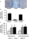
Background and purpose: abnormal vascular remodeling triggered by hemodynamic stresses and inflammation is believed to be a key process in the pathophysiology of intracranial aneurysms. Numerous studies have shown infiltration of inflammatory cells, especially macrophages, into intracranial aneurysmal walls in humans. Using a mouse model of intracranial aneurysms, we tested whether macrophages play critical roles in the formation of intracranial aneurysms.
Methods: intracranial aneurysms were induced in adult male mice using a combination of a single injection of elastase into the cerebrospinal fluid and angiotensin II-induced hypertension. Aneurysm formation was assessed 3 weeks later. Roles of macrophages were assessed using clodronate liposome-induced macrophage depletion. In addition, the incidence of aneurysms was assessed in mice lacking monocyte chemotactic protein-1 (CCL2) and mice lacking matrix metalloproteinase-12 (macrophage elastase).
Results: intracranial aneurysms in this model showed leukocyte infiltration into the aneurysmal wall, the majority of the leukocytes being macrophages. Mice with macrophage depletion had a significantly reduced incidence of aneurysms compared with control mice (1 of 10 versus 6 of 10; P<0.05). Similarly, there was a reduced incidence of aneurysms in mice lacking monocyte chemotactic protein-1 compared with the incidence of aneurysms in wild-type mice (2 of 10 versus 14 of 20, P<0.05). There was no difference in the incidence of aneurysms between mice lacking matrix metalloproteinase-12 and wild-type mice.
Conclusions: these data suggest critical roles of macrophages and proper macrophage functions in the formation of intracranial aneurysms in this model.
Conflict of interest statement
None
Figures


Similar articles
-
Elastase-induced intracranial aneurysms in hypertensive mice.Hypertension. 2009 Dec;54(6):1337-44. doi: 10.1161/HYPERTENSIONAHA.109.138297. Epub 2009 Nov 2. Hypertension. 2009. PMID: 19884566 Free PMC article.
-
Myeloperoxidase is increased in human cerebral aneurysms and increases formation and rupture of cerebral aneurysms in mice.Stroke. 2015 Jun;46(6):1651-6. doi: 10.1161/STROKEAHA.114.008589. Epub 2015 Apr 28. Stroke. 2015. PMID: 25922506 Free PMC article. Clinical Trial.
-
Prostaglandin E2-EP2-NF-κB signaling in macrophages as a potential therapeutic _target for intracranial aneurysms.Sci Signal. 2017 Feb 7;10(465):eaah6037. doi: 10.1126/scisignal.aah6037. Sci Signal. 2017. PMID: 28174280
-
Flow-induced, inflammation-mediated arterial wall remodeling in the formation and progression of intracranial aneurysms.Neurosurg Focus. 2019 Jul 1;47(1):E21. doi: 10.3171/2019.5.FOCUS19234. Neurosurg Focus. 2019. PMID: 31261126 Free PMC article. Review.
-
[Roles of macrophages in formation and progression of intracranial aneurysms].Zhejiang Da Xue Xue Bao Yi Xue Ban. 2019 Apr 25;48(2):204-213. doi: 10.3785/j.issn.1008-9292.2019.04.13. Zhejiang Da Xue Xue Bao Yi Xue Ban. 2019. PMID: 31309760 Free PMC article. Review. Chinese.
Cited by
-
Aspirin--another type of headache it prevents.J Am Heart Assoc. 2013 Mar 8;2(2):e000111. doi: 10.1161/JAHA.113.000111. J Am Heart Assoc. 2013. PMID: 23537809 Free PMC article. No abstract available.
-
TNF-α induces phenotypic modulation in cerebral vascular smooth muscle cells: implications for cerebral aneurysm pathology.J Cereb Blood Flow Metab. 2013 Oct;33(10):1564-73. doi: 10.1038/jcbfm.2013.109. Epub 2013 Jul 17. J Cereb Blood Flow Metab. 2013. PMID: 23860374 Free PMC article.
-
Integrated Transcriptional Profiling Analysis and Immune-Related Risk Model Construction for Intracranial Aneurysm Rupture.Front Neurosci. 2021 Apr 1;15:613329. doi: 10.3389/fnins.2021.613329. eCollection 2021. Front Neurosci. 2021. PMID: 33867914 Free PMC article.
-
Dysregulated Expression Profiles of MicroRNAs of Experimentally Induced Cerebral Aneurysms in Rats.J Korean Neurosurg Soc. 2013 Feb;53(2):72-6. doi: 10.3340/jkns.2013.53.2.72. Epub 2013 Feb 28. J Korean Neurosurg Soc. 2013. PMID: 23560169 Free PMC article.
-
Sustained expression of MCP-1 by low wall shear stress loading concomitant with turbulent flow on endothelial cells of intracranial aneurysm.Acta Neuropathol Commun. 2016 May 9;4(1):48. doi: 10.1186/s40478-016-0318-3. Acta Neuropathol Commun. 2016. PMID: 27160403 Free PMC article.
References
-
- Chyatte D, Bruno G, Desai S, Todor DR. Inflammation and intracranial aneurysms. Neurosurgery. 1999;45:1137–1146. - PubMed
-
- Kataoka K, Taneda M, Asai T, Kinoshita A, Ito M, Kuroda R. Structural fragility and inflammatory response of ruptured cerebral aneurysms. A comparative study between ruptured and unruptured cerebral aneurysms. Stroke. 1999;30:1396–1401. - PubMed
-
- Shi C, Awad IA, Jafari N, Lin S, Du P, Hage ZA, Shenkar R, Getch CC, Bredel M, Batjer HH, Bendok BR. Genomics of human intracranial aneurysm wall. Stroke. 2009;40:1252–1261. - PubMed
-
- Inoue K, Mineharu Y, Inoue S, Yamada S, Matsuda F, Nozaki K, Takenaka K, Hashimoto N, Koizumi A. Search on chromosome 17 centromere reveals tnfrsf13b as a susceptibility gene for intracranial aneurysm: A preliminary study. Circulation. 2006;113:2002–2010. - PubMed
Publication types
MeSH terms
Substances
Grants and funding
LinkOut - more resources
Full Text Sources
Medical
Research Materials

