. 2011 Apr 20;30(8):1536-48.
doi: 10.1038/emboj.2011.69. Epub 2011 Mar 11.

p38MAPK is a novel DNA damage response-independent regulator of the senescence-associated secretory phenotype

Affiliations

p38MAPK is a novel DNA damage response-independent regulator of the senescence-associated secretory phenotype

Adam Freund et al. EMBO J. .

Abstract

Cellular senescence suppresses cancer by forcing potentially oncogenic cells into a permanent cell cycle arrest. Senescent cells also secrete growth factors, proteases, and inflammatory cytokines, termed the senescence-associated secretory phenotype (SASP). Much is known about pathways that regulate the senescence growth arrest, but far less is known about pathways that regulate the SASP. We previously showed that DNA damage response (DDR) signalling is essential, but not sufficient, for the SASP, which is restrained by p53. Here, we delineate another crucial SASP regulatory pathway and its relationship to the DDR and p53. We show that diverse senescence-inducing stimuli activate the stress-inducible kinase p38MAPK in normal human fibroblasts. p38MAPK inhibition markedly reduced the secretion of most SASP factors, constitutive p38MAPK activation was sufficient to induce an SASP, and p53 restrained p38MAPK activation. Further, p38MAPK regulated the SASP independently of the canonical DDR. Mechanistically, p38MAPK induced the SASP largely by increasing NF-κB transcriptional activity. These findings assign p38MAPK a novel role in SASP regulation--one that is necessary, sufficient, and independent of previously described pathways.


Conflict of interest statement

The authors declare that they have no conflict of interest.

Figures

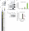
Figure 1
Figure 1
p38MAPK is activated with slow kinetics during genotoxic stress-induced senescence and is required for the SASP. (A) p38MAPK phosphorylation increases during DNA damage-induced senescence. Cells were irradiated and whole cell lysates collected at the indicated days thereafter. +: p38MAPK was continuously inhibited by 10 μM SB203580 (SB) beginning 48 h before irradiation. Left: western blot analysis of indicated proteins. Right: western blot quantitation, normalized to PRE levels. p38-P, phosphorylated p38MAPK; Hsp27-P, phosphorylated heat shock protein 27. (B) p38MAPK inhibition decreases intracellular IL-6 in SEN(XRA) cells. PRE and SEN(XRA) cells immunostained for IL-6. +SB: p38MAPK was inhibited by SB023580 (SB) for 48 h before fixation. (C) p38MAPK inhibition decreases IL-6, IL-8, and GM-CSF secreted by SEN(XRA) cells. Conditioned media (CM) were collected from PRE and SEN(XRA) cells and analysed by ELISA. +SB: p38MAPK was inhibited by SB023580 (SB) for 48 h before CM collection. (D) p38MAPKα depletion decreases secreted IL-6. Cells were infected with lentivirus expressing either of two shRNAs against p38MAPKα (shp38α) or a control shRNA (shGFP) and selected. Cells were irradiated and allowed to senesce (SEN(XRA)). CM were analysed by ELISA. (E) p38MAPK inhibition suppresses the SEN(XRA) SASP. CM from PRE and SEN(XRA) cells, with (SB) or without p38MAPK inhibition, were analysed by antibody arrays. Shown are factors for which the SEN(XRA) level was significantly increased (P<0.05) over PRE. For each protein, signals from all conditions were averaged to generate the baseline. Signals above baseline are yellow; signals below baseline are blue. The heat map key shows log2-fold changes from baseline. The hierarchical clustering relationship between sample profiles is shown graphically as a dendrogram (left). *: Factors significantly decreased by p38MAPK inhibition (P<0.05). (F) p38MAPK depletion decreases the ability of senescent cells to stimulate cancer cell invasiveness. CM from cells described in (D) were analysed for the ability to stimulate invasiveness of MDA-MB-231 breast cancer cells in a Boyden chamber invasion assay.
Figure 2
Figure 2
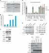
p38MAPK drives the amplified SASPs induced by RAS or p53 inactivation. (A) p38MAPK is phosphorylated during RASV12-induced senescence. PRE cells were infected with lentivirus expressing oncogenic RASV12, selected, and allowed to senesce (SEN(RAS)) for 10 days. Whole cell lysates were then collected and analysed by western blot. Presenescent controls (PRE) were infected with insertless vector. (B) p38MAPK inhibition suppresses the SEN(RAS) SASP. Cells were infected as described in (A). CM from PRE and SEN(RAS) cells were analysed by antibody arrays. +SB: p38MAPK was inhibited by SB203580 for 48 h before CM collection. Shown are the top 40 factors for which the SEN(RAS) level was significantly increased over PRE. PRE and SEN(RAS) values were averaged to generate the baseline. Heat map and dendrogram were generated as in Figure 1E. *: Factors significantly decreased by p38MAPK inhibition. (C) Amplified p38MAPK phosphorylation in SEN(RAS) cells and SEN(XRA) cells lacking functional p53 (SEN(XRA)+GSE). Cells were infected with lentivirus expressing GSE22 (GSE) or an insertless vector, selected, then irradiated (XRA) or infected with lentivirus expressing oncogenic RASV12 (RAS) and allowed to senesce. Whole cell lysates were analysed by western blot. (D) SEN(RAS) and SEN(XRA) cells lacking functional p53 secrete amplified IL-6 levels. Cells were treated as in (C), then CM were collected and analysed by ELISA. (E) p53 inactivation accelerates p38MAPK phosphorylation after XRA. Cells were infected with lentivirus lacking insert (Vector) or expressing GSE22 (GSE), selected, and irradiated. Whole cell lysates were collected at specified time points and analysed by western blot. (F) GSE-amplified levels of IL-6, IL-8, and GM-CSF are p38MAPK dependent. Cells were infected as in (E) and irradiated. CM were collected 3 days later and analysed by ELISA. SB: p38MAPK was inhibited by SB203580 for 48 h before CM collection.
Figure 3
Figure 3
Constitutive p38MAPK activation is sufficient to induce an SASP. (A) PRE cells were infected with lentivirus expressing a constitutively active MAP kinase kinase 6 mutant (MKK6EE). Whole cell lysates were collected 8 days after infection and analysed by western blot. Presenescent controls (PRE) were infected with insertless vector. (B) MKK6EE induces p38MAPK-dependent growth arrest. Cells were infected as in (A) and counted at the indicated intervals thereafter. +SB: p38MAPK was continuously inhibited by SB203580 beginning 48 h before infection. (C) MKK6EE induces an SASP. Cells were infected as described in (A); CM were collected 8 days after infection. +SB: p38MAPK was inhibited by SB203580 for 48 h before CM collection. Secreted proteins were analysed using antibody arrays. Shown are proteins for which the MKK6EE level was significantly increased (P<0.05) over PRE. PRE and MKK6EE values were averaged to generate the baseline. Heat map and dendrogram were generated as in Figure 1E. *: Factors significantly decreased by p38MAPK inhibition (P<0.05). (D) The MKK6EE SASP resembles SEN(XRA) and SEN(RAS) SASPs. Shown are the 10 most upregulated factors in the SEN(XRA) and SEN(RAS) SASPs. +: Factors significantly increased by MKK6EE expression.
Figure 4
Figure 4
p38MAPK induces an SASP independent of the DNA damage response. (A) p38MAPK inhibition does not prevent the DDR. Whole cell lysates were collected at specified intervals after irradiation and analysed by western blot. Where indicated, p38MAPK was continuously inhibited by SB203580(+) beginning 48 h before irradiation. ATM-P, Ser 1981 phosphorylated ATM; CHK2-P, Thr 68 phosphorylated CHK2. (B) Constitutive p38MAPK activation does not induce 53BP1 foci. Cells were fixed 8 days after MKK6EE expression (MKK6EE), 10 days after RAS expression (SEN(RAS)), 8 days after irradiation (SEN(XRA)), or after replicative senescence (69 population doublings) (SEN(REP)) and immunostained for 53BP1. Foci were quantified using CellProfiler to count cells with ⩾3 53BP1 foci per nucleus. Error bars indicate 95% confidence interval. (C) Constitutive p38MAPK activation does not induce a DDR. Whole cell lysate was collected 8 days after MKK6EE infection and analysed by western blot. Presenescent controls (PRE) were infected with insertless vector. ATM-P, Ser 1981 phosphorylated ATM; CHK2-P, Thr 68 phosphorylated CHK2. (D) Neither ATM nor CHK2 depletion prevents the SASP induced by constitutive p38MAPK activation. PRE cells were irradiated (SEN(XRA)), infected with RAS lentivirus (SEN(RAS)), or infected with MKK6EE lentivirus (MKK6EE). Simultaneously, cells were infected with lentivirus expressing shRNAs against ATM (shATM #12), CHK2 (shChk2 #2, shChk2 #12), or GFP (shGFP; control) and selected. CM from 8 days after infection/irradiation were analysed by ELISA. (E) ATM or CHK2 depletion does not prevent p38MAPK phosphorylation at senescence. Top: cells were irradiated; 6 days later cells were infected with lentivirus expressing shRNAs against ATM (shATM #12), CHK2 (shChk2 #2, shChk2 #12), or GFP (shGFP; control) and selected. Bottom: cells were simultaneously infected with RAS lentivirus, lentivirus expressing an shRNA against ATM (shATM #12), CHK2 (shChk2 #2, shChk2 #12), or GFP (shGFP; control) and selected. Whole cell lysates from 8 days after irradiation/infection were analysed by western blot. p38-P: phosphorylated p38.
Figure 5
Figure 5
p38MAPK induces SASP mRNAs by increasing NF-κB activity. (A) p38MAPK inhibition decreases SASP mRNA levels. Total RNA was extracted from PRE and SEN(XRA) HCA2 cells; mRNA levels for indicated genes were analysed by qRT–PCR. +SB: p38MAPK was inhibited with SB203580 for 48 h before sample collection. For each gene, the four signals were averaged to generate the baseline. Signals above baseline are red; signals below baseline are green. The heat map key shows log2-fold changes from baseline. p38MAPK inhibition significantly decreased (P<0.05) mRNA levels for all genes assayed. (B) Transcription factor (TF) binding sites (BS) in MKK6EE-induced genes. Genes encoding SASP proteins upregulated by MKK6EE expression (Figure 3C) were analysed for statistically overrepresented TFBS in the 200 bp upstream of the transcriptional start site. ‘% of sequences’ indicates percentage of sequences with ⩾1 binding site for each indicated weight matrix. TFBS are sorted by P-value. (C) RelA partially localizes to the nucleus during damage-induced senescence. PRE and SEN(XRA) cells were immunostained for RelA; representative images are shown. (D) Increased RelA binding to the promoters of SASP genes. Cells were irradiated, allowed to senesce (SEN(XRA)), and lysates were analysed by chromatin immunoprecipitation using an antibody against RelA. RelA binding to the promoters of the indicated genes is represented relative to Histone H3 binding and is normalized to SEN(XRA) binding for each gene. (E) p38MAPK inhibition and ATM depletion reduce NF-κB transcriptional activity in senescent cells. Cells were infected with lentivirus expressing an NF-κB luciferase reporter construct, irradiated, and allowed to senesce (SEN(XRA)). Cells were lysed and luciferase activity was measured. SB: p38MAPK was inhibited with SB203580 for 48 h before lysis. shATM: cells were infected with lentivirus expressing shRNA against ATM 5 days before lysis. (F) RelA depletion suppresses the SASP of SEN(XRA) cells. Cells were infected with lentivirus expressing either of two shRNAs against RelA (shRelA) or GFP (shGFP; control), selected, irradiated, and allowed to senesce (SEN(XRA)). Secreted proteins were detected by antibody arrays as in Figure 1. Shown are factors for which the SEN(XRA) level was significantly increased (P<0.05) over PRE. For each protein, the six signals were averaged to generate the baseline. Heat map and dendrogram were generated as in Figure 1E. Asterisks indicate factors that are significantly decreased by both RelA shRNAs (P<0.05). (G) Most p38MAPK-dependent SASP proteins are NF-κB dependent. Left: proportional Venn diagram displaying the overlap between p38MAPK-dependent factors (red), RelA-dependent factors (blue), and the SEN(XRA) SASP (yellow). In all, 76% of p38MAPK-dependent factors are also RelA-dependent (dashed area). Right: the 10 most upregulated SEN(XRA) SASP factors from Figure 1E. +: Proteins dependent on RelA or p38MAPK. (H) SEN(RAS)-induced IL-6, IL-8, and GM-CSF are RelA-dependent. Cells were infected with lentivirus expressing either of two shRNAs against RelA (shRelA) or GFP (shGFP; control) and selected. Cells were then infected with lentivirus lacking an insert (PRE) or expressing RASV12 and allowed to senesce (SEN(RAS)). CM were analysed by ELISA.

Similar articles

Cited by

References

    1. Acosta JC, O’Loghlen A, Banito A, Guijarro MV, Augert A, Raguz S, Fumagalli M, Da Costa M, Brown C, Popov N, Takatsu Y, Melamed J, d’Adda di Fagagna F, Bernard D, Hernando E, Gil J (2008) Chemokine signalling via the CXCR2 receptor reinforces senescence. Cell 133: 1006–1018 - PubMed
    1. Bavik C, Coleman I, Dean JP, Knudsen B, Plymate S, Nelson PS (2006) The gene expression program of prostate fibroblast senescence modulates neoplastic epithelial cell proliferation through paracrine mechanisms. Cancer Res 66: 794–802 - PubMed
    1. Beausejour CM, Krtolica A, Galimi F, Narita M, Lowe SW, Yaswen P, Campisi J (2003) Reversal of human cellular senescence: roles of the p53 and p16 pathways. EMBO J 22: 4212–4222 - PMC - PubMed
    1. Beyaert R, Cuenda A, Vanden Berghe W, Plaisance S, Lee JC, Haegeman G, Cohen P, Fiers W (1996) The p38/RK mitogen-activated protein kinase pathway regulates interleukin-6 synthesis response to tumor necrosis factor. EMBO J 15: 1914–1923 - PMC - PubMed
    1. Bhaumik D, Scott G, Schokrpur S, Patil CK, Orjalo AV, Rodier F, Lithgow G, Campisi J (2009) MicroRNAs miR-146a/b negatively modulate the senescence-associated inflammatory mediators IL-6 and IL-8. Aging 1: 402–411 - PMC - PubMed

Publication types

  NODES
twitter 2