Transplantation of mesenchymal stem cells from young donors delays aging in mice
- PMID: 22355586
- PMCID: PMC3216554
- DOI: 10.1038/srep00067
Transplantation of mesenchymal stem cells from young donors delays aging in mice
Abstract
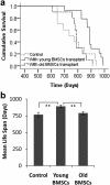
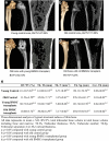
Increasing evidence suggests that the loss of functional stem cells may be important in the aging process. Our experiments were originally aimed at testing the idea that, in the specific case of age-related osteoporosis, declining function of osteogenic precursor cells might be at least partially responsible. To test this, aging female mice were transplanted with mesenchymal stem cells from aged or young male donors. We find that transplantation of young mesenchymal stem cells significantly slows the loss of bone density and, surprisingly, prolongs the life span of old mice. These observations lend further support to the idea that age-related diminution of stem cell number or function may play a critical role in age-related loss of bone density in aging animals and may be one determinant of overall longevity.
Figures

Similar articles
-
[Anti-aging effect of transplantation of mouse fetus-derived mesenchymal stem cells].Sheng Li Xue Bao. 2010 Feb 25;62(1):79-85. Sheng Li Xue Bao. 2010. PMID: 20179893 Chinese.
-
Distribution pattern following systemic mesenchymal stem cell injection depends on the age of the recipient and neuronal health.Stem Cell Res Ther. 2017 Apr 18;8(1):85. doi: 10.1186/s13287-017-0533-2. Stem Cell Res Ther. 2017. PMID: 28420415 Free PMC article.
-
Aging alters bone-fat reciprocity by shifting in vivo mesenchymal precursor cell fate towards an adipogenic lineage.Bone. 2016 Apr;85:29-36. doi: 10.1016/j.bone.2016.01.014. Epub 2016 Jan 19. Bone. 2016. PMID: 26805026 Free PMC article.
-
Transplantation of Adipose Tissue-Derived Mesenchymal Stem Cell (ATMSC) Expressing Alpha-1 Antitrypsin Reduces Bone Loss in Ovariectomized Osteoporosis Mice.Hum Gene Ther. 2017 Feb;28(2):179-189. doi: 10.1089/hum.2016.069. Epub 2016 Nov 1. Hum Gene Ther. 2017. PMID: 27802778 Review.
-
Pineal cross-transplantation (old-to-young and vice versa) as evidence for an endogenous "aging clock".Ann N Y Acad Sci. 1994 May 31;719:456-60. doi: 10.1111/j.1749-6632.1994.tb56850.x. Ann N Y Acad Sci. 1994. PMID: 8010614 Review.
Cited by
-
A Periodic Diet that Mimics Fasting Promotes Multi-System Regeneration, Enhanced Cognitive Performance, and Healthspan.Cell Metab. 2015 Jul 7;22(1):86-99. doi: 10.1016/j.cmet.2015.05.012. Epub 2015 Jun 18. Cell Metab. 2015. PMID: 26094889 Free PMC article. Clinical Trial.
-
Effect of Polymeric Matrix Stiffness on Osteogenic Differentiation of Mesenchymal Stem/Progenitor Cells: Concise Review.Polymers (Basel). 2021 Aug 31;13(17):2950. doi: 10.3390/polym13172950. Polymers (Basel). 2021. PMID: 34502988 Free PMC article. Review.
-
Recent clinical trials with stem cells to slow or reverse normal aging processes.Front Aging. 2023 Apr 6;4:1148926. doi: 10.3389/fragi.2023.1148926. eCollection 2023. Front Aging. 2023. PMID: 37090485 Free PMC article. Review.
-
S-nitrosylation and MSC-mediated body composition.Onco_target. 2015 Oct 6;6(30):28517-8. doi: 10.18632/onco_target.5672. Onco_target. 2015. PMID: 26415218 Free PMC article. No abstract available.
-
Mixing old and young: enhancing rejuvenation and accelerating aging.J Clin Invest. 2019 Jan 2;129(1):4-11. doi: 10.1172/JCI123946. Epub 2019 Jan 2. J Clin Invest. 2019. PMID: 30601138 Free PMC article. Review.
References
-
- Mauck K. F. & Clarke B. L. Diagnosis, screening, prevention, and treatment of osteoporosis. Mayo Clin Proc 81, 662–672 (2006). - PubMed
-
- Reginster J. Y. & Burlet N. Osteoporosis: a still increasing prevalence. Bone 38, S4–9 (2006). - PubMed
-
- Harvey N., Dennison E. & Cooper C. Osteoporosis: impact on health and economics. Nat Rev Rheumatol 6, 99–105 (2010). - PubMed
-
- Llorens R. A review of osteoporosis: diagnosis and treatment. Mo Med 103, 612–615; quiz 615–616 (2006). - PubMed
Publication types
MeSH terms
Grants and funding
LinkOut - more resources
Full Text Sources
Other Literature Sources
Medical

