. 2013 May 21:4:55.
doi: 10.3389/fendo.2013.00055. eCollection 2013.

Alternative Splicing of the Pituitary Adenylate Cyclase-Activating Polypeptide Receptor PAC1: Mechanisms of Fine Tuning of Brain Activity

Affiliations

Alternative Splicing of the Pituitary Adenylate Cyclase-Activating Polypeptide Receptor PAC1: Mechanisms of Fine Tuning of Brain Activity

Janna Blechman et al. Front Endocrinol (Lausanne). .

Abstract

Alternative splicing of the precursor mRNA encoding for the neuropeptide receptor PAC1/ADCYAP1R1 generates multiple protein products that exhibit pleiotropic activities. Recent studies in mammals and zebrafish have implicated some of these splice isoforms in control of both cellular and body homeostasis. Here, we review the regulation of PAC1 splice variants and their underlying signal transduction and physiological processes in the nervous system.

Keywords: ADCYAP1R1; PACAP receptor; activity-dependent gene regulation; homeostasis; hypothalamic hormones; post-traumatic; stress disorders; zebrafish model system.


Figures
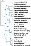
Figure 1
Figure 1
Tree topology of the vertebrate PAC1 receptor family. Phylogenetic relationships of PAC1 receptors analyzed using the zebrafish (Danio rerio) Vpac1b gene as an out-group.
Figure 2
Figure 2
A diagram showing genomic organization of the PAC1 (ADCYAP1R1) gene. Exons 2–18 encode the open reading frame. Exons undergoing the alternative splicing discussed in this review are marked with blue asterisks. Exon 4, 5, and 6 encode parts of the N-terminal domain, whereas exons 14 and 15 encode the third intracellular loop. TM, transmembrane.
Figure 3
Figure 3
A scheme depicting the topology of the PAC1 receptor. The relative location of amino acids encoded by PAC1 exons are depicted by the blue and gray beads. Predicted PAC1 structural motifs known to be involved in protein-protein interactions determining G-protein mediated PAC1 signaling are marked on the protein diagram with regard to putative motifs formed due to insertions of hop and hip cassettes into the IC3 loop sequence. EC, extracellular loop; IC, intracellular loop; TM, transmembrane domain.
Figure 4
Figure 4
A scheme depicting the current knowledge on PAC1-mediated signaling cascades resulting in a variety of neuronal outcomes. PKA was shown to induce ERK1/2 thereby contributing to PACAP neuroprotective (Villalba et al., ; Vaudry et al., ; Falluel-Morel et al., ; Stumm et al., 2007) and neurotrophic (Ravni et al., ; Botia et al., ; Monaghan et al., 2008) functions. Action potential (AP) firing induces a CRCT1/CREB mediated neuroprotective effect, presumably through NMDA receptor activation and was shown to be initiated by PKA activation (Baxter et al., 2011). NMDA receptor was also shown to be indirectly activated by PACAP (Llansola et al., 2004) or cAMP/PKA signaling (Costa et al., 2009). PAC1/PKA signaling controls cellular apoptosis through inhibition of potassium channels (Mei et al., ; Castel et al., ; Pugh and Margiotta, 2006) or induction of calcium channels (Pugh and Margiotta, 2006). PAC1/PKA-mediated activation of ion channels leads to activity-dependent neuronal differentiation and synaptic plasticity (Lee et al., ; Maturana et al., ; Grumolato et al., ; Nishimoto et al., 2007). The tumor suppressor gene Lot1 known as a negative regulator of neuronal precursor proliferation was shown to be controlled by PAC1/cAMP/ERK signaling pathway (Fila et al., 2009). PAC1/PKA-dependent phosphorylation of Tau is involved in the control of granule cell migration during cerebellar development (Falluel-Morel et al., 2006). PAC1-mediated cAMP/ERK-dependent neurite outgrowth was shown to be regulated via a novel neuritogenic factor NCS (Emery and Eiden, 2012) or Epac/ERK (Gerdin and Eiden, 2007) pathways. PAC1/Epac was shown to regulate neuronal differentiation via activation of p38 kinase along with mobilization of Ca2+ from intracellular stores (Ster et al., 2007) as well as Epac/Rit-dependent pathway involving CREB signaling (Shi et al., 2006). PAC1 was demonstrated to induce Rit through TrkA/Shc/SOS signaling initiated by Src activation via dual Gs/Epac and Gi stimulation (Shi et al., 2010). PACAP also signals through Gq-linked PLC > IP3 > Ca2+/DAG > PKC, and PLD > phosphatidic acid (PA) pathways (Journot et al., ; Makhlouf and Murthy, ; Dejda et al., 2006). PAC1 is a mediator of gene transcription, neuronal differentiation, and synaptic development (Masmoudi-Kouki et al., ; Vaudry et al., ; Andero and Ressler, 2012). As an example of functional diversity caused by PAC1 alternative splicing two additional PAC1-hop1 signaling pathways are presented on the scheme. They depict PAC1-hop1/ARF dependent PLD activation (McCulloch et al., 2001) and internalization-dependent engagement of PI3Kγ/Akt activation (May et al., 2010). AC, adenylate cyclase; AGS, activator of G protein signaling; Akt, serine-threonine protein kinase PKB; cAMP, cyclic adenosine monophosphate; ARF, ADP(adenosine diphosphate) ribosylation factor; Caln, calcineurin; CaMK, calcium calmodulin kinase; CBP, creb binding protein; CRCT1, cysteine-rich C-terminal 1; CREB, cAMP responsive element-binding protein; DAG, diacyl glycerol; Epac, exchange factor directly activated by cAMP; ERK, extracellular signal-regulate kinase; G, guanine nucleotide-binding regulatory protein; IP3, inositol-1,4,5-triphosphate; JNK, c-Jun oncogene N-terminal kinase 1; Lot1, lost on transformation 1; NCS, neuritogenic cAMP sensor; NGF, nerve growth factor; NMDAR, N-methyl-d-aspartate receptor; p38, p38 mitogen-activated protein kinase; PA, phosphatidic acid; PI3K, phosphatidylinositol 3′ OH kinase; PKA, protein kinase A; PKC, protein kinase C;PLC, phospholipase; Raf, B-Raf proto-oncogene serine/threonine kinase;Ras, retrovirus-associated DNA sequences; Rap1, Rit, small GTPases of the RAS oncogene family; Sos, son of sevenless homolog 1; Src, sarcoma viral oncogen homolog; Tau, neuron-specific microtubule-associated protein; TrkA, tropomyosin-related kinase.
Figure 5
Figure 5
Regulation of PAC1 splicing. (A) A chart depicting the predicted binding sites for neuronal-specific RNA-binding proteins that potentially regulate PAC1 gene splicing by binding in proximity to PAC1 exons. Consensus cis-acting binding DNA elements for the respective RNA-binding protein are depicted with respect to their position in the PAC1 genes of rat, mouse, human, and zebrafish PAC1. We analyzed the presence of putative consensus binding sites located within 300 base-pairs upstream (upst) or downstream (dnst) and inside the exons. (B) Schematic representation of the PAC1 gene in which the location of the analyzed cis-acting elements is color-coded as above. Corresponding exons numbers and sizes (bp) are indicated.
Figure 6
Figure 6
A model illustrating the role of alternative splicing of PAC1 hop cassette that serves as an ON/OFF stress switch. In response to various stressors, the so called PAC1-null splice variant (i.e., no deletion in the N-terminal domain and no addition to the intracellular loops) modulate transcriptional activation of CRH and stress behaviors to adapt to the changes in homeostasis. Termination of CRH-mediated stress response is mediated by means of regulation of PAC1 gene splicing and inclusion of an altered exon (hop1) encoding to 28 amino acids of the third intracellular loop leading to the formation of the PAC1-hop1 splice variant. Generation of the PAC1-hop isoform terminates stress response by means yet to be uncovered (see text and Amir-Zilberstein et al., 2012).

Similar articles

Cited by

References

    1. Abu-Hamdan M. D., Drescher M. J., Ramakrishnan N. A., Khan K. M., Toma V. S., Hatfield J. S., et al. (2006). Pituitary adenylyl cyclase-activating polypeptide (PACAP) and its receptor (PAC1-R) in the cochlea: evidence for specific transcript expression of PAC1-R splice variants in rat microdissected cochlear subfractions. Neuroscience 140, 147–16110.1016/j.neuroscience.2006.01.019 - DOI - PubMed
    1. Acampora D., Postiglione M. P., Avantaggiato V., Di Bonito M., Vaccarino F. M., Michaud J., et al. (1999). Progressive impairment of developing neuroendocrine cell lineages in the hypothalamus of mice lacking the Orthopedia gene. Genes Dev. 13, 2787–280010.1101/gad.13.21.2787 - DOI - PMC - PubMed
    1. Agarwal A., Halvorson L. M., Legradi G. (2005). Pituitary adenylate cyclase-activating polypeptide (PACAP) mimics neuroendocrine and behavioral manifestations of stress: evidence for PKA-mediated expression of the corticotropin-releasing hormone (CRH) gene. Brain Res. Mol. Brain Res. 138, 45–5710.1016/j.molbrainres.2005.03.016 - DOI - PMC - PubMed
    1. Ahren B. (2008). Role of pituitary adenylate cyclase-activating polypeptide in the pancreatic endocrine system. Ann. N. Y. Acad. Sci. 1144, 28–3510.1196/annals.1418.003 - DOI - PubMed
    1. Aino H., Hashimoto H., Ogawa N., Nishino A., Yamamoto K., Nogi H., et al. (1995). Structure of the gene encoding the mouse pituitary adenylate cyclase-activating polypeptide receptor. Gene 164, 301–30410.1016/0378-1119(95)00391-I - DOI - PubMed

LinkOut - more resources

  NODES
INTERN 1
twitter 2