Review
. 2015 Jan;11(1):38-59.
doi: 10.1039/c4mb00443d. Epub 2014 Oct 31.

Fatty acid biosynthesis revisited: structure elucidation and metabolic engineering

Affiliations
Review

Fatty acid biosynthesis revisited: structure elucidation and metabolic engineering

Joris Beld et al. Mol Biosyst. 2015 Jan.

Abstract

Fatty acids are primary metabolites synthesized by complex, elegant, and essential biosynthetic machinery. Fatty acid synthases resemble an iterative assembly line, with an acyl carrier protein conveying the growing fatty acid to necessary enzymatic domains for modification. Each catalytic domain is a unique enzyme spanning a wide range of folds and structures. Although they harbor the same enzymatic activities, two different types of fatty acid synthase architectures are observed in nature. During recent years, strained petroleum supplies have driven interest in engineering organisms to either produce more fatty acids or specific high value products. Such efforts require a fundamental understanding of the enzymatic activities and regulation of fatty acid synthases. Despite more than one hundred years of research, we continue to learn new lessons about fatty acid synthases' many intricate structural and regulatory elements. In this review, we summarize each enzymatic domain and discuss efforts to engineer fatty acid synthases, providing some clues to important challenges and opportunities in the field.


Figures

Figure 1
Figure 1
Comparison of FAS domain organizations. a) Mammalian type I synthase (PDB: 2VZ8) structure with a diagram below of the domain organization. Domains that were not observed in the crystal structure are denoted by hashed borders. b) E. coli type II discrete structures with a comparative diagram below denoting multimeric states of the proteins. (PDB: KR, 1Q7B; DH, 1MKB; ER, 1DFI; ACP, 2FAD; KS, 2VB9; MAT, 2G2Z) c) Two fungal type I synthases demonstrating increased scaffolding elements and organization compared to the mammalian type I synthase in (a). At left, top-view cross-section of FAS from yeast Saccharomyces cerevisiae (PDB: 2UV8). At right, thermophilic fungus Thermomyces lanuginosus (PDB: 2UVB, 2UVC). d) Domain organization for the ACP-independent type II-like FAS discovered in Archaea, from which no structures are currently available.
Figure 2
Figure 2
Cartoon of cargo sequestration and chain-flipping mechanism by acyl carrier proteins. a) the acyl carrier protein (ACP, grey protein cartoon structure) is post-translationally modified with a 4′-phosphopantetheine arm, bearing a terminal thiol. b) when a fatty acid intermediate (yellow rectangle) is loaded onto the 4′-phosphopantetheine moiety, it is protected from the environment by sequestration in the inner core of the ACP. c) a partner protein (orange) binds to the ACP. d) the protein-protein binding event induces a conformational change in the carrier protein, allowing the cargo to flip out of the inner core into the active site of the partner enzyme (the “chain-flipping mechanism”). e) chemical transformation of the fatty acid intermediate (yellow > red) occurs in the active site of the partner enzyme. f) after the reaction is complete the partner enzyme leaves, and another partner protein (blue) can bind to the ACP.
Figure 3
Figure 3
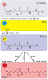
Acyl carrier protein post-translational manipulations and modifications. Carrier proteins (CPs) come from the ribosome in its naked apo-form (blue circle). By the action of a PPTase, utilizing coenzyme A, a highly conserved serine residue of the CP is post-translationally modified with a 4′-phosphopantetheine arm forming holo-ACP (yellow circle). This active form of the carrier protein is used to carry the growing fatty acid chain, by action of the FAS enzymes (acyl-ACP, red circle). A thioesterase (TE) or acyltransferase (AT) can cleave off the fatty acid, regenerating holo-CP. The adenylate-forming enzyme acyl-ACP synthetase (AasS) can directly load a fatty acid onto holo-CP, resulting in acyl-ACP (red circle). In the laboratory, it is possible to attach pantetheine-probes, in a CoaA, -D, -E and PPTase Sfp dependent fashion, to apo-CP, leading to crypto-CP (pink circle). Acyl carrier protein hydrolase or phosphodiesterase, AcpH, can regenerate the apo-form of the carrier protein from acyl-, crypto- and holo-ACP.
Figure 4
Figure 4
Malonyl-CoA acyltransferases (MCAT/FabD). A–D) Similar to Arthur et al. we used several protein-protein docking servers to visualize the tentative interaction between ACP and MCAT. Using Patchdock, Grammx and Cluspro, E. coli ACP (PDB: 1T8K) was docked onto E. coli FabD (PDB: 2G1H) and compared with the modeled structure of ScACP and ScFabD, PDB: 1NNZ, shown in A. B) Low energy state observed by all three methods, while C and D were observed by two. E) reaction catalyzed by MCAT/FabD. F) Published structures of malonyl-CoA acyltransferases (MCAT/FabD) show very close homology. Here, we docked using Cluspro to various MCAT X-ray crystal structures (in dark brown) E. coli ACP, showing the top 5 hits (protein bundles in multicolor). From top left to bottom right: 1NM2 (from Streptomyces coelicolor), 2G1H (from E. coli), 2H1Y (from Helicobacter pylori), 3TQE (from Coxiella burnetii), 3PTW (from Clostridium perfringens), 2QC3 (from Mycobacterium tuberculosis), 3EZO (from Burkholderia pseudomallei) and 3IM8 (from Streptococcus pneumoniae).
Figure 5
Figure 5
Ketoacyl synthases. The ketoacyl synthase dimer is highly conserved throughout all branches of life. Top: FabH (KSIII, PDB: 1EBL) and FabD (MCAT) are responsible for initiating fatty acid biosynthesis. The acetyl group of acetyl-CoA is transferred to the active site cysteine residue of KSIII. Malonyl-ACP (the product of FabD/MCAT) binds to KSIII and a new carbon-carbon bond is formed, while CO2 is released. Bottom: FabB (KSI, PDB: 1G5X) and FabF (KSII, PDB: 2GFW) extend the chain further by first transfer of a fatty acid from an alkyl-ACP species to a conserved cysteine residue on the ketoacyl synthase. A different carrier protein loaded with a malonyl moiety binds to the loaded KS and a new carbon-bond is formed, while CO2 is released. The type I FAS KS from pig is shown as insert, showcasing the highly conserved fold and dimer structure of these enzymes.
Figure 6
Figure 6
Dehydratases. a) The bacterium E. coli has only one way to produce unsaturated fatty acids: FabA can not only reduce 3-hydroxydecanoyl-ACP, but also isomerize the double bond from trans-2-decenoyl-ACP to cis-3-decenoyl-ACP. Subsequent chain elongation by action of the other fatty acid synthase enzymes leads to the fatty acid C16:1. b) Other organisms (including cyanobacteria, plants, algae) have elongases and desaturases (green panel) that can extend and desaturate the saturated C16:0 or C18:0 fatty acids, as either acyl-ACP, acyl-CoA, acyl-lipid or free fatty acids. c) Mechanistic crosslinking between ACP and DH using a sulfonyl alkynyl pantetheinamide probe. d) The mechanistically crosslinked X-ray crystal structure of E. coli AcpP with FabA (PDB: 4KEH).
Figure 7
Figure 7
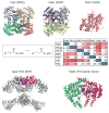
Structures of enoyl-ACP reductases. Top: FabI from E. coli (PDB: 2FHS), FabL from B. subtilis (PDB: 3OIC) and FabV from X. oryzae (PDB: 3S8M) are all members of the SDR superfamily, utilizing NAD(P)H. Whereas FabI and FabL are tetramers, FabV is a monomer. ClustalOmega was used to generate a sequence identity matrix, including FAS1y (the yeast FAS ER), FAS1h (the human FAS ER) and MER (the human mitochondrial ER). Bottom: The ER dimer in type I FAS from pig (2VZ8) is shown in pink/purple, and FabK from S. pneumoniae (2Z6I).
Figure 8
Figure 8
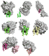
Comparison of all eight ACP-partner protein X-ray crystal structures deposted in the PDB, from the perspective of the ACP, showing the similar binding orientation of the ACP, but the different binding motifs of the partner proteins. From top to bottom and left to right: PDB: 1F80, E. coli PPTase AcpS with E. coli holo-AcpP, PDB: 2FHS, E. coli FabI with E. coli AcpP, PDB: 2XZ0, Ricinus communis stearoyl desaturase with R. communis ACP bearing a phosphoserine, PDB: 3EJB, B. subtilis P450BioI with E. coli AcpP bearing tetradecanoic acid, PDB: 3NY7, STAS domain of E. coli YchM with E. coli AcpP bearing a terminal acid propionic acid thioester, PDB: 4ETW, E. coli BioH with E. coli pimeloyl-AcpP, PDB: 4KEH, E. coli FabA mechanistically crosslinked with E. coli AcpP bearing a non-hydrolyzable pantetheinamide crosslinker (in light pink surface the second ACP) and PDB: 2CG5, Homo sapiens AASDHPPT with human FAS apo-ACP excised from the human type I FAS.
Figure 9
Figure 9
Fatty acid biosynthesis termination by thioesterases or acyltransferases. Top: a) In plants, green algae and some bacteria, dedicated hotdog-fold acyl-ACP thioesterases (e.g. CrTE, Chlamydomonas reinhardtii Fat1) are responsible for hydrolyzing fatty acids, with a certain chain-length off the ACP (e.g. CrACP, C. reinhardtii ACP). TesA is a multifunctional enzyme that can hydrolyze fatty acids off the ACP, but this is presumably not its primary function in E. coli. In bacteria, dedicated acyl-transferases, like PlsB, trans-esterify lipid headgroups with a fatty acid, directly from the ACP. Alternatively, E. coli uses PlsX to synthesize phospho-fatty acids, which are loaded onto lipids using PlsY. b) Mechanistic crosslinking of ACP to TE using α-bromo acid pantetheinamide probe. Bottom: structures of TesA (PDB: 1IVN), PlsB (PDB: 1K30) and a docked structure of chloroplastic C. reinhardtii ACP (model) with C. reinhardtii TE FAT1 (model).
Figure 10
Figure 10
Fatty acid metabolism in bacteria. In the fatty acid synthase (yellow circle): ACCase is the acetyl-CoA carboxylase; MCAT, malonyl-CoA acyltransferase; KSIII (FabH), ketoacyl synthase; KR (FabG), ketoreductase; DH (FabA/FabZ), dehydratase; ER (FabI/K/L/V), enoyl-ACP reductase; KSI (FabB), KSII (FabF), ketoacyl synthases and PlsB, acyltransferase. In green the thioesterase (TE) pathway present in plants, algae and some bacteria. In orange the acyl-ACP synthetase (AasS) found in some organisms. The dotted arrow in the bottom right corner represent diffusion of fatty acids into the cell, as well as active FadL-mediated transport. Fatty acid catabolism (blue circle): FadD, CoA-ligase; FadE, acyl-CoA dehydrogenase; FadB, dual-function enoyl-CoA hydratase and hydroxyacyl-CoA dehydrogenase; FadA, acetyl-CoA acetyltransferase. FadR and FabR are the master regulators of fatty acid degradation and fatty acid biosynthesis, whereas ppGpp is an alarmone.

Similar articles

Cited by

References

    1. Howarth RW, Ingraffea A, Engelder T. Nature. 2011;477:271–275. - PubMed
    1. Mayfield S. Genome. 2013;56:551–555. - PubMed
    1. McCarthy AD, Hardie DG. Trends Biochem Sci. 1984;9:60–63.
    1. Wada H, Shintani D, Ohlrogge J. Proc Natl Acad Sci USA. 1997;94:1591–1596. - PMC - PubMed
    1. Lombard J, López-García P, Moreira D. Archaea. 2012;2012 - PMC - PubMed

Publication types

MeSH terms

LinkOut - more resources

  NODES
Note 1
twitter 2