Radiation response of mesenchymal stem cells derived from bone marrow and human pluripotent stem cells
- PMID: 25425005
- PMCID: PMC4380046
- DOI: 10.1093/jrr/rru098
Radiation response of mesenchymal stem cells derived from bone marrow and human pluripotent stem cells
Abstract
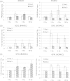
Mesenchymal stem cells (MSCs) isolated from human pluripotent stem cells are comparable with bone marrow-derived MSCs in their function and immunophenotype. The purpose of this exploratory study was comparative evaluation of the radiation responses of mesenchymal stem cells derived from bone marrow- (BMMSCs) and from human embryonic stem cells (hESMSCs). BMMSCs and hESMSCs were irradiated at 0 Gy (control) to 16 Gy using a linear accelerator commonly used for cancer treatment. Cells were harvested immediately after irradiation, and at 1 and 5 days after irradiation. Cell cycle analysis, colony forming ability (CFU-F), differentiation ability, and expression of osteogenic-specific runt-related transcription factor 2 (RUNX2), adipogenic peroxisome proliferator-activated receptor gamma (PPARγ), oxidative stress-specific dismutase-1 (SOD1) and Glutathione peroxidase (GPX1) were analyzed. Irradiation arrested cell cycle progression in BMMSCs and hESMSCs. Colony formation ability of irradiated MSCs decreased in a dose-dependent manner. Irradiated hESMSCs showed higher adipogenic differentiation compared with BMMSCs, together with an increase in the adipogenic PPARγ expression. PPARγ expression was upregulated as early as 4 h after irradiation, along with the expression of SOD1. More than 70% downregulation was found in Wnt3A, Wnt4, Wnt 7A, Wnt10A and Wnt11 in BMMSCs, but not in hESMSCs. hESMSCs are highly proliferative but radiosensitive compared with BMMSCs. Increased PPARγ expression relative to RUNX2 and downregulation of Wnt ligands in irradiated MSCs suggest Wnt mediated the fate determination of irradiated MSCs.
Keywords: adipogenesis; human pluripotent stem cell; mesenchymal stem cell; osteogenesis; radiation effect.
© The Author 2014. Published by Oxford University Press on behalf of The Japan Radiation Research Society and Japanese Society for Radiation Oncology.
Figures


Similar articles
-
Dysregulated systemic lymphocytes affect the balance of osteogenic/adipogenic differentiation of bone mesenchymal stem cells after local irradiation.Stem Cell Res Ther. 2017 Mar 20;8(1):71. doi: 10.1186/s13287-017-0527-0. Stem Cell Res Ther. 2017. PMID: 28320453 Free PMC article.
-
[Spermidine enhances osteogenic differentiation and inhibits adipogenic differentiation of bone marrow-derived mesenchymal stem cells from ovariectomized mice].Xi Bao Yu Fen Zi Mian Yi Xue Za Zhi. 2015 Jun;31(6):787-91. Xi Bao Yu Fen Zi Mian Yi Xue Za Zhi. 2015. PMID: 26062423 Chinese.
-
Crif1 Promotes Adipogenic Differentiation of Bone Marrow Mesenchymal Stem Cells After Irradiation by Modulating the PKA/CREB Signaling Pathway.Stem Cells. 2015 Jun;33(6):1915-26. doi: 10.1002/stem.2019. Stem Cells. 2015. PMID: 25847389
-
[Regulation of the differentiation of bone marrow mesenchymal stem cells into osteoblastic and adipogenic lineage].Chir Narzadow Ruchu Ortop Pol. 2011 Mar-Apr;76(2):96-8. Chir Narzadow Ruchu Ortop Pol. 2011. PMID: 21853910 Review. Polish.
-
Potential mechanisms underlying the Runx2 induced osteogenesis of bone marrow mesenchymal stem cells.Am J Transl Res. 2015 Dec 15;7(12):2527-35. eCollection 2015. Am J Transl Res. 2015. PMID: 26885254 Free PMC article. Review.
Cited by
-
Indirect effects of X-irradiation on proliferation and osteogenic potential of bone marrow mesenchymal stem cells in a local irradiated rat model.Mol Med Rep. 2017 Jun;15(6):3706-3714. doi: 10.3892/mmr.2017.6464. Epub 2017 Apr 12. Mol Med Rep. 2017. PMID: 28440500 Free PMC article.
-
In vitro cellular and proteome assays identify Wnt pathway and CDKN2A-regulated senescence affected in mesenchymal stem cells from mice after a chronic LD gamma irradiation in utero.Radiat Environ Biophys. 2021 Aug;60(3):397-410. doi: 10.1007/s00411-021-00925-7. Epub 2021 Jul 21. Radiat Environ Biophys. 2021. PMID: 34287697 Free PMC article.
-
Radio-resistant mesenchymal stem cells: mechanisms of resistance and potential implications for the clinic.Onco_target. 2015 Aug 14;6(23):19366-80. doi: 10.18632/onco_target.4358. Onco_target. 2015. PMID: 26203772 Free PMC article. Review.
-
Resistance to energy metabolism - _targeted therapy of AML cells residual in the bone marrow microenvironment.Cancer Drug Resist. 2023 Mar 14;6(1):138-150. doi: 10.20517/cdr.2022.133. eCollection 2023. Cancer Drug Resist. 2023. PMID: 37065866 Free PMC article. Review.
-
3-D Cell Culture Systems in Bone Marrow Tissue and Organoid Engineering, and BM Phantoms as In Vitro Models of Hematological Cancer Therapeutics-A Review.Materials (Basel). 2020 Dec 9;13(24):5609. doi: 10.3390/ma13245609. Materials (Basel). 2020. PMID: 33316977 Free PMC article. Review.
References
-
- Hui S, Verneris M, Froelich J, et al. Multimodality image guided total marrow irradiation and verification of the dose delivered to the lung, PTV, and thoracic bone in a patient: a case study. Technol Cancer Res Treat. 2009;8:23–28. - PubMed
Publication types
MeSH terms
Grants and funding
LinkOut - more resources
Full Text Sources
Other Literature Sources
Miscellaneous

