. 2015 Jul;16(7):727-42.
doi: 10.1111/tra.12283. Epub 2015 Apr 30.

Recruitment of VPS33A to HOPS by VPS16 Is Required for Lysosome Fusion with Endosomes and Autophagosomes

Affiliations

Recruitment of VPS33A to HOPS by VPS16 Is Required for Lysosome Fusion with Endosomes and Autophagosomes

Lena Wartosch et al. Traffic. 2015 Jul.

Abstract

The mammalian homotypic fusion and vacuole protein sorting (HOPS) complex is comprised of six subunits: VPS11, VPS16, VPS18, VPS39, VPS41 and the Sec1/Munc18 (SM) family member VPS33A. Human HOPS has been predicted to be a tethering complex required for fusion of intracellular compartments with lysosomes, but it remains unclear whether all HOPS subunits are required. We showed that the whole HOPS complex is required for fusion of endosomes with lysosomes by monitoring the delivery of endocytosed fluorescent dextran to lysosomes in cells depleted of individual HOPS proteins. We used the crystal structure of the VPS16/VPS33A complex to design VPS16 and VPS33A mutants that no longer bind each other and showed that, unlike the wild-type proteins, these mutants no longer rescue lysosome fusion with endosomes or autophagosomes in cells depleted of the endogenous proteins. There was no effect of depleting either VIPAR or VPS33B, paralogs of VPS16 and VPS33A, on fusion of lysosomes with either endosomes or autophagosomes and immunoprecipitation showed that they form a complex distinct from HOPS. Our data demonstrate the necessity of recruiting the SM protein VPS33A to HOPS via its interaction with VPS16 and that HOPS proteins, but not VIPAR or VPS33B, are essential for fusion of endosomes or autophagosomes with lysosomes.

Keywords: CORVET; HOPS; SM protein; VPS16; VPS33A; autophagy; endocytosis; lysosomes; tethering factor.


Figures

Figure 1
Figure 1
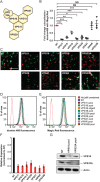
siRNA depletion of HOPS components but not VIPAR or VPS33B inhibits the fusion of endosomes with lysosomes. HeLaM cells were transfected twice with a single siRNA oligonucleotide or a pool of four siRNA oligonucleotides at 100 nm. A) Schematic model of the human HOPS complex 44. B) Colocalisation of dextran Alexa Fluor® 488 with Magic Red® using images captured by live‐cell confocal microscopy after knock‐down of HOPS proteins with siRNA oligonucleotide pools (NT: non‐_targeting siRNA). Mean ± SEM of three independent experiments with five fields each, ≥30 cells total per condition. *p < 0.01, **p < 0.003, ***p < 0.0004, NS, not significant, using two‐tailed unpaired t‐test. C) Representative live‐cell confocal microscopy images of cells quantified in (B) that had been loaded with dextran Alexa Fluor 488 10,000 MW (green) for 2 h, chased for 1 h and then stained with Magic Red® (red) for lysosomes. Scale bar 5 µm. D) FACS analysis of dextran Alexa Fluor 488 fluorescence after uptake as in (B) and (C). E) FACS analysis of Magic Red® fluorescence of cells treated as in (B) and (C). For both (D) and (E), 30,000 cells were measured per condition, with representative traces shown for each condition from one of three independent experiments. Data were analysed using flowjo software and histogram overlays are displayed as %Max, scaling each curve to mode = 100%. F) RNA was purified from cells and transcribed into cDNA. The knock‐down efficiency was evaluated by quantitative real‐time PCR with gene‐specific primers (Table S1). Detection of Actin transcripts served as a reference. ΔΔCt values were calculated and relative transcript levels of three independent experiments are shown as mean ± SD. G) Protein lysates were analysed by immunoblotting with specific VPS18 or VPS33A antibodies to confirm knock‐down efficiencies. Actin served as loading control.
Figure 2
Figure 2
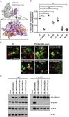
The association of VPS33A with HOPS via VPS16 is essential for endosome–lysosome fusion. Analysis of HeLaM cells or HeLaM cells stably expressing N‐terminally haemagglutinin (HA)‐tagged siRNA oligonucleotide 3 (oligo3)‐resistant VPS16 wild type (WT), single mutant (A669D, A725E), or double mutant A669D/R725E (A/R, two independent clonal lines, #1 and #2). A) Structure of VPS33A in complex with VPS16 residues 642–736 44. VPS33A and VPS16 are shown as white and purple molecular surfaces, respectively. Inset shows the binding footprint of VPS33A on VPS16 in darker purple, with residues essential for robust VPS33A binding highlighted in orange. Image was prepared using PyMOL. B and C) Cells were transfected with non‐_targeting (NT) siRNA oligonucleotide or VPS16 siRNA oligo3 at 100 nm and subjected to live cell microscopy. Quantification of colocalisation of dextran Alexa Fluor® 488 (green) with Magic Red® (red) shown in (B). Mean ± SEM of three independent experiments with five fields each, ≥30 cells total per condition. *p < 0.01, **p < 0.003, ***p < 0.001, NS, not significant, using two‐tailed unpaired t‐test. Representative confocal microscopy images of cells quantified in (B) are shown in (C). Scale bar: 2.5 µm. D) Protein lysates were subjected to immunoprecipitation (IP) with anti‐HA affinity matrix and immunoblotting. Overexpressed HA‐VPS16 was detected with an anti‐HA antibody and endogenous VPS33A and VPS18 with specific antibodies. Actin served as loading control.
Figure 3
Figure 3
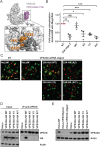
VPS33A mutated at its binding interface with VPS16 cannot support endosome‐lysosome fusion. Analysis of HeLaM cells or HeLaM cells stably expressing C‐terminally haemagglutinin (HA)‐tagged siRNA oligonucleotide 2 (oligo2)‐resistant VPS33A wild type (WT) or the double mutants K429D/I441K (K/I), Y438D/I441K (Y/I) or K429D/Y438D (K/Y). A) Structure of VPS33A in complex with VPS16 residues 642–736 44, coloured as in Figure 2A. Inset shows the binding footprint of VPS16 on VPS33A in darker grey, with residues essential for robust VPS16 binding highlighted in orange. Image was prepared using PyMOL. B and C) Cells were transfected with non‐_targeting (NT) siRNA oligonucleotide or VPS33A siRNA oligo2 at 100 nm and subjected to live cell microscopy. Quantification of colocalisation of dextran Alexa Fluor® 488 (green) with Magic Red® (red) shown in (C). Mean ± SEM of three independent experiments with five fields each, ≥30 cells total per condition. *p < 0.02, **p < 0.002, ***p < 0.001, NS, not significant, using two‐tailed unpaired t‐test. Representative confocal microscopy images of cells quantified in (B) are shown in (C). Scale bar 2.5 µm. D) Protein lysates were subjected to immunoprecipitation (IP) with an anti‐VPS18 antibody and immunoblotting. Overexpressed HA‐VPS33A was detected with an anti‐HA antibody and endogenous VPS18 with a specific antibody. Actin served as loading control. E) Immunoblot of cell lysates from HeLaM cells, or from HeLaM cells stably overexpressing VPS33A‐HA(WT), VPS33A‐HA(K/I), VPS33A‐HA(Y/I) or VPS33A‐HA(K/Y), transfected twice with non‐_targeting (NT) siRNA oligonucleotide as a control or a specific siRNA pool or single oligo2 to deplete endogenous VPS33A. VPS33A was detected with a specific VPS33A antibody. Actin served as a loading control. Expression levels, calculated as an increase compared to endogenous VPS33A in HeLaM cells, were: WT: ninefold, K/I: fourfold, Y/I: 14‐fold, K/Y: ninefold as measured by densitometric analysis of X‐ray films using imagej software.
Figure 4
Figure 4
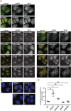
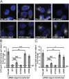
siRNA depletion of the HOPS proteins VPS33A or VPS16 inhibits the fusion of autophagosomes with lysosomes. Endogenous VPS16, VPS33A, VIPAR or VPS33B were depleted by transfection with specific siRNA oligonucleotides at 100 nm. Transfection with non‐_targeting (NT) siRNA oligonucleotide served as control. A) Confocal images of HeLaM cells stably expressing the autophagy marker mRFP‐GFP‐LC3. Cells were treated with dimethyl sulphoxide (DMSO) (control) or 400 nm bafilomycinA1 (BafA1) to block autophagosome–lysosome fusion. Scale bar: 10 µm. B) Representative confocal images of fixed HeLa cells that were immunostained for endogenous LC3 (white). DNA (blue) was labelled with Hoechst 33258. Scale bar: 10 µm. C) For each condition, 10 fields (≥5 cells per field) were imaged for each of three independent experiments and the endogenous LC3 fluorescence signal per area measured using imagej. Mean ± SEM. *p < 0.04, **p < 0.01, NS, not significant calculated using two‐tailed unpaired t‐test.
Figure 5
Figure 5
Association of VPS33A with the HOPS complex is required for autophagosome‐lysosome fusion. HeLaM cells or HeLaM cells stably expressing HA‐tagged siRNA oligo3‐resistant VPS16 wild type (WT), single mutant (A669D, A725E) or double mutant A669D/R725E (A/R, independent clonal cell lines #1 and #2) were transfected with non‐_targeting (NT), VPS16 pool or VPS16 oligo3 siRNA oligonucleotides at 100 nm. A) Representative confocal images of fixed cells that were immunostained for endogenous LC3 (white). DNA (blue) was labelled with Hoechst 33258. Scale bar: 10 µm. B and C) Cells were subjected to automated measurement of LC3 signal intensity using a Cellomix® ArrayScan™ VTi microscope. B) LC3 spot total intensity per object. C) LC3 spot total area per object. For (B) and (C) fold changes were calculated between NT and siRNA oligo3 transfected cells (depletion of endogenous VPS16) of the same genotype. Dotted line indicates levels of NT‐transfected controls. Means ± SD of at least 4500 cells per condition from three independent experiments performed in triplicate are shown. *p < 0.02, **p < 0.01, ***p < 0.008, NS, not significant using two‐tailed unpaired t‐test.
Figure 6
Figure 6
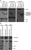
VIPAR and VPS33B are not part of the HOPS complex. Immunoprecipitation followed by SDS PAGE and immunoblotting. A) Lysates of HeLaM cells transiently expressing GFP or stably expressing VPS16‐GFP, VPS33A‐GFP, VIPAR‐GFP or VPS33B‐GFP were subjected to immunoprecipitation with anti‐VPS18 antibody (middle) or GFP‐TRAP® agarose beads (right). Dotted line indicates separate images arising from different exposure times of membrane. *Heavy chain of anti‐VPS18 antibody used for immunoprecipitation detected by cross reactivity with HRP conjugated, goat‐anti‐rabbit IgG secondary antibody. B) Lysates of HeLaM cells stably expressing VPS33B‐HA that were transiently transfected with empty GFP vector or stably coexpressing both VPS33B‐HA and VIPAR‐GFP were subjected to immunoprecipitation with anti‐HA affinity matrix (middle) or GFP‐TRAP agarose beads (right).

Similar articles

Cited by

References

    1. Huotari J, Helenius A. Endosome maturation. EMBO J 2011;30:3481–3500. - PMC - PubMed
    1. Luzio JP, Hackmann Y, Dieckmann NM, Griffiths GM. The biogenesis of lysosomes and lysosome‐related organelles. Cold Spring Harb Perspect Biol 2014;6:a016840. - PMC - PubMed
    1. Shen HM, Mizushima N. At the end of the autophagic road: an emerging understanding of lysosomal functions in autophagy. Trends Biochem Sci 2014;39:61–71. - PubMed
    1. Yang Z, Klionsky DJ. An overview of the molecular mechanism of autophagy. Curr Top Microbiol Immunol 2009;335:1–32. - PMC - PubMed
    1. Brocker C, Kuhlee A, Gatsogiannis C, Balderhaar HJ, Honscher C, Engelbrecht‐Vandre S, Ungermann C, Raunser S. Molecular architecture of the multisubunit homotypic fusion and vacuole protein sorting (HOPS) tethering complex. Proc Natl Acad Sci USA 2012;109:1991–1996. - PMC - PubMed

Publication types

Substances

  NODES
Association 6
twitter 2