. 2016 Jan 19;113(3):E396-405.
doi: 10.1073/pnas.1512779113. Epub 2015 Dec 30.

Calcium-dependent oligomerization of CAR proteins at cell membrane modulates ABA signaling

Affiliations

Calcium-dependent oligomerization of CAR proteins at cell membrane modulates ABA signaling

Maira Diaz et al. Proc Natl Acad Sci U S A. .

Abstract

Regulation of ion transport in plants is essential for cell function. Abiotic stress unbalances cell ion homeostasis, and plants tend to readjust it, regulating membrane transporters and channels. The plant hormone abscisic acid (ABA) and the second messenger Ca(2+) are central in such processes, as they are involved in the regulation of protein kinases and phosphatases that control ion transport activity in response to environmental stimuli. The identification and characterization of the molecular mechanisms underlying the effect of ABA and Ca(2+) signaling pathways on membrane function are central and could provide opportunities for crop improvement. The C2-domain ABA-related (CAR) family of small proteins is involved in the Ca(2+)-dependent recruitment of the pyrabactin resistance 1/PYR1-like (PYR/PYL) ABA receptors to the membrane. However, to fully understand CAR function, it is necessary to define a molecular mechanism that integrates Ca(2+) sensing, membrane interaction, and the recognition of the PYR/PYL interacting partners. We present structural and biochemical data showing that CARs are peripheral membrane proteins that functionally cluster on the membrane and generate strong positive membrane curvature in a Ca(2+)-dependent manner. These features represent a mechanism for the generation, stabilization, and/or specific recognition of membrane discontinuities. Such structures may act as signaling platforms involved in the recruitment of PYR/PYL receptors and other signaling components involved in cell responses to stress.

Keywords: abiotic stress; ion transport; membrane biology; signaling.


Conflict of interest statement

The authors declare no conflict of interest.

Figures

Fig. 1.
Fig. 1.
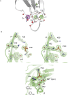
The crystal structures of CAR4 and CAR1. (A) A ribbon representation of CAR4 in complex with Ca2+ and PSF. (B) The ribbon representation of the Ca2+-dependent lipid binding sites of CAR4 (Left) and CAR1 (Right). The key structural features of the CAR fold are labeled and highlighted in different colors. The Ca2+ atoms are shown as gray spheres. Other nonprotein atoms and key residues at the Ca2+ binding sites are displayed in a ball-and-stick representation.
Fig. S1.
Fig. S1.
The lipid binding sites of CAR4. (A) The analysis of the anomalous difference density maps computed with data collected with crystals of CAR4 Ca2+ and Zn2+ at wavelength 1.7 (green) and 0.93 A (magenta) allows the unambiguous identification of the nature of the CAR4 metal centers, as Zn2+ and Ca2+ absorb radiation differentially at these wavelengths. The maps are contoured at 7 σ. (B) The polybasic binding site of CAR4 and three sections of the 2Fo-Fc simulated annealed omit map at the polybasic lipid binding site of CAR4 for the complex between CAR4 and PSF, the complex between CAR4 and POC, and the complex between CAR4, Zn, and POC.
Fig. S2.
Fig. S2.
The ribbon representation of the crystal structures of CAR4 (Left) and CAR1 (Right).
Fig. 2.
Fig. 2.
The polybasic lipid binding site of CAR4. (A) Structure of CAR4 in complex with POC and (B) with PSF. (C) Structure of the C2 domain of PKC-α in complex with PI(4,5)P2 is shown for comparison. The C2 domains are represented as green ribbons. Residues involved in ligand binding are represented as sticks. (D) Schematic representation of the polybasic lipid binding site highlighting the similarities and differences between CAR4 and PKC-α. Key CAR4 amino acids are labeled in black, and the corresponding amino acids in PKC-α are indicated in blue.
Fig. S3.
Fig. S3.
Structural comparison of the metal-dependent POC binding sites of (A) CAR4, (B) the C2 domain of PKC-e (PDB ID code 1GMI), (C) phosphoryl choline esterase (PDB ID code 2BIB), and (D) C reactive protein (PDB ID code 1B09).
Fig. 3.
Fig. 3.
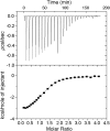
Calorimetric titration of CAR4 with Ca2+. (Upper) Representative thermogram obtained by the addition of 2 mM Ca2+ (1 × 1-μL, 10 × 5-μL, and 10 × 10-μL injections) to a solution of 64.4 μM CAR4 at 25 °C. (Lower) Dependence of the heat released per mol of Ca2+ injected as a function of the Ca2+:CAR4 molar ratio. The solid line corresponds to the best fit of the experimental data based on a one-set-of-sites model.
Fig. 4.
Fig. 4.
In vitro membrane binding and subcellular localization of CAR proteins. (A) Calcium-dependent lipid cosedimentation assays with CAR4. The protein was incubated with liposomes in the presence of increasing concentrations of Ca2+. (B) Comparative analyses of phospholipid binding of wild-type CAR4 and mutants at the Ca2+-dependent lipid binding site CAR4-DADA and the polybasic lipid binding site CAR4-KAKA. Protein quantifications of the soluble fraction after lipid pelleting on A and B were performed by measuring the optical density at 280 nm on a spectrophotometer. Lipid binding activity is expressed as the percentage of the unbound protein to lipids. The differences in the percentage of soluble protein in A and B are explained by the different protein concentration used in the experiments (Materials and Methods). Error bars indicate the SD calculated from three independent measurements. (C) A representative Coomassie blue-stained SDS/PAGE corresponding to the experiments shown in B. (D) Quantification of the subcellular location of CAR4 in the presence or absence of free Ca2+; S, C, and M represent the nonnuclear protein fraction, the cytosolic fraction, and the microsomal fraction, respectively. Immunoblot signals obtained in D were captured using the image analyzer LAS3000, and quantification of the protein signal was done using Image Guache version 4.0 software.
Fig. 5.
Fig. 5.
PYR/PYL ABA receptors do not interact with CAR4 in solution. Size exclusion chromatography profiles of CAR4, dimeric PYL1, monomeric PYL6, and their equimolecular mixtures as described in Materials and Methods.
Fig. 6.
Fig. 6.
(A) CAR4 forms oligomeric structures in solution. (Right) Chemical cross-linking assays for self-association of CAR4 and CAR4-DADA. A Coomassie blue-stained SDS/PAGE showing the cross-linked species by 0.05% glutaraldehyde at different times in minutes. Cross-linked species as well as the monomer migrate aberrantly and are highlighted with arrows. (Left) Analytical ultracentrifugation analysis for CAR4 in native conditions and sedimentation coefficient c(s) distribution. The Inset corresponds to a zoomed area of the distribution. The position and the area under each peak reveal the molecular weight and the relative abundance of the CAR4 oligomers. (B) CAR4 binds to the periphery of liposomes and generates membrane tubules in a Ca2+-dependent manner. Negative-stain transmission electron micrographs of 12.5 μM liposomes (Upper Left) incubated with 8 μM CAR4 (Upper Right), 0.08 μM CAR4 plus 1 mM Ca2+ (Lower Left), and 8 μM CAR4 plus 1 mM Ca2+ (Lower Right). Peripherally membrane-bound CAR4 produces roughness on the surface of the liposomes and is indicated with black arrows, liposome tubules with white arrows, and protein aggregates as black asterisks. (Scale bar, 200 nm.)
Fig. S4.
Fig. S4.
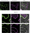
Interaction of PYR1 and CAR1 generates punctate/globular structures in plasma membrane. Confocal images of transiently transformed tobacco epidermal cells coexpressing CAR1-YFPN/YFPC-PYR1 interacting proteins and a key cellular component for membrane trafficking. Asterisks indicate the presence of CAR1-PYR1 in membrane complexes decorated by At1g20110. To investigate the interaction of CAR1 and PYR1, we used the pSPYNE-35S and pYFPC43 vectors, respectively. At1g20110 was recombined by LR reaction into pH7WGR2 vector to coexpress RFP-At1g20110 together with CAR1-YFPN/YFPC-PYR1 using Agrobacterium-mediated transfection of tobacco leaves.
Fig. 7.
Fig. 7.
Overexpression of CAR1-DADA leads to a dominant-negative phenotype with respect to ABA sensitivity. (A) CAR1-DADA OE lines show reduced sensitivity to ABA compared with Col WT (Upper Left). Expression of HA-tagged CAR1-DADA was verified by immunoblot analysis of the transgenic lines (Upper Right). Photographs are shown of representative seedlings from Col WT and two CAR1-DADA OE lines grown for 12 d on MS medium either lacking or supplemented with 0.5 μM ABA. Seedlings were rearranged on agar plates, and longitudinal or zenital photographs were taken to measure root length or the maximum rosette radius. The histograms show the quantification of root length (Lower Left) or the maximum rosette radius (Lower Right). (B) Quantification of ABA-mediated inhibition of seedling establishment of Col WT compared with hab1-1abi1-2 double mutant, CAR1 OE lines, and HAB1 OE and CAR1D22 D27A OE lines. Approximately 100 seeds of each genotype were sown on MS plates lacking or supplemented with either 0.5 or 1 mM ABA and scored for the presence of green expanded cotyledons 7 d later. *P < 0.05 (Student’s t test) when comparing data of each genetic background to Col WT plants in the same assay conditions. (C) The CAR1-DADA mutation abolishes the interaction of CAR1 with PYR1 in the plasma membrane. Confocal images of transiently transformed tobacco epidermal cells coexpressing the indicated constructs and increasing amounts of CAR1-DADA. The ratio of the relative concentration of agrobacteria in the different coinfiltrations is indicated by numbers (1×, 2×, or 4×).
Fig. 8.
Fig. 8.
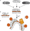
A schematic representation of the mechanism for CAR membrane interaction and PYR/PYL receptor recruitment. (1) Oligomeric CAR protein is recruited to the membrane. In the absence of a Ca2+ signal, the membrane interaction of CAR proteins is promoted but restricted both by a structural Ca2+ binding site (site I) and by the positively charged polybasic binding site. The dominant-negative CAR-DADA mutant precludes the anchoring of the oligomer to the membrane. (2) Abiotic stress induces an increase of the physiological Ca2+ concentration to the μM range. This triggers Ca2+ binding to site II and CAR membrane insertion. CAR molecules would act as molecular wedges into the membrane that have to overcome the lateral membrane energy barrier. (3) The bulkier side chain of lipids at membrane nanodomains generates membrane curvature. This facilitates CAR membrane insertion, as the membrane lateral pressure is relieved. The accumulation of CAR molecules favors the recruitment of PYR/PYL receptors. Additionally, this situation would help to recruit lipidated proteins for the control of ion homeostasis.

Similar articles

Cited by

References

    1. Serrano R, Rodriguez-Navarro A. Ion homeostasis during salt stress in plants. Curr Opin Cell Biol. 2001;13(4):399–404. - PubMed
    1. Bassil E, Blumwald E. The ins and outs of intracellular ion homeostasis: NHX-type cation/H(+) transporters. Curr Opin Plant Biol. 2014;22:1–6. - PubMed
    1. Batistič O, Kudla J. Analysis of calcium signaling pathways in plants. Biochim Biophys Acta. 2012;1820(8):1283–1293. - PubMed
    1. Cutler SR, Rodriguez PL, Finkelstein RR, Abrams SR. Abscisic acid: Emergence of a core signaling network. Annu Rev Plant Biol. 2010;61:651–679. - PubMed
    1. McAinsh MR, Brownlee C, Hetherington AM. Abscisic acid-induced elevation of guard cell cytosolic Ca2+ precedes stomatal closure. Nature. 1990;343(6254):186–188.

Publication types

MeSH terms

  NODES
Association 1
INTERN 1
twitter 2