Review
. 2016 Jun 20;8(1):69.
doi: 10.1186/s13073-016-0324-x.

Roles of telomeres and telomerase in cancer, and advances in telomerase-_targeted therapies

Affiliations
Review

Roles of telomeres and telomerase in cancer, and advances in telomerase-_targeted therapies

Mohammad A Jafri et al. Genome Med. .

Abstract

Telomeres maintain genomic integrity in normal cells, and their progressive shortening during successive cell divisions induces chromosomal instability. In the large majority of cancer cells, telomere length is maintained by telomerase. Thus, telomere length and telomerase activity are crucial for cancer initiation and the survival of tumors. Several pathways that regulate telomere length have been identified, and genome-scale studies have helped in mapping genes that are involved in telomere length control. Additionally, genomic screening for recurrent human telomerase gene hTERT promoter mutations and mutations in genes involved in the alternative lengthening of telomeres pathway, such as ATRX and DAXX, has elucidated how these genomic changes contribute to the activation of telomere maintenance mechanisms in cancer cells. Attempts have also been made to develop telomere length- and telomerase-based diagnostic tools and anticancer therapeutics. Recent efforts have revealed key aspects of telomerase assembly, intracellular trafficking and recruitment to telomeres for completing DNA synthesis, which may provide novel _targets for the development of anticancer agents. Here, we summarize telomere organization and function and its role in oncogenesis. We also highlight genomic mutations that lead to reactivation of telomerase, and mechanisms of telomerase reconstitution and trafficking that shed light on its function in cancer initiation and tumor development. Additionally, recent advances in the clinical development of telomerase inhibitors, as well as potential novel _targets, will be summarized.


Figures
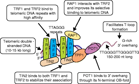
Fig. 1
Fig. 1
Schematic representation of telomeric DNA and components of the shelterin complex. Telomeres comprise a specialized nucleoprotein-capping structure consisting of DNA and shelterin protein complexes. Telomeric DNA contains a variable number of G-rich, non-coding, tandem repeats of the double-stranded DNA sequence 5′-(TTAGGG)n-3′, followed by a terminal 3′ G-rich single-stranded overhang (150–200 nucleotides (nt) long). The 3′ G-rich overhang facilitates telomeric DNA in forming a higher-order structure in which the 3′ single-stranded overhang folds back and invades the homologous double-stranded TTAGGG region, forming a telomeric loop (T-loop) that provides 3′-end protection by sequestering it from recognition by the DNA damage response machinery. The proteins associated with telomeres form the shelterin complex, which consists of three core shelterin subunits, TRF1 and TRF2, which directly recognize and bind duplex TTAGGG repeats, and POT1, which recognizes and binds single-stranded TTAGGG overhangs. These three proteins are interconnected by three additional shelterin proteins, TIN2, TPP1 and RAP1, forming a complex that enables the DNA damage response surveillance machinery to distinguish telomere DNA from sites of genomic DNA damage
Fig. 2
Fig. 2
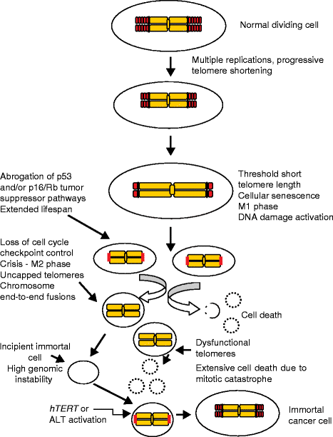
Cellular senescence and crisis. Telomeres protect chromosome ends from undergoing fusions and recombination by masking telomeric DNA with shelterin protein protective caps, preventing the ends from being recognized by the DNA damage surveillance pathways. Telomere shortening is a natural consequence of cell division due to the “end replication problem” whereby lagging strand DNA synthesis cannot be completed all the way to the very end, and increased cell divisions lead to critically shortened telomeres which elicit DNA damage responses that trigger cellular senescence. In the cells undergoing replicative senescence, the p53 and p16–RB pathways are often activated leading to essentially irreversible growth arrest. Cells that gain additional oncogenic changes (p53 loss) can bypass senescence and continue to divide until multiple critically shortened telomeres initiate crisis, a period of increased chromosome end-to-end fusions and extensive cell death. Only a rare human cell (one in 105 to 107) can engage a mechanism to bypass crisis and become immortal. This is almost universally accomplished by the upregulation or reactivation of telomerase. A rarer telomerase negative immortalization pathway, termed ALT (alternative lengthening of telomeres), involves DNA recombination to maintain telomeres
Fig. 3
Fig. 3
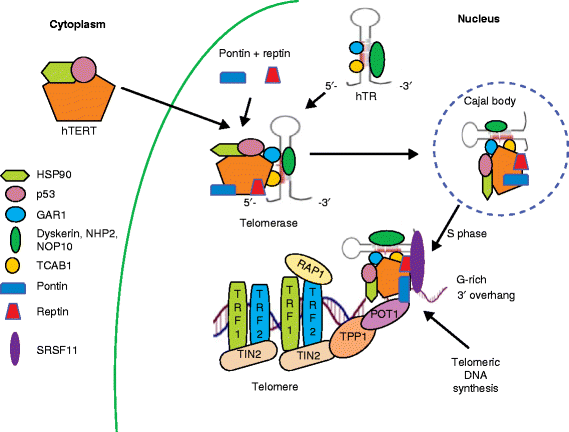
Telomerase assembly, recruitment to the telomere, and telomeric DNA synthesis. Telomerase is the cellular ribonucleoprotein enzyme complex that catalyzes the extension of telomeric DNA in eukaryotic organisms. Telomerase action involves multiple steps including assembly of the telomerase complex, its intracellular trafficking and finally recruitment to telomeres. Human telomerase is composed of hTR (hTERC—a template functional RNA), hTERT (the catalytic protein component with reverse transcriptase activity), and the accessory proteins dyskerin, NOP10, NHP2, and GAR1. hTERT protein associates with p23 and HSP90 in the cytoplasm, and moves to the nucleus. Nascent hTR transcripts complex with dyskerin, NHP2, NOP10 and GAR1. This complex then undergoes Reptin and Pontin (ATPases)-mediated binding to hTERT + p23 + HSP90 complex. Then TCAB1 attaches to this assembling complex and guides it to Cajal bodies in the nucleus. Telomerase recruitment to telomeres takes place during the S phase of the cell cycle through interactions between the shelterin complex components TPP1 and POT1 and the DAT domain of hTERT. SRSF11 stabilizes the association of the telomerase enzyme complex with the telomere overhang for DNA synthesis
Fig. 4
Fig. 4
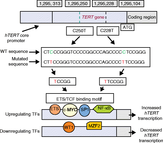
hTERT transcription and promoter mutations. The hTERT gene is tightly repressed in almost all normal cells and tissues. Specific hTERT promoter mutations as part of cancer progression occur leading to increased transcription of hTERT. The transcription of hTERT is regulated by a series of transcription factors (TFs). hTERT promoter mutations create ETS/TCF binding motifs. Each mutation generates a new ETS/TCF binding site. Upregulating TFs such as ETS, c-MYC, SP1 and NF-kB bind to their respective sites and can promote hTERT transcription. Although binding of TFs is essential for hTERT transcription, in addition a permissive chromatin microenvironment is required. Binding of downregulating transcription factors decreases transcription. WT wild type; WT1 Wilms tumor protein1; MZF2 myeloid zinc finger protein 2
Fig. 5
Fig. 5
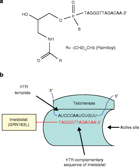
Structure and action of imetelstat (GRN163L). a Structure of imetelstat. Imetelstat is a lipid-conjugated 13-mer oligonucleotide sequence with a thio-phosphoramidate backbone. The oligonucleotide sequence is complementary to the hTR component of telomerase and is responsible for the inhibitory activity of imetelstat, whereas the thio-phosphoramidate backbone imparts resistance to the action of plasma and cellular nucleases. b Action of imetelstat. Imetelstat binds to the hTR template region at the hTERT active site with high affinity and blocks its recruitment to telomeric DNA. Imetelstat is a competitive telomerase template antagonist (not antisense that _targets mRNA). Binding of imetelstat to hTR results in telomerase inhibition leading to progressively shortened telomeres
Fig. 6
Fig. 6
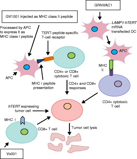
Anti-telomerase immunotherapy. Several telomerase-based vaccines have been developed, which sensitize immune cells to cancer cells expressing hTERT peptides as surface antigens via the human leukocyte antigen (HLA) class I and class II pathways. This results in an expansion of telomerase-specific CD4+ and CD8+ cytotoxic T lymphocytes (CTLs) in cancer patients leading T cells to _target and kill telomerase-positive tumor cells. GV1001 is an MHC class II-restricted hTERT peptide that is further processed by antigen presenting cells (APCs) to present as an MHC class I peptide, and it produces both CD4+- and CD8+-based immune responses. GRNVAC1 stimulates CD4+ T cells to _target and kill hTERT-expressing tumor cells. Vx001 action is mediated by CD8+ T cells

Similar articles

Cited by

References

    1. Akincilar SC, Unal B, Tergaonkar V. Reactivation of telomerase in cancer. Cell Mol Life Sci. 2016;73:1659–70. doi: 10.1007/s00018-016-2146-9. - DOI - PMC - PubMed
    1. Shay JW. Are short telomeres predictive of advanced cancer? Cancer Discov. 2013;3:1096–8. doi: 10.1158/2159-8290.CD-13-0506. - DOI - PMC - PubMed
    1. Horn S, Figl A, Rachakonda PS, Fischer C, Sucker A, Gast A, et al. TERT promoter mutations in familial and sporadic melanoma. Science. 2013;339:959–61. doi: 10.1126/science.1230062. - DOI - PubMed
    1. Huang FW, Hodis E, Xu MJ, Kryukov GV, Chin L, Garraway LA. Highly recurrent TERT promoter mutations in human melanoma. Science. 2013;339:957–9. doi: 10.1126/science.1229259. - DOI - PMC - PubMed
    1. Vinagre J, Almeida A, Popula H, Batista R, Lyra J, Pinto V, et al. Frequency of TERT promoter mutations in human cancers. Nat Commun. 2013;4:2185. doi: 10.1038/ncomms3185. - DOI - PubMed

Publication types

MeSH terms

  NODES
Association 1
coding 1
HOME 3
Javascript 1
mac 9
OOP 4
os 24
text 12
twitter 2
web 4