Review
. 2016 Oct 31;5(11):93.
doi: 10.3390/jcm5110093.

Air Embolism: Practical Tips for Prevention and Treatment

Affiliations
Review

Air Embolism: Practical Tips for Prevention and Treatment

Colin J McCarthy et al. J Clin Med. .

Abstract

Air embolism is a rarely encountered but much dreaded complication of surgical procedures that can cause serious harm, including death. Cases that involve the use of endovascular techniques have a higher risk of air embolism; therefore, a heightened awareness of this complication is warranted. In particular, central venous catheters and arterial catheters that are often placed and removed in most hospitals by a variety of medical practitioners are at especially high risk for air embolism. With appropriate precautions and techniques it can be preventable. This article reviews the causes of air embolism, clinical management and prevention techniques.

Keywords: Air embolism; catheter; embolization; endovascular.


Conflict of interest statement

The authors declared no conflicts of interest and have no financial disclosures.

Figures

Figure 1
Figure 1
A 35-year-old man was transferred from an outside hospital with altered mental status following the placement of a peripheral intravenous line. A CT of the brain was performed in the emergency room. Axial (a) and coronal (b) CT revealed a moderate amount of air in the cavernous sinuses bilaterally (arrows) as well as multiple small foci of air in the region of the pterygoid venous plexuses (c), scattered in the soft tissues and in the extradural space at the level of the upper cervical spine (arrowheads). A follow-up CT (d) was performed the next day, which revealed resolution of the previously seen air. In this case, the patient made a full recovery.
Figure 1
Figure 1
A 35-year-old man was transferred from an outside hospital with altered mental status following the placement of a peripheral intravenous line. A CT of the brain was performed in the emergency room. Axial (a) and coronal (b) CT revealed a moderate amount of air in the cavernous sinuses bilaterally (arrows) as well as multiple small foci of air in the region of the pterygoid venous plexuses (c), scattered in the soft tissues and in the extradural space at the level of the upper cervical spine (arrowheads). A follow-up CT (d) was performed the next day, which revealed resolution of the previously seen air. In this case, the patient made a full recovery.
Figure 2
Figure 2
A 61-year-old male underwent an initial femoral angiogram (a), which revealed a small amount of air in the tubing of the side port (arrowhead) that had traveled into the superficial femoral artery (arrows). As the lower extremity arteriogram (b) revealed no visible distal emboli, it was presumed the air was resorbed. There were no adverse sequelae.
Figure 3
Figure 3
A 69-year-old man who was critically ill with sepsis, pneumonia, and Cerebral Vascular Accident (CVA). Immediately after his pulmonary arterial line was pulled, he developed sudden cardiopulmonary arrest, suspected to be related to air embolism. A post-mortem CT (ac) was performed 18 h later, revealing intravascular air in the ascending aorta (a, arrow), pulmonary artery (b, arrowhead), right ventricle (c, *) and the liver (arrow). Given the extent of air, it was considered most likely that the air was related to post-mortem state. Despite these findings, even when the heart was opened under water at autopsy, no air escaped, highlighting the difficulty in diagnosis of air embolism in some cases.
Figure 4
Figure 4
Following the placement of right internal jugular tunneled Hickman line, a 76-year-old man developed an episode of desaturation, tachycardia, and non-responsiveness. Subsequently, an immediate CT of brain, neck and chest was performed and gas was found in the left cavernous sinus (a,b) and within a left parietal sulcus (c); it was felt to represent air in a vessel, possibly a pial vein. There was also a large amount of air surrounding the right internal jugular vein (d); with a partially visualized central venous catheter (*). A brain MRI was performed showing FLAIR hyperintensity in multiple regions (e) with evidence of restricted diffusion (f); and corresponding low signal (arrowheads) on the apparent diffusion coefficient (ADC) images (g). The patient received a single treatment of hyperbaric oxygen but died 8 days later.
Figure 4
Figure 4
Following the placement of right internal jugular tunneled Hickman line, a 76-year-old man developed an episode of desaturation, tachycardia, and non-responsiveness. Subsequently, an immediate CT of brain, neck and chest was performed and gas was found in the left cavernous sinus (a,b) and within a left parietal sulcus (c); it was felt to represent air in a vessel, possibly a pial vein. There was also a large amount of air surrounding the right internal jugular vein (d); with a partially visualized central venous catheter (*). A brain MRI was performed showing FLAIR hyperintensity in multiple regions (e) with evidence of restricted diffusion (f); and corresponding low signal (arrowheads) on the apparent diffusion coefficient (ADC) images (g). The patient received a single treatment of hyperbaric oxygen but died 8 days later.
Figure 5
Figure 5
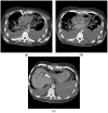
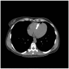
A 35-year-old woman had an incidental finding of air in the right ventricle on non-contrast CT abdomen performed for renal stone evaluation. The patient was asymptomatic and an IV line had been placed approximately 1 hour earlier.
Figure 6
Figure 6
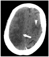
Clinical illustration of an arterial air embolism in a patient with patent foramen ovale (PFO) can be seen in the following example in which a 57-year-old female underwent liver resection. At the end of the case, the patient became profoundly hypotensive, and an air embolism was suspected. Approximately 20 cc of air was aspirated from an indwelling central venous catheter, an Extracorporeal membrane oxygenation (ECMO) was initiated and the patient was transferred to ICU where CT brain was performed. An axial CT shows multiple areas of low attenuation representing infarction of the right (arrow) and left (arrowhead) cerebral hemispheres, consistent with underlying embolic source of infarct. Furthermore, the CT shows loss of gray-white differentiation consistent with cerebral edema. Transesophageal echocardiography was performed (not shown), revealing a large amount of air bubbles in the right and left chambers heart chambers. This, together with a patent foramen ovale, resulted in paradoxical embolism.
Figure 7
Figure 7
By ensuring the syringe is held with the plunger upright (A), there is a significant decrease in the risk of inadvertent injection of any air that may be contained within the syringe (B).
Figure 8
Figure 8
A 27-year-old woman with encephalitis who suffered a respiratory arrest. Two images from axial CT brain (a,b) performed shortly afterward revealed extensive, serpiginous hypodensity in the sulcal distribution overlying the cerebral hemispheres bilaterally (arrowheads), representing intravascular air embolization. The patient made a full recovery with prompt 100% oxygen administration and supportive measures alone.
Figure 9
Figure 9
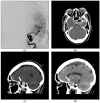
A 68-year-old woman underwent cerebral angiogram (a) for evaluation of 3 cm left cavernous internal carotid artery aneurysm (*). Post procedure, she developed transient aphasia, right facial droop, and right arm brachyplegia likely secondary to air embolism. This was presumed to be related to faulty occlusion balloon. The patient was immediately placed in a cervical collar to minimize head movements and distal air embolization as well as started on 100% oxygen therapy. Hyperbaric chamber therapy was discussed but declined due to concerns about patient movement during transport. CT angiogram (b,c) was performed immediately. Post-procedure revealed air within the large left cavernous segment internal carotid artery aneurysm (arrow). A CT performed 2 days later (d) showed the embolism had resolved and this patient made a full recovery.
Figure 10
Figure 10
The successful use of a hyperbaric oxygen therapy (HBOT) is outlined in this case, in which a 69-year-old female with a history of suspected lung cancer underwent a CT-guided lung biopsy at an outside facility (a). During the procedure, the patient suffered a cardiorespiratory arrest. An immediate CT (b) revealed a right pneumothorax (arrow), together with air in the right pulmonary vein (arrowhead). Additional images from the CT scan (c,d), revealed air in the right coronary artery (black arrow), the ascending thoracic aorta (white arrow) and the epidural veins (arrowhead). The patient was initially unresponsive and required cardiopulmonary resuscitation. A non-contrast CT brain (e) was performed, revealing air scattered in the vessels overlying the right cerebral hemisphere (arrowheads). Subsequent MRI of the brain confirmed multiple areas of acute infarction in the right cerebral and cerebellar hemispheres. Axial T2 (f) and DWI (g) images of the brain demonstrated acute areas of infarction in the right cerebellum. The patient required ICU management and HBOT.
Figure 10
Figure 10
The successful use of a hyperbaric oxygen therapy (HBOT) is outlined in this case, in which a 69-year-old female with a history of suspected lung cancer underwent a CT-guided lung biopsy at an outside facility (a). During the procedure, the patient suffered a cardiorespiratory arrest. An immediate CT (b) revealed a right pneumothorax (arrow), together with air in the right pulmonary vein (arrowhead). Additional images from the CT scan (c,d), revealed air in the right coronary artery (black arrow), the ascending thoracic aorta (white arrow) and the epidural veins (arrowhead). The patient was initially unresponsive and required cardiopulmonary resuscitation. A non-contrast CT brain (e) was performed, revealing air scattered in the vessels overlying the right cerebral hemisphere (arrowheads). Subsequent MRI of the brain confirmed multiple areas of acute infarction in the right cerebral and cerebellar hemispheres. Axial T2 (f) and DWI (g) images of the brain demonstrated acute areas of infarction in the right cerebellum. The patient required ICU management and HBOT.

Similar articles

Cited by

References

    1. Mirski M.A., Lele A.V., Fitzsimmons L., Toung T.J. Diagnosis and treatment of vascular air embolism. Anesthesiology. 2007;106:164–177. doi: 10.1097/00000542-200701000-00026. - DOI - PubMed
    1. Muth C.M., Shank E.S. Gas embolism. N. Engl. J. Med. 2000;342:476–482. doi: 10.1056/NEJM200002173420706. - DOI - PubMed
    1. Vesely T.M. Air embolism during insertion of central venous catheters. J. Vasc. Int. Radiol. 2001;12:1291–1295. doi: 10.1016/S1051-0443(07)61554-1. - DOI - PubMed
    1. Wysoki M.G., Covey A., Pollak J., Rosenblatt M., Aruny J., Denbow N. Evaluation of various maneuvers for prevention of air embolism during central venous catheter placement. J. Vasc. Int. Radiol. 2001;12:764–766. doi: 10.1016/S1051-0443(07)61451-1. - DOI - PubMed
    1. Toung T.J., Rossberg M.I., Hutchins G.M. Volume of air in a lethal venous air embolism. Anesthesiology. 2001;94:360–361. doi: 10.1097/00000542-200102000-00031. - DOI - PubMed

LinkOut - more resources

  NODES
admin 1
INTERN 8
twitter 2