. 2016 Dec 5:6:38550.
doi: 10.1038/srep38550.

Ubiquitin Ligase NEDD4 Regulates PPARγ Stability and Adipocyte Differentiation in 3T3-L1 Cells

Affiliations

Ubiquitin Ligase NEDD4 Regulates PPARγ Stability and Adipocyte Differentiation in 3T3-L1 Cells

Jing Jing Li et al. Sci Rep. .

Abstract

Peroxisome proliferator-activated receptor-γ (PPARγ) is a ligand-activated nuclear receptor which controls lipid and glucose metabolism. It is also the master regulator of adipogenesis. In adipocytes, ligand-dependent PPARγ activation is associated with proteasomal degradation; therefore, regulation of PPARγ degradation may modulate its transcriptional activity. Here, we show that neural precursor cell expressed developmentally down-regulated protein 4 (NEDD4), an E3 ubiquitin ligase, interacts with the hinge and ligand binding domains of PPARγ and is a bona fide E3 ligase for PPARγ. NEDD4 increases PPARγ stability through the inhibition of its proteasomal degradation. Knockdown of NEDD4 in 3T3-L1 adipocytes reduces PPARγ protein levels and suppresses adipocyte conversion. PPARγ correlates positively with NEDD4 in obese adipose tissue. Together, these findings support NEDD4 as a novel regulator of adipogenesis by modulating the stability of PPARγ.


Figures

Figure 1
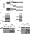
Figure 1. NEDD4 interacts with PPARγ.
(A) Interaction between NEDD4 and PPARγ in 3T3-L1 adipocytes. Total lysates (500 μg) were immunoprecipitated with 1 μg of anti-PPARγ antibody (clone E-8). Normal mouse IgG served as negative control. (B) In vivo interaction between NEDD4 and PPARγ. Epididymal adipose tissue lysates (500 μg) were immunoprecipitated with 1 μg of anti-PPARγ antibody (clone E-8) and immunoblotted with anti-NEDD4 antibody. PPARγ immunoblot was performed with clone T.647.5. (C) Co-immunoprecipitation of NEDD4 and PPARγ in HEK293 cells transiently expressing plasmids containing T7-tagged NEDD4 and FLAG-tagged PPARγ2. The rabbit monoclonal anti-PPARγ antibody (clone T.647.5) was used for immunoprecipitation, and the mouse monoclonal anti-PPARγ antibody (clone E-8) was used for immunoblotting. Lysates pulled down with normal rabbit IgG served as negative control. Full-length blots are presented in Supplementary Fig. S4.
Figure 2
Figure 2. NEDD4 associates with the hinge/ligand binding domain of PPARγ.
(A) Schematic drawings of PPARγ protein domains and domain-deleted mutants. GAL4DBD-HA plasmid was fused with AF-1, DBD, Hinge, or LBD domain of mouse PPARγ2. (B,C) The PPxY motif is not required for NEDD4-PPARγ interaction. HEK293 cells were transiently co-transfected with T7‐tagged NEDD4 and FLAG‐tagged PPARγ2 or its mutants. The mouse anti-PPARγ antibody was used for immunoprecipitation. For immunoblotting, the mouse anti-PPARγ (clone E-8) antibody was used in panel B, while the rabbit anti-PPARγ (clone T.647.5) antibody was used in panel C. The IgG heavy chain was thus not detected in panel C. (D) NEDD4 interacts with the hinge and LBD domains of PPARγ. GAL4DBD-HA plasmids containing AF-1, DBD, Hinge, or LBD domain of PPARγ2 were co-expressed in HEK293 cells with T7‐NEDD4. Lysates were immunoprecipitated with anti-HA antibody. (E) Interaction between NEDD4 and domain-deleted FLAG-PPARγ2. HEK293 cells were transfected with T7-NEDD4 alone, or together with FLAG-PPARγ2, FLAG-PPARγ2∆Hinge (without amino acids 206–280), FLAG-PPARγ2∆LBD (without amino acids 281–505), or FLAG-PPARγ2∆∆ (without amino acids 206–505). Cells were harvested 48 hours after transfection. MG132 (10 μM) was added to the cells 16 hr before harvesting. Lysates were immunoprecipitated with anti-FLAG antibody. The rabbit anti-PPARγ (clone H-100) antibody against N-terminal PPARγ was used for immunoblotting. Arrowhead indicates expected size of the ∆∆ mutant. Full-length blots are presented in Supplementary Fig. S4.
Figure 3
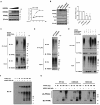
Figure 3. NEDD4 mediates lysine-48 independent ubiquitination of PPARγ.
(A) Dose-dependent expression of NEDD4 does not reduce steady-state PPARγ protein abundance. Quantification of NEDD4 and PPARγ levels is on the right. Variable amounts of pRc-CMV-T7-NEDD4 expression vector from 1 to 3 μg were transfected with the PPARγ into HEK293 cells. NEDD4 plasmid expression saturates at 2 μg per well of a 6-well plate. (B) 1 μg of either FLAG-tagged SIAH1 or HA-tagged SIAH2, together with 1 μg of T7-tagged NEDD4, were transfected into CHO cells stably expressing FLAG-tagged PPARγ2. The relative PPARγ levels are quantified on the right. (C) NEDD4 overexpression enhances in vivo ubiquitination of PPARγ. T7-tagged NEDD4, FLAG-tagged PPARγ2, and HA-tagged ubiquitin (Ub) were transiently expressed in HEK293 cells at a ratio of 1.5:1:2 in combinations as indicated above the blots. (D) NEDD4 knockdown reduces in vivo ubiquitination of PPARγ. HEK293 cells seeded on 6-well plates were transfected with 1 μg of FLAG-tagged PPARγ2, 2 μg of HA-tagged Ub, along with 1.5 μg of plasmid containing NEDD4-_targeting shRNA or non-_targeting control shRNA per well. Cells were harvested for Western analysis 48 hr after the transfection. (E) NEDD4 overexpression enhances lysine-48 independent ubiquitination of PPARγ. T7-tagged NEDD4, FLAG-tagged PPARγ2, along with HA-tagged wild-type (WT) Ub or HA-tagged K48R Ub were transfected into HEK293 cells at a ratio of 1.5:1:2 in combinations as indicated above the blots. Western assays were performed at 48 hr after the transfection. (F) In vitro ubiquitination assay between NEDD4 and escalating amounts of recombinant His-GST tagged PPARγ2. (G) In vitro PPARγ2 ubiquitination by NEDD4 in the presence of wild-type ubiquitin (WT-Ub), K48-only ubiquitin (K48O-Ub), or K63-only (K63O-Ub). Data represent mean ± SEM of 3 independent experiments; *P < 0.05, **P < 0.01. Full-length blots are presented in Supplementary Fig. S4.
Figure 4
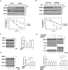
Figure 4. NEDD4 protects against rapid degradation of PPARγ.
(A,B) NEDD4 regulates PPARγ protein half-life. Representative western blot image of PPARγ protein levels during cycloheximide-chase experiment. CHO cells stably expressing FLAG-tagged PPARγ2 were transfected with indicated plasmids. Two days after transfection, cells were treated with cycloheximide for the indicated times. Quantification of data is shown below. (C) The human (h) or rat (r) NEDD4 cDNAs were expressed in CHO cells stably expressing FLAG-tagged PPARγ2. Cycloheximide was added to the media 2 hr before cell harvesting. Quantification of data is on the right. (D,E) CHO cells stably expressing FLAG-tagged PPARγ2 were co-transfected with the indicated plasmids and treated with or without the indicated reagents. Cycloheximide and MG132 were added to the media 15 hr before cell harvesting. Cycloheximide was used at 20 μM. MG132 was used at 10 μM. Data represent mean ± SEM of 3–4 independent experiments; *P < 0.05, **P < 0.01, ***P < 0.001. CHX: cycloheximide. Full-length blots are presented in Supplementary Fig. S4.
Figure 5
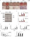
Figure 5. NEDD4 knockdown inhibits adipogenic response.
(A) Photographs and micrographs of oil red O staining of the differentiated adipocytes. Either DMSO, 10 μM of rosiglitazone, or 10 μM of GW 9662 were added to the 3T3-L1 cells at the beginning of adipocyte differentiation and were replaced with culture media every 2 days. The 3T3-L1 cells were infected with AAV virus for expressing non-_targeting shRNA control (shControl) or NEDD4-_targeting shRNA (shNEDD4) 2 days before adipocyte differentiation. Photographs and micrographs were taken 5 days or 10 days after differentiation. Scale bar represents 40 μm. (B) Endogenous PPARγ and C/EBPα expression levels during 3T3-L1 adipocyte differentiation in the presence of AAV virus were measured by Western analysis. (C) Quantification of the endogenous PPARγ1 and PPARγ2 protein expression from panel B. (DG) The mRNA levels of (D) aP2, (E) C/EBPα, (F) PPARγ, and (G) NEDD4 during 3T3-L1 adipocyte differentiation in the presence of AAV virus. (H) NEDD4 has no direct effect on the transcriptional activity of PPARγ. HEK293 cells were transiently co-transfected with 3 × PPRE-Luc, FLAG-tagged PPARγ2, Renilla luciferase control reporter vector pRL-SV40 and plasmids indicated under each column. After 32 hr, cells were treated with or without rosiglitazone (10 μM) for 16 hr prior to luciferase assay. Luciferase activity was normalized to Renilla activity as a control for transfection efficiency. IDM: 0.5 mM IBMX, 1 μM dexamethasone, and 1.5 μg/mL insulin. Data represent mean ± SEM of 3 independent experiments; *P < 0.05, **P < 0.01, n.s., no significance. Full-length blots are presented in Supplementary Fig. S4.
Figure 6
Figure 6. NEDD4 promotes PPARγ ubiquitination but is not required for ligand-dependent PPARγ degradation in 3T3-L1 adipocytes.
(A) Representative western blot image of PPARγ expression in differentiated 3T3-L1 cells in the presence of ligand. The 3T3-L1 cells were differentiated for 8 days and treated with 5 μM of rosiglitazone for 16 hr with or without the pretreatment of lysosome inhibitor chloroquine (25 μM) or proteasome inhibitor MG132 (10 μM). Quantification figure is on the right. (B) Effect of NEDD4 knockdown on PPARγ expression in differentiated 3T3-L1 cells in the presence or absence of ligand. The 3T3-L1 cells infected with shControl or shNEDD4 AAV were differentiated for 6 days and treated with or without 5 μM of rosiglitazone for 16 hr. Representative western blot image is shown and its quantification figure is shown below. (C) NEDD4 knockdown reduces PPARγ ubiquitination in 3T3-L1 adipocytes in the presence or absence of ligand. The 3T3-L1 cells infected with shControl or shNEDD4 AAV were differentiated for 6 days and treated with or without 5 μM of rosiglitazone for 16 hr. MG132 (10 μM) was added to the media 6 hr before harvesting the cells. Arrows indicate NEDD4, PPARγ1 and γ2. (D) Representative western blot image of time-dependent expression changes of PPARγ in AAV infected 3T3-L1. The 3T3-L1 cells infected with shControl or shNEDD4 AAV were differentiated for 2–3 days and treated with or without 5 μM of rosiglitazone for 8–24 hr. Quantification figures are shown on the right. Data represent mean ± SEM of 3 independent experiments; *P < 0.05, **P < 0.01, ***P < 0.001. Full-length blots are presented in Supplementary Fig. S4.
Figure 7
Figure 7. Positive correlation between NEDD4 and PPARγ levels in obese adipose tissue.
(A,B) Pearson’s R correlation coefficient between steady-state NEDD4 and PPARγ2 protein abundance in epididymal fat in 24 male HFD-fed wild-type (WT) and Nedd4+/− (Het) mice (WT: n = 12; Het: n = 12) or 28 aged (18–20 months) WT and Het mice (WT: n = 14, 12 males and 2 females; Het: n = 14, 12 males and 2 females). (C,D) Representative western blot image of NEDD4 and PPARγ expression in epididymal fat in HFD-fed or aged WT and Het mice. The HFD-fed mice at 6-week of age were fed a HFD (TD.06414, Teklad, Harlan Laboratories), containing 60% calories from fat, for 16 weeks. Samples were arranged in increasing order of NEDD4 expression. Full-length blots are presented in Supplementary Fig. S4.

Similar articles

Cited by

References

    1. Krey G. et al.. Fatty acids, eicosanoids, and hypolipidemic agents identified as ligands of peroxisome proliferator-activated receptors by coactivator-dependent receptor ligand assay. Mol. Endocrinol. 11, 779–791, doi: 10.1210/mend.11.6.0007 (1997). - DOI - PubMed
    1. Guan H. P. et al.. A futile metabolic cycle activated in adipocytes by antidiabetic agents. Nat. Med. 8, 1122–1128, doi: 10.1038/nm780 (2002). - DOI - PubMed
    1. Delerive P., Fruchart J. C. & Staels B. Peroxisome proliferator-activated receptors in inflammation control. J. Endocrinol. 169, 453–459 (2001). - PubMed
    1. Chawla A., Schwarz E. J., Dimaculangan D. D. & Lazar M. A. Peroxisome proliferator-activated receptor (PPAR) gamma: adipose-predominant expression and induction early in adipocyte differentiation. Endocrinology 135, 798–800, doi: 10.1210/endo.135.2.8033830 (1994). - DOI - PubMed
    1. Semple R. K., Chatterjee V. K. & O’Rahilly S. PPAR gamma and human metabolic disease. J. Clin. Invest. 116, 581–589, doi: 10.1172/JCI28003 (2006). - DOI - PMC - PubMed

Publication types

MeSH terms

  NODES
chat 1
twitter 2