. 2017 Mar;133(3):381-393.
doi: 10.1007/s00401-016-1661-y. Epub 2016 Dec 23.

Brain-to-stomach transfer of α-synuclein via vagal preganglionic projections

Affiliations

Brain-to-stomach transfer of α-synuclein via vagal preganglionic projections

Ayse Ulusoy et al. Acta Neuropathol. 2017 Mar.

Abstract

Detection of α-synuclein lesions in peripheral tissues is a feature of human synucleinopathies of likely pathogenetic relevance and bearing important clinical implications. Experiments were carried out to elucidate the relationship between α-synuclein accumulation in the brain and in peripheral organs, and to identify potential pathways involved in long-distance protein transfer. Results of this in vivo study revealed a route-specific transmission of α-synuclein from the rat brain to the stomach. Following _targeted midbrain overexpression of human α-synuclein, the exogenous protein was capable of reaching the gastric wall where it was accumulated into preganglionic vagal terminals. This brain-to-stomach connection likely involved intra- and inter-neuronal transfer of non-fibrillar α-synuclein that first reached the medulla oblongata, then gained access into cholinergic neurons of the dorsal motor nucleus of the vagus nerve and finally traveled via efferent fibers of these neurons contained within the vagus nerve. Data also showed a particular propensity of vagal motor neurons and efferents to accrue α-synuclein and deliver it to peripheral tissues; indeed, following its midbrain overexpression, human α-synuclein was detected within gastric nerve endings of visceromotor but not viscerosensory vagal projections. Thus, the dorsal motor nucleus of the vagus nerve represents a key relay center for central-to-peripheral α-synuclein transmission, and efferent vagal fibers may act as unique conduits for protein transfer. The presence of α-synuclein in peripheral tissues could reflect, at least in some synucleinopathy patients, an ongoing pathological process that originates within the brain and, from there, reaches distant organs innervated by motor vagal projections.

Keywords: Adeno-associated virus; Enteric nervous system; Parkinson’s disease; Rat; Synucleinopathies; Vagus nerve.


Figures
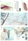
Fig. 1
Fig. 1
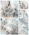
Accumulation of hα-synuclein in the DMnX, nodose ganglion and gastric wall after injections of hα-synuclein-AAVs into the vagus nerve. a–c Rats (n = 5) received a single injection of hα-synuclein-carrying AAVs into the left vagus nerve. Analyses were performed at 2–3 weeks post-treatment. a A representative image shows a section of the medulla oblongata immunostained with anti-hα-synuclein; the left DMnX is delineated by dashed lines, and the area postrema (AP) and central canal (cc) are indicated. Scale bar = 100 μm. b Medulla oblongata sections were used for stereological counting of Nissl-stained neurons (empty bar) and hα-synuclein-immunoreactive cells (solid bar) in the left DMnX. Values (means ± SEM) are expressed as percent of the total number of Nissl-stained neurons. c The representative image shows neurons robustly labeled with anti-hα-synuclein in a section of the left nodose ganglion. Scale bar = 100 μm. d–f Rats (n = 5) were killed 6 to 12 months after a single injection of hα-synuclein-carrying AAVs into the left vagus nerve. Stomach whole mounts were stained with anti-hα-synuclein and counterstained with Cuprolinic Blue. Representative images show immunoreactive fibers and nerve terminals: long intramuscular arrays (IMAs) of rectilinear terminals (d), a single vagal afferent terminating as highly arborizing intraganglionic laminar endings (IGLEs) (e), and varicosity-rich fibers with morphological features of preganglionic vagal efferents (f). Scale bars = 100 μm in (d), 25 μm in (e) and 20 μm in (f).
Fig. 2
Fig. 2
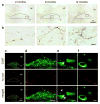
Widespread brain distribution of hα-synuclein after midbrain AAV injections. Rats received a single intraparenchymal injection of hα-synuclein-AAVs into the right midbrain. a Analyses were made in 6 rats killed at 2 months. Representative images show brain sections at different Bregma levels stained with anti-hα-synuclein. A midbrain section containing the substantia nigra is at Bregma −5.20 mm (arrow). For illustration purposes, different scale factors were used to resize each section. b, c Rats (n ≥ 5/time point) were killed at 2, 6 and 12 months post-treatment. Sections of the forebrain and pons containing the striatum (b) and locus coeruleus (LC) (c), respectively, were stained with anti-hα-synuclein. Representative images are from an animal killed at 2 months. Scale bars = 250 μm.
Fig. 3
Fig. 3
Human α-synuclein immunoreactivity in the gastric wall after midbrain AAV injections. Rats received a single intraparenchymal injection of hα-synuclein-AAVs into the right midbrain. a–f Stomach whole mounts from animals killed at 6 (a–c, n = 6) or 12 (d–f, n = 10) months post-treatment were stained with anti-hα-synuclein and counterstained with Cuprolinic Blue. Images from 5 of these animals (3 at 6 and 2 at 12 months) show a labeled swollen axon (arrow in a) and immunoreactive nerve endings around ganglionic cells (b, c, f) or groups of cells (d, e) of the myenteric plexus. Scale bars = 20 μm.
Fig. 4
Fig. 4
Fibers of the vagus nerve containing hα-synuclein. Longitudinal sections of right vagus nerves collected from the rat neck at 6 (n = 6) or 12 (n = 7) months post-AAV-injection were stained with anti-hα-synuclein. Images from 4 of these animals, 2 at 6 and 2 at 12 months, show labeled portions of vagal fibers (arrows). Scale bars = 10 μm.
Fig. 5
Fig. 5
Markers of transduction in the medulla oblongata and midbrain of AAV-injected rats. a Rats (n = 6) received midbrain injections of hα-synuclein-AAVs and were killed after 2 months. Non-injected (NI) animals provided control samples. Hα-synuclein (hα-syn), WPRE or hypoxantine phosphoribosyltransferase 1 (HPRT) mRNA was assayed by RT-PCR in samples of the right (AAV-injected) ventral mesencephalon (VM) or dorso-medial medulla oblongata (MO). Specific bands were at 79 (hα-syn), 204 (WPRE), and 61 (HPRT) bp. b, c Medulla oblongata sections from rats injected with hα-synuclein-AAVs and killed at 2 months (n = 4) were processed for fluorescent in situ hybridization coupled with immunofluorescence to detect WPRE mRNA (white), ChAT (green) and DAPI (blue). Representative images show ChAT-positive cells (b) in the absence (no white signal) of WPRE hybridization (c). Insets show higher magnification images of the right DMnX. Scale bars = 100 μm (large panels) and 50 μm (insets). d, e mRNA expression of WPRE (white) was evaluated in 4 rats killed 3 weeks after a single midbrain AAV injection. Midbrain sections were processed for fluorescent in situ hybridization and DAPI (blue). Representative images show right (AAV-injected) and left hemispheres (d, Bregma = −5.64 mm) or only the right hemisphere (e, Bregma = −4.92 mm). Scale bars = 1 mm. f Rats (n = 5) received midbrain injections of GFP-AAVs and were killed after 2 months. Non-injected (NI) animals provided control samples. GFP, WPRE, or hypoxantine phosphoribosyltransferase 1 (HPRT) mRNA was assayed by RT-PCR in samples of the right ventral mesencephalon (VM) or dorso-medial medulla oblongata (MO). Specific bands were at 94 (GFP), 204 (WPRE), and 61 (HPRT) bp.
Fig. 6
Fig. 6
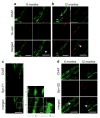
Human α-synuclein protein in the DMnX of AAV-injected rats. a, b Medulla oblongata sections from rats injected with hα-synuclein-AAVs (n ≥ 5/time point) were immunostained with anti-hα-synuclein. Representative images at lower magnification (a) show labeled neuronal projections in the right medulla oblongata; the DMnX is delineated by dashed lines, and the area postrema (AP) and central canal (cc) are indicated. Rectangular boxes encompass portions of the right DMnX that are also shown at higher magnification (b). Scale bars = 200 μm in (a) and 20 μm in (b). c–f Rats (n = 5) received a single midbrain injection of hα-synuclein-AAVs and were killed 6 months later. Sections of the medulla oblongata were double-stained with anti-ChAT and anti-hα-synuclein (hα-syn). The right DMnX is delineated by dashed lines at low magnification (c) and shown in its entireness in (d). Images at high magnification (e and f) show DMnX axons co-labeled with hα-syn and ChAT (e; white arrows in the merged panel), or immunoreactive for hα-syn but not ChAT (f). Scale bars = 200 μm in (c), 100 μm in (d) and 10 μm in (e and f).
Fig. 7
Fig. 7
Human α-synuclein-positive vagal axons in the rat medulla oblongata. a, b Rats (n = 5/time point) received a single midbrain injection of hα-synuclein-AAVs. Sections of the lower medulla oblongata (between Bregma −13.92 and −14.04 mm) were double-stained with anti-ChAT and anti-hα-synuclein. Images at lower magnification (left panels) show bundles of fibers that cross the right medulla oblongata converging into the vagus nerve. Higher magnification panels (right) show intramedullary vagal fibers co-labeled for ChAT and hα-synuclein (some of these fibers are indicated by white arrows). Scale bars = 200 μm (lower magnification) and 10 μm (higher magnification). c Rats (n = 5) were killed at 12 months after a single midbrain injection of hα-synuclein-AAVs. Sections of the lower medulla oblongata were double-stained with anti-ChAT plus an antibody that specifically recognizes mature α-synuclein fibrils (Syn-F1). Images show co-labeled punctae within a vagal axon crossing the reticular formation of the right medulla oblongata. Co-localization was confirmed in the enlarged images showing orthogonal cross-sections in the x–y, x–z and y–z axes. Scale bars = 10 μm (lower magnification) and 2 μm (higher magnification). d Rats (n = 5/time point) were killed at 6 and 12 months after a single midbrain injection of hα-synuclein-AAVs. Sections of the lower medulla oblongata were double-stained with anti-ChAT plus an antibody that recognizes oligomeric and fibrillar forms of α-synuclein (Syn-O2). Images show co-labeled vagal axons (white arrows in the merged panels) in the right medulla oblongata. Scale bar = 10 μm.
Fig. 8
Fig. 8
Schematic representations of patterns of hα-synuclein accumulation after vagal or intra-parenchymal injections of hα-synuclein-AAVs. a Following AAV injections into the vagus nerve, neuronal cell bodies in the DMnX and nodose ganglion produce hα-synuclein. The exogenous protein is then accumulated within efferent DMnX projections and afferent vagal fibers, reaching both visceromotor and viscerosensory nerve endings in the gastric wall. Afferent fibers also terminate onto neurons of the nucleus of the tractus solitaries (NTS) in the medulla oblongata (MO). b Following AAV injections into the ventral mesencephalon (VM), hα-synuclein is overexpressed within neurons that project toward lower brainstem regions. Traveling rostro-caudally through these axons, the exogenous protein reaches the medulla oblongata (MO) and gains access into DMnX neurons. The red arrow underscores the fact that passage into these cells would require a neuron-to-neuron jump. Hα-synuclein then uses efferent projections stemming from the DMnX and contained in the vagus nerve to reach preganglionic vagal nerve endings in the stomach wall.

Similar articles

Cited by

References

    1. Agostoni E, Chinnock JE, De Daly MB, Murray JG. Functional and histological studies of the vagus nerve and its branches to the heart, lungs and abdominal viscera in the cat. J Physiol. 1957;135:182–205. - PMC - PubMed
    1. Annerino DM, Arshad S, Taylor GM, Adler CH, Beach TG, Greene JG. Parkinson’s disease is not associated with gastrointestinal myenteric ganglion neuron loss. Acta Neuropathol. 2012;124:665–680. doi: 10.1007/s00401-012-1040-2. - DOI - PMC - PubMed
    1. Beach TG, Adler CH, Sue LI, Vedders L, Lue L, White CL, III, Akiyama H, Caviness JN, Shill HA, Sabbagh MN, Walker DG. Multi-organ distribution of phosphorylated α-synuclein histopathology in subjects with Lewy body disorders. Acta Neuropathol. 2010;119:689–702. doi: 10.1007/s00401-010-0664-3. - DOI - PMC - PubMed
    1. Berthoud HR, Jedrzejewska A, Powley TL. Simultaneous labeling of vagal innervation of the gut and afferent projections from the visceral forebrain with dil injected into the dorsal vagal complex in the rat. J Comp Neurol. 1990;301:65–79. - PubMed
    1. Braak H, de Vos RA, Bohl J, Del Tredici K. Gastric α-synuclein immunoreactive inclusions in Meissner’s and Auerbach’s plexuses in cases staged for Parkinson’s disease-related brain pathology. Neurosci Lett. 2006;396:67–72. doi: 10.1016/j.neulet.2005.11.012. - DOI - PubMed

MeSH terms

LinkOut - more resources

  NODES
chat 9
Project 10
twitter 2