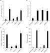
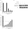
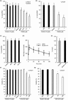
. 2017 Jun 8;7(1):3043.
doi: 10.1038/s41598-017-02934-3.

RCAN-11R peptide provides immunosuppression for fully mismatched islet allografts in mice

Affiliations

RCAN-11R peptide provides immunosuppression for fully mismatched islet allografts in mice

Hirofumi Noguchi et al. Sci Rep. .

Abstract

Calcineurin inhibitors have been used for transplant therapy. However, the inhibition of calcineurin outside the immune system has a number of side effects. We previously developed a cell-permeable inhibitor of NFAT (nuclear factor of activated T cells) using the polyarginine peptide delivery system. This peptide (11R-VIVIT) selectively interferes with calcineurin-NFAT interaction without affecting the activity of calcineurin phosphatase and provides immunosuppression for fully mismatched islet allografts in mice. However, our recent study showed that 11R-VIVIT affected cell viability in vitro when it was used at higher concentration because of the VIVIT sequence. The aim of this study is to develop a safer NFAT inhibitor (RCAN-11R) that does not affect cell viability, and which is less toxic than calcineurin inhibitors. The minimal sequence of the protein family of regulators of calcineurin (RCAN) that is responsible for the inhibition of calcineurin-NFAT signaling was recently characterized. The peptide could selectively interfere with the calcineurin-NFAT interaction without affecting the activity of calcineurin phosphatase, similar to 11R-VIVIT. RCAN-11R did not affect cell viability when it was used at a higher concentration than the toxic concentration of 11R-VIVIT. RCAN-11R could therefore be useful as a therapeutic agent that is less toxic than current drugs or 11R-VIVIT.


Conflict of interest statement

The authors declare that they have no competing interests.

Figures
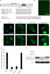
Figure 1
Figure 1
The transduction of RCAN-11R into lymphocytes and the inhibition of NFAT nuclear translocation. (a) The CIC (Calcineurin inhibitor of Calcipressin) motif of the human RCAN protein family (upper schematic illustration) and the sequence of RCAN-11R and the scramble RCAN-11R peptides (lower schematic illustration). (b) Transduction of RCAN-11R into lymphocytes. Jurkat cells were incubated with 10 μM FITC-RCAN-11R and examined using an Olympus confocal microscope. Scale bar = 100 µm. (c) The inhibition of NFAT nuclear translocation. HEK 293 cells that had been transfected with GFP-NFAT-1 plasmid were incubated with 1 μM FK506 or 20 μM RCAN-11R/scRCAN-11R for 2 h and then 500 nM ionomycin was added to the culture medium. Scale bar = 20 µm. (d) The percentage of NFAT nuclear translocation. GFP-NFAT-1 nuclear translocation was quantified by microscopy. *p < 0.01 in comparison to control (no treatment). The values represent the mean ± SE of five independent experiments. (e) Western blot of phosphorylated NFAT1 (Phospho-NFAT) and the faster-migrating dephosphorylated forms (Dephospho-NFAT).
Figure 2
Figure 2
The inhibition of NFAT reporter activity and the production of IL2. (a,b) NFAT (a) and NF-κB (b) reporter activity. Jurkat cells were electroporated with 5 μg of pNFAT-SEAP or pNF-κB-SEAP, respectively. The cells were incubated with 1 μM FK506 or 20 μM RCAN-11R/scRCAN-11R for 1 h, and stimulated with 200 nM PMA and 4 μM ionomycin for 12 h. (c) The inhibition of IL-2 transcription. Jurkat cells were treated with 1 μM FK506 or 20 μM RCAN-11R/scRCAN-11R for 1 h, then with 200 nM PMA and 4 μM ionomycin for an additional 12 h. The cells were subjected to a quantitative RT-PCR. (d) The inhibition of IL-2 production. Jurkat cells were treated with 1 μM FK506 or 20 μM RCAN-11R/scRCAN-11R for 1 h and then incubated with 200 nM PMA and 4 μM ionomycin for an additional 12 h. *p < 0.01 in comparison to stimulation (control) without treatment of FK506 or peptides. The values represent the mean ± SE of five independent experiments.
Figure 3
Figure 3
The dose-response and half-life of RCAN-11R. (a) The dose-response of RCAN-11R. Jurkat cells were treated with 0.1–100 μM RCAN-11R or 1 μM FK506 for 1 h, then with 200 nM PMA and 4 μM ionomycin for an additional 12 h. The cells were subjected to a quantitative RT-PCR. (b) Dephosphorylation of NFAT. Jurkat cells were treated with 0.1, 1, or 10 μM RCAN-11R for 1 h and with 20 nM PMA and 300 nM ionomycin for an additional 15 min and then western blot was performed. (c) The half-life of RCAN-11R. Jurkat cells were treated with 20 μM RCAN-11R for periods of 1–72 h or with 1 μM FK506 for 1 h, then with 200 nM PMA and 4 μM ionomycin for an additional 12 h. The cells were then subjected to a quantitative RT-PCR. The values represent the mean ± SE of five independent experiments.
Figure 4
Figure 4
The effects of RCAN-11R on islet transplantation. (a) The differences in the duration of graft survival between the RCAN-11R group (n = 10) and the scRCAN-11R group (n = 10), as evaluated by a Kaplan-Meier log-rank test. (b) Intraperitoneal glucose tolerance testing (IPGTT). IPGTT was performed ten days after transplantation. Glucose (2.0 g/kg body weight) was intraperitoneally injected. (c,d) The inhibition of IL-2 (c) or IFNγ (d) transcription in vivo. Ten days after transplantation, islet allografts with lymphocyte (n = 3 each) were surgically obtained from kidneys and the cells were subjected to a quantitative RT-PCR. *p < 0.01 in comparison to scRCAN-11R at the same time. The values represent the mean ± SE.
Figure 5
Figure 5
The effects of RCAN-11R on insulin secretion and cell viability. (a) Insulin secretion in β-cell line, βTC6 cells. βTC6 cells were added to 96-well plates (50,000 cells/well) with FK506 or with RCAN-11R in complete medium. The medium with FK506 or with RCAN-11R was changed every 24 hours. After 96 hours, the medium was changed and the samples for investigating the release of insulin were taken 1 hour later. The values represent the mean ± SE of five independent experiments. (b) Insulin secretion in isolated islets from BALB/c mice. Isolated islets from BALB/c mice were added to 24-well plates (250 IE) with FK506 or with RCAN-11R in complete medium. The medium with FK506 or with RCAN-11R was changed every 24 hours. After 96 hours, the medium was changed and the samples for investigating the release of insulin were taken 1 hour later. The values represent the mean ± SE of five independent experiments. (c) Insulin secretion in βTC6 cells with RCAN-11R treatment for 48, 96, or 144 hours. βTC6 cells were added to 96-well plates (50,000 cells/well) with RCAN-11R for 48, 96, or 144 hours and then the medium was changed and the samples for investigating the release of insulin were taken 1 hour later. The values represent the mean ± SE of five independent experiments. (d) IPGTT. IPGTT was carried out on normal BALB/c mice (n = 5 each) after treatment with 50 mg/kg of RCAN-11R or saline for ten days. Glucose (2.0 g/kg body weight) was intraperitoneally injected. (e) Blood insulin level in mice after RCAN-11R treatment. Blood insulin level in mice after RCAN-11R treatment was evaluated ten days after treatment with 50 mg/kg of RCAN-11R or saline. The mice were fasted overnight and the blood samples were assayed with a mouse insulin ELISA kit. (f) Cell viability in β-cell line, βTC6 cells. The cell viability after treatment with RCAN-11R or 11R-VIVIT for 24 h was assessed using FDA/PI staining to visualize the living and dead cells simultaneously. Ten sets of one hundred βTC6 cells (total 1,000 cells) were inspected, and their individual viabilities were visually determined. The average viability was then calculated. *p < 0.01/**p < 0.05 in comparison to control (No treatment). (g) Cell viability in isolated islets from BALB/c mice. The cell viability after treatment with RCAN-11R or 11R-VIVIT for 24 h was assessed using FDA/PI staining to visualize the living and dead cells simultaneously. Fifty islets were inspected, and their individual viabilities were visually determined. The average viability was then calculated. *p < 0.01 in comparison to control (No treatment). The values represent the mean ± SE.

Similar articles

Cited by

References

    1. Rao A, Luo C, Hogan PG. Transcription factors of the NFAT family: regulation and function. Annu Rev Immunol. 1997;15:707–747. doi: 10.1146/annurev.immunol.15.1.707. - DOI - PubMed
    1. Crabtree GR. Generic signals and specific outcomes: signaling through Ca2+, calcineurin, and NF-AT. Cell. 1999;96:611–614. doi: 10.1016/S0092-8674(00)80571-1. - DOI - PubMed
    1. Sigal NH, et al. Is cyclophilin involved in the immunosuppressive and nephrotoxic mechanism of action of cyclosporin A? J Exp Med. 1991;173:619–628. doi: 10.1084/jem.173.3.619. - DOI - PMC - PubMed
    1. Platz KP, et al. Nephrotoxicity following orthotopic liver transplantation. A comparison between cyclosporine and FK506. Transplantation. 1994;58:170–178. doi: 10.1097/00007890-199407270-00007. - DOI - PubMed
    1. Pascual M, et al. Chronic rejection and chronic cyclosporin toxicity in renal allografts. Immunol Today. 1998;19:514–519. doi: 10.1016/S0167-5699(98)01324-3. - DOI - PubMed

Publication types

MeSH terms

  NODES
INTERN 2
twitter 2