The Crucial Role of Biofilms in Cryptococcus neoformans Survival within Macrophages and Colonization of the Central Nervous System
- PMID: 29371529
- PMCID: PMC5715963
- DOI: 10.3390/jof3010010
The Crucial Role of Biofilms in Cryptococcus neoformans Survival within Macrophages and Colonization of the Central Nervous System
Abstract
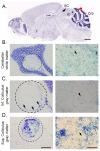
Cryptococcus neoformans is an encapsulated yeast-like fungus capable of causing life threatening meningoencephalitis in patients with impaired immunity. This microbe primarily infects the host via inhalation but has the ability to disseminate to the central nervous system (CNS) either as a single cell or inside of macrophages. Upon traversing the blood brain barrier, C. neoformans has the capacity to form biofilm-like structures known as cryptococcomas. Hence, we will discuss the C. neoformans elements contributing to biofilm formation including the fungus' ability to survive in the acidic environment of a macrophage phagosome and inside of the CNS. The purpose of this mini-review is to instill fresh interest in understanding the importance of biofilms on fungal pathogenesis.
Keywords: CNS; Cryptococcus neoformans; biofilms; cryptococcomas; macrophages.
Conflict of interest statement
The authors declare no conflict of interest.
Figures


Similar articles
-
Phospholipase B Is Critical for Cryptococcus neoformans Survival in the Central Nervous System.mBio. 2023 Apr 25;14(2):e0264022. doi: 10.1128/mbio.02640-22. Epub 2023 Feb 14. mBio. 2023. PMID: 36786559 Free PMC article.
-
Invasion of the central nervous system by Cryptococcus neoformans requires a secreted fungal metalloprotease.mBio. 2014 Jun 3;5(3):e01101-14. doi: 10.1128/mBio.01101-14. mBio. 2014. PMID: 24895304 Free PMC article.
-
Cryptococcus neoformans Infection in the Central Nervous System: The Battle between Host and Pathogen.J Fungi (Basel). 2022 Oct 12;8(10):1069. doi: 10.3390/jof8101069. J Fungi (Basel). 2022. PMID: 36294634 Free PMC article. Review.
-
Biofilm Formation by Cryptococcus neoformans.Microbiol Spectr. 2015 Jun;3(3). doi: 10.1128/microbiolspec.MB-0006-2014. Microbiol Spectr. 2015. PMID: 26185073
-
Mechanisms and Virulence Factors of Cryptococcus neoformans Dissemination to the Central Nervous System.J Fungi (Basel). 2024 Aug 17;10(8):586. doi: 10.3390/jof10080586. J Fungi (Basel). 2024. PMID: 39194911 Free PMC article. Review.
Cited by
-
Cryptococcal Traits Mediating Adherence to Biotic and Abiotic Surfaces.J Fungi (Basel). 2018 Jul 29;4(3):88. doi: 10.3390/jof4030088. J Fungi (Basel). 2018. PMID: 30060601 Free PMC article. Review.
-
Geometrical Distribution of Cryptococcus neoformans Mediates Flower-Like Biofilm Development.Front Microbiol. 2017 Dec 19;8:2534. doi: 10.3389/fmicb.2017.02534. eCollection 2017. Front Microbiol. 2017. PMID: 29312225 Free PMC article.
-
Biology and function of exo-polysaccharides from human fungal pathogens.Curr Clin Microbiol Rep. 2020 Mar;7(1):1-11. doi: 10.1007/s40588-020-00137-5. Epub 2020 Jan 17. Curr Clin Microbiol Rep. 2020. PMID: 33042730 Free PMC article.
-
Fungal immunity and pathogenesis in mammals versus the invertebrate model organism Galleria mellonella.Pathog Dis. 2021 Mar 20;79(3):ftab013. doi: 10.1093/femspd/ftab013. Pathog Dis. 2021. PMID: 33544836 Free PMC article. Review.
-
Delineating the Biofilm Inhibition Mechanisms of Phenolic and Aldehydic Terpenes against Cryptococcus neoformans.ACS Omega. 2019 Oct 15;4(18):17634-17648. doi: 10.1021/acsomega.9b01482. eCollection 2019 Oct 29. ACS Omega. 2019. PMID: 31681870 Free PMC article.
References
-
- Singh N., Alexander B.D., Lortholary O., Dromer F., Gupta K.L., John G.T., del Busto R., Klintmalm G.B., Somani J., Lyon G.M., et al. Pulmonary cryptococcosis in solid organ transplant recipients: Clinical relevance of serum cryptococcal antigen. Clin. Infect. Dis. 2008;46:e12–e18. doi: 10.1086/524738. - DOI - PMC - PubMed
-
- Lindell D.M., Ballinger M.N., McDonald R.A., Toews G.B., Huffnagle G.B. Immunologic homeostasis during infection: Coexistence of strong pulmonary cell-mediated immunity to secondary Cryptococcus neoformans infection while the primary infection still persists at low levels in the lungs. J. Immunol. 2006;177:4652–4661. doi: 10.4049/jimmunol.177.7.4652. - DOI - PubMed
Publication types
Grants and funding
LinkOut - more resources
Full Text Sources
Other Literature Sources

