. 2019 Jun 13;10(1):175.
doi: 10.1186/s13287-019-1265-2.

Elevated level of mitochondrial reactive oxygen species via fatty acid β-oxidation in cancer stem cells promotes cancer metastasis by inducing epithelial-mesenchymal transition

Affiliations

Elevated level of mitochondrial reactive oxygen species via fatty acid β-oxidation in cancer stem cells promotes cancer metastasis by inducing epithelial-mesenchymal transition

Caihua Wang et al. Stem Cell Res Ther. .

Abstract

Background: Cancer stem cells (CSCs) play a critical role in tumor development and progression and are involved in cancer metastasis. The role of reactive oxygen species (ROS) in CSCs and cancer metastasis remains controversial. The aim of the present study was to investigate the correlation between ROS level of CSCs and cancer metastasis and to explore the possible underlying molecular mechanisms.

Methods: Four different cell lines were used to isolate tumor spheres and to analyze intrinsic properties of tumor sphere cells including proliferation, self-renewal potential, differentiation, drug-resistance and cancer metastasis in vitro and in vivo. ROS assays were used to detect the intracellular ROS level of tumor spheres cells. Gene expression analysis and western blot were used to investigate the underlying mechanisms of ROS in regulating cancer metastasis.

Results: Tumor spheres possessed the characteristic features of CSCs, and ROS-high tumor spheres (RH-TS) displayed elevated mitochondrial ROS level exclusively drove metastasis formation. The gene expression analysis showed elevated fatty acid β-oxidation, downregulation of epithelial marker upregulation of mesenchymal markers, and the activation of MAP kinase cascades. Furthermore, 14 up-regulated genes in RH-TS cells were associated with reduced overall survival of different cancer patients.

Conclusions: Our findings demonstrate that CSCs characterized by elevated mitochondrial ROS level potentiate cancer metastasis. Mechanistically, elevated mitochondrial ROS via fatty acid β-oxidation, activates the MAPK cascades, resulting in the epithelial-mesenchymal transition (EMT) process of RH-TS cells, thereby potentiating caner invasion and metastasis. Therefore, _targeting mitochondrial ROS might provide a promising approach to prevent and alleviate cancer metastasis induced by RH-TS cells.

Keywords: Cancer metastasis; Cancer stem cells; Epithelial-mesenchymal transition; Mitochondria; ROS; Tumor sphere.


Conflict of interest statement

The authors declare that they have no competing interests.

Figures
Fig. 1
Fig. 1
Tumor sphere cells possessed CSCs features. a Tumor spheres were isolated from 4 T1 cells, HCT116 cells, SW480cells, and HT29 cells by using cancer stem cell culture method as described in Experimental Procedures. Scale bars represent 100 μm. b The relative proportion of tumor sphere cells remained stable after 3 generations of replating. Results are shown as mean values ± SEM from three independent experiments. c The surface markers of 4 T1 parental cells and primary tumor sphere cells isolated from 4 T1 parental cells. Scale bars represent 50 μm. d The percentage of CD44 + CD24- cells in 4 T1 parental cells and tumor sphere cells. Results are from three independent experiments. e 4 T1 tumor sphere cells grown in 3D in matrigel formed acinar-like colony (a representative image under bright field, phase contrast microscopy) and differentiation markers (TP63, myoepithelial cell marker; EpCAM, epithelial cell marker). Data shown are representative of three independent experiments. f Tumors from tumor sphere cells containing glandular tubules stained with hematoxylin/eosin (H&E) and differentiation markers (TP63, myoepithelial cell marker; EpCAM, epithelial cell marker). Data shown are representative of three independent experiments. g Co-culture of tumor sphere cells (red fluorescence) and 4 T1 cells (green fluorescence) with drugs including cisplatin, methotrexate (MTX), doxorubicin (Dox), taxel, etoposide, or vincristine (VCR). After 2 days of incubation, much more tumor sphere cells survived than 4 T1 cells. Results are representative of three independent experiments. Scale bars =100 μm
Fig. 2
Fig. 2
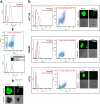
Isolation of RH-TS and RL-TS cells. a RH-TS and RL-TS cells were isolated from 4 T1 tumor spheres by flow cytometry and intracellular ROS concentrations were measured by DCFH-DA staining. RH-TS and RL-TS cells were re-analyzed by FACS and confocal microscopy. b Tumor spheres were isolated from HCT116 cells, SW480 cells, and HT29 cells. ROS profile representing by DCFH-DA fluorescence was measured by FACS and confocal microscopy. Scale bars =100 μm
Fig. 3
Fig. 3
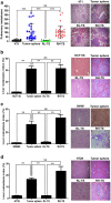
RH-TS contributed to In Vivo cancer metastasis. a The pulmonary metastases of tumor spheres and RH-TS were much more than that of 4 T1 cells and RL-TS when orthotopically inoculated into Balb/c mouse. Five independent experiments. NS, not significant; ***, p < 0.001. b The liver metastases of tumor spheres and RH-TS were much more than that of HCT116 cells and RL-TS when inoculated subcutaneously into NOD/SCID mouse. Two independent experiments performed. NS, not significant; **, p < 0.01. c The liver metastases of tumor spheres and RH-TS were much more than that of SW480 cells and RL-TS when inoculated subcutaneously into NOD/SCID mouse. Two independent experiments performed. NS, not significant; **, p < 0.01. d The liver metastases of tumor spheres and RH-TS were much more than that of HT29 cells and RL-TS when inoculated subcutaneously into NOD/SCID mouse. Two independent experiments performed. NS, not significant; **, p < 0.01
Fig. 4
Fig. 4
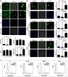
Mitochondrial ROS elevated significantly in RH-TS. a After isolation of RH-TS and RL-TS cells from 4 T1 tumor spheres, the ROS composition was measured by DCFH-DA, DAF-FM, DHE, and MitoSox Red as described in Materials and Methods. Scale bar = 50 μm. *** p < 0.001. b The ROS composition of RH-TS and RL-TS cells in HCT116, SW480, and HT29 cells was measured by DCFH-DA, DHE and MitoSOX Red. Results are shown as mean ± SEM from three independent experiments. Scale bar = 50 μm. *** p < 0.001. c RH-TS cells increased MitoSox Red level significantly. MitoSox Red levels of parental cells (blue), RL-TS (green), RH-TS (red) were analyzed by flow cytometry. Three independent experiments performed
Fig. 5
Fig. 5
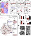
Metabolic remodeling in RH-TS. a Heatmap of differentially expressed genes among parental cells, RH-TS cells, and RL-TS cells. b Expression changes of metabolic enzymes between RH-TS cells and RL-TS cells. * p < 0.05, ** p < 0.01, *** p < 0.001; unmarked, not significant. c A diagram depicting the changes in major metabolic pathways in RH-TS cells compared to RL-TS cells. Red objects and blue objects represent significant upregulation or downregulation in RH-TS cells, respectively. d The cytosolic free NAD/NADH ratio in RH-TS cells was significantly less than that in RL-TS cells. e Representative photographs of cell mitochondria by transmission electron microscopy
Fig. 6
Fig. 6
MAPK signaling and genes for cell invasion and metastasis in RH-TS. a The GO enrichment analysis of differentially expressed genes between RH-TS cells and RL-TS cells. b The reactome analysis of differentially biological pathways between the RH-TS cells and RL-TS cells. c The metastasis genes expression of RH-TS and RL-TS. * p < 0.05, ** p < 0.01, *** p < 0.001
Fig. 7
Fig. 7
Elevated mitochondrial ROS level promoted the epithelial mesenchymal transition via activation of MAPK signaling pathway. a RT-PCR analysis of redox-sensitive signaling pathways, including MEK1, MEK2, P38, and PI3K, and EMT related genes including PTGS2, MMP1, MMP2, LOX, ANGPTL4, CCL5, TWIST1, SNAI1, SNAI2, MET, ID1, and DARC in RH-TS cells and RL-TS cells. Results are shown as mean ± SEM from three independent experiments. * p < 0.05, ** p < 0.01, *** p < 0.001. b Analysis of redox-sensitive signaling pathways and EMT related proteins. RH-TS cells had downregulation of an epithelial marker E-cadherin, and upregulation of mesenchymal markers vimentin and keratin-4. Redox-sensitive signaling pathways, including MEK1, MEK2, P38, and PI3K were significantly overexpressed in RH-TS compared to RL-TS cells. c Proposed mechanism of the mitochondrial ROS promoted cancer metastasis of RH-TS cells. Elevated ROS of tumor sphere activated the p38 MAPK signaling pathway and promoted EMT process thus enforced the metastases formation
Fig. 8
Fig. 8
Fourteen up-regulated genes in RH-TS correlated with patient survival negatively. The caner survival analysis of 14 up-regulated genes in RH-TS cells in different cancer patients in the Human Protein Atlas database. a Overall survival of patients with renal cancer, urothelial cancer, cervical cancer, pancreatic cancer, colorectal cancer, glioma, liver cancer, ovarian cancer, lung cancer, stomach cancer, thyroid cancer, or endometrial cancer was determined on expression of 14 genes (Spred3, Nptx1, Angptl2, Adamts14, Meis3, Dlg4, Col6a1, Ltbp2, Cmtm3, Antxr1, Ptges, Fam114a1, Sema4b, or Sned1). Color code represents the P value for each gene for 5-year survival from different cancer patients. Blue cells indicate that high expression of a certain gene correlated with cancer survival negatively, while yellow cells represent positive correlation. White cells indicate no clear correlation between the gene and cancer survival. b The 14 up-regulated genes in RH-TS cells correlated with renal cancer survival negatively

Similar articles

Cited by

References

    1. Chaffer CL, Weinberg RA. A perspective on cancer cell metastasis. Science. 2011;331:1559–1564. doi: 10.1126/science.1203543. - DOI - PubMed
    1. Korolev KS, Xavier JB, Gore J. Turning ecology and evolution against cancer. Nat Rev Cancer. 2014;14:371–380. doi: 10.1038/nrc3712. - DOI - PubMed
    1. Valastyan S, Weinberg RA. Tumor metastasis: molecular insights and evolving paradigms. Cell. 2011;147:275–292. doi: 10.1016/j.cell.2011.09.024. - DOI - PMC - PubMed
    1. Nassar D, Blanpain C. Cancer stem cells: basic concepts and therapeutic implications. Annu Rev Pathol. 2016;11:47–76. doi: 10.1146/annurev-pathol-012615-044438. - DOI - PubMed
    1. Nguyen LV, Vanner R, Dirks P, Eaves CJ. Cancer stem cells: an evolving concept. Nat Rev Cancer. 2012;12:133–143. doi: 10.1038/nrc3184. - DOI - PubMed

Publication types

MeSH terms

Substances

LinkOut - more resources

  NODES
twitter 2