. 2018 Apr 23;19(4):1258.
doi: 10.3390/ijms19041258.

Liu Jun Zi Tang-A Potential, Multi-Herbal Complementary Therapy for Chemotherapy-Induced Neurotoxicity

Affiliations

Liu Jun Zi Tang-A Potential, Multi-Herbal Complementary Therapy for Chemotherapy-Induced Neurotoxicity

Chun-Tang Chiou et al. Int J Mol Sci. .

Abstract

Liu Jun Zi Tang (LJZT) has been used to treat functional dyspepsia and depression, suggesting its effects on gastrointestinal and neurological functions. LJZT is currently used as a complementary therapy to attenuate cisplatin-induced side effects, such as dyspepsia. However, its effect on chemotherapy-induced neuropathic pain or neurotoxicity has rarely been studied. Thus, we explored potential mechanisms underlying LJZT protection against cisplatin-induced neurotoxicity. We observed that LJZT attenuated cisplatin-induced thermal hyperalgesia in mice and apoptosis in human neuroblastoma SH-SY5Y cells. Furthermore, it also attenuated cisplatin-induced cytosolic and mitochondrial free radical formation, reversed the cisplatin-induced decrease in mitochondrial membrane potential, and increased the release of mitochondrial pro-apoptotic factors. LJZT not only activated the peroxisome proliferator-activated receptor gamma coactivator 1-alpha (PGC-1α) promoter region, but also attenuated the cisplatin-induced reduction of PGC-1α expression. Silencing of the PGC-1α gene counteracted the protection of LJZT. Taken together, LJZT mediated, through anti-oxidative effect and mitochondrial function regulation, to prevent cisplatin-induced neurotoxicity.

Keywords: Liu Jun Zi Tang; PGC-1α; cisplatin; mitochondria; neurotoxicity.


Conflict of interest statement

The authors declare no conflict of interest.

Figures

Figure 1
Figure 1
Liu Jun Zi Tang attenuated cisplatin-induced thermal hyperalgesia in mice. Eight-week-old Balb/c mice were housed in a cage and habituated for two weeks to avoid stress-induced analgesia. Cisplatin was freshly prepared in normal saline. Mice were treated with daily intraperitoneal injections for 5 days, followed by 9 days of rest, for two cycles. Liu Jun Zi Tang (LJZT) was administered 1 h prior to cisplatin administration. After the treatments, a tail-flick assay was performed to monitor tolerance time in response to heat. Data points represented the mean tolerance time ± standard deviation. Differences among multiple groups were evaluated using one-way analysis of variance. Differences between means were calculated using the Student-Newman-Keuls method and were considered significant at p < 0.05. * p < 0.05, compared with controls (n = 6–8).
Figure 2
Figure 2
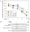
Effects of Liu Jun Zi Tang on cisplatin-induced cell cytotoxicity in human neuroblastoma (SH-SY5Y) cells. (A) Different doses of cisplatin were administered for another 24 h with or without pretreatment of different doses of LJZT for 1 h. Cells were then subjected to a neutral red assay. Viability was expressed as a percentage of controls, and data are presented as the mean ± standard deviation. Differences among multiple groups were evaluated through two-way ANOVA. Differences between means were calculated using the Student-Newman-Keuls method with significance set at p < 0.05. * p < 0.05, compared with controls (n = 3–6). # p < 0.05, compared with the cisplatin-only treatment group (n = 3–6). (B) After pretreatment with or without LJZT, cells were treated with or without cisplatin for another 24 h. Cells were then harvested and subjected to a Western blot analysis.
Figure 3
Figure 3
Effects of Liu Jun Zi Tang on cisplatin-induced cytosolic reactive oxygen species formation in human neuroblastoma (SH-SY5Y) cells. Cisplatin was administered for another 24 h with or without pretreatment with LJZT for 1 h. Cells were then stained with (A) di-hydroxyethidium (DHE) and (B) CO3-H2DCFDA and subjected to image acquisition and quantitation. At least 10 frames were randomly acquired for each group. Fluorescence intensity is expressed as a percentage of controls. Data are presented as the mean ± standard deviation. Differences among multiple groups were evaluated using one-way analysis of variance and those between means were calculated using the Student-Newman-Keuls method with significance set at p < 0.05. * p < 0.05, compared with controls (n = 3–6). # p < 0.05, compared with the cisplatin-only treatment group (n = 3–6). The bar represents 50 µm. These data represent one out of three independent experiments that provided similar results.
Figure 4
Figure 4
Effects of Liu Jun Zi Tang on cisplatin-induced mitochondrial reactive oxygen species formation and membrane potential in human neuroblastoma (SH-SY5Y) cells. Cisplatin was administered for another 24 h with or without pretreatment with LJZT for 1 h. Cells were then stained with (A) MitoSOX and (B) tetramethylrhodamine, ethyl ester (TMRE), and subjected to image acquisition and quantitation. At least 10 frames were randomly acquired for each group. Fluorescence intensity is expressed as a percentage of controls. Data are presented as the mean ± standard deviation. Differences among multiple groups were evaluated using one-way analysis of variance, and those between means were calculated using the Student-Newman-Keuls method with significance set at p < 0.05. * p < 0.05, compared with controls (n = 3–6). # p < 0.05, compared with the cisplatin-only treatment group (n = 3–6). The bar represents 50 µm. These data represent one out of three independent experiments that provided similar results.
Figure 5
Figure 5
U74389G attenuated cisplatin-induced cell death in human SH-SY5Y cells. Cells were treated with or without 10 µM of cisplatin for 24 h and with or without U74389G in different doses for 1 h. Subsequently, cells were subjected to a neutral red assay. Viability is expressed as a percentage of controls. Data are presented as the mean ± standard deviation. Differences among multiple groups were evaluated using one-way analysis of variance. Differences between means were calculated using the Student-Newman-Keuls method and were considered significant at p < 0.05. * p < 0.05, compared with controls (n = 3–6).
Figure 6
Figure 6
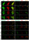
Effects of Liu Jun Zi Tang on cisplatin-induced mitochondrial pro-apoptotic factor release in human neuroblastoma (SH-SY5Y) cells. After co-transfection with pGFP/TagRFP (upper left panels), pGFP-cytochrome C/pDsRed-Mito (upper right panels), pHtrA2/Omi-GFP/pDsRed-Mito (lower left panels), or pSmac/Diablo-GFP/pDsRed-Mito (lower right panels) for 24 h, cells were pretreated with or without LJZT (100 µg/mL) for 1 h, then 10 µM of cisplatin (Cisp.) was administered for another 24 h. Cells were fixed and subjected to confocal analysis. In each treatment, 10–15 cells were randomly acquired. The bar represents 5 µm. These data represent one out of at least three independent experiments that provided similar results.
Figure 7
Figure 7
Effects of Liu Jun Zi Tang on peroxisome proliferator-activated receptor gamma coactivator 1-alpha (PGC-1α) expression during cisplatin intoxication in human neuroblastoma (SH-SY5Y) cells. (A) After transient transfection with pEGFP-N1 (expressed as a non-_targeting green fluorescent protein)/pDsRed-Mito (expressed as a mitochondria-_targeting red fluorescent protein) or pPGC-1α-GFP (expressed as a PGC-1α conjugating with green fluorescent protein)/pDsRed-Mito for 24 h, cells were pretreated with or without LJZT for 1 h, followed by the addition of cisplatin and incubation for another 24 h. Cells were then fixed, and images were acquired using a confocal microscope. At least three different fields were investigated for each treatment, and at least 10 cells per field were acquired. The bar represents 5 µm. (B) Cisplatin was administered at different intervals with or without LJZT pretreatment. Cells were then harvested and subjected to a Western blot analysis. The relative optical densities of the bands were quantified through densitometry relative to actin, and normalized to the levels of the control condition. Data points represent the mean ± standard deviation. Differences among multiple groups were evaluated using one-way analysis of variance, and those between means were calculated using the Student-Newman-Keuls method with significance set at p < 0.05. * p < 0.05, compared with the control group. # p < 0.05, compared with the group treated with cisplatin treatment for 24 h. (C) After the transient transfection of the PGC-1α promoter region (−2 Kb) was ligated with the luciferase gene for 24 h, cells were treated with or without LJZT for 4 h. Subsequently, cells were directly lysed, and their luciferase activities were measured. The Student’s t-test was used to compare differences between the two groups (n = 4/group). * p < 0.05, compared with the control group. These data represent one out of at least three independent experiments that provided similar results.
Figure 8
Figure 8
Effects of PGC-1α on the protection of Liu Jun Zi Tang in preventing cisplatin-induced cytotoxicity in human neuroblastoma (SH-SY5Y) cells. (A) Cells were harvested and subjected to a Western blot analysis with or without knockdown of the PGC-1α gene by siRNA (Si-PGC-1α) for 48 h. A control siRNA (Si-Control) was also used as a negative control. (B) Cells silenced by the control siRNA (Si-Control) or PGC-1α siRNA (Si-PGC-1α) were pretreated with LJZT for 1 h and then cisplatin for another 24 h. Subsequently, cells were subjected to a neutral red assay. Viability was calculated as a percentage with respect to its control group. In each gene-silenced group, the relative survival ratio was calculated from the viability of cells subtracted from that of the cells treated with cisplatin alone. Data are presented as the mean ± standard deviation. Differences among multiple groups were evaluated using two-way analysis of variance, and those between means were calculated using the Student-Newman-Keuls method with significance set at p < 0.05. * p < 0.05, compared with its control group respectively (n = 3–6). # p < 0.05, compared with its Si-Control-treated group (n = 3–6).

Similar articles

Cited by

References

    1. Yang J., Huang H., Zhu L.J. World Century Compendium to TCM. Volume 5 World Centry Publishing Corporation; Hackensack, NJ, USA: 2013. Introduction to Formulae of Traditional Chinese Medicine.
    1. Xiao Y., Liu Y.Y., Yu K.Q., Ouyang M.Z., Luo R., Zhao X.S. Chinese herbal medicine liu jun zi tang and xiang sha liu jun zi tang for functional dyspepsia: Meta-analysis of randomized controlled trials. Evid. Based Complement. Alternat. Med. 2012;2012:936459. doi: 10.1155/2012/936459. - DOI - PMC - PubMed
    1. Ohnishi S., Takeda H. Herbal medicines for the treatment of cancer chemotherapy-induced side effects. Front. Pharmacol. 2015;6:14. doi: 10.3389/fphar.2015.00014. - DOI - PMC - PubMed
    1. Chao T.H., Fu P.K., Chang C.H., Chang S.N., Chiahung Mao F., Lin C.H., Evidence-based Chinese Medicine Research Group Prescription patterns of Chinese herbal products for post-surgery colon cancer patients in Taiwan. J. Ethnopharmacol. 2014;155:702–708. doi: 10.1016/j.jep.2014.06.012. - DOI - PubMed
    1. Lam W., Bussom S., Guan F., Jiang Z., Zhang W., Gullen E.A., Liu S.H., Cheng Y.C. The four-herb Chinese medicine PHY906 reduces chemotherapy-induced gastrointestinal toxicity. Sci. Transl. Med. 2010;2:45ra59. doi: 10.1126/scitranslmed.3001270. - DOI - PubMed

MeSH terms

Substances

LinkOut - more resources

  NODES
admin 7
twitter 2