. 2020 May 25;21(10):3714.
doi: 10.3390/ijms21103714.

An Evaluation of the In Vitro Roles and Mechanisms of Silibinin in Reducing Pyrazinamide- and Isoniazid-Induced Hepatocellular Damage

Affiliations

An Evaluation of the In Vitro Roles and Mechanisms of Silibinin in Reducing Pyrazinamide- and Isoniazid-Induced Hepatocellular Damage

Zhang-He Goh et al. Int J Mol Sci. .

Abstract

Tuberculosis remains a significant infectious lung disease that affects millions of patients worldwide. Despite numerous existing drug regimens for tuberculosis, drug-induced liver injury is a major challenge that limits the effectiveness of these therapeutics. Two drugs that form the backbone of the commonly administered quadruple antitubercular regimen, that is, pyrazinamide (PZA) and isoniazid (INH), are associated with such hepatotoxicity. Yet, we lack safe and effective alternatives to the antitubercular regimen. Consequently, current research largely focuses on exploiting the hepatoprotective effect of nutraceutical compounds as complementary therapy. Silibinin, a herbal product widely believed to protect against various liver diseases, potentially provides a useful solution given its hepatoprotective mechanisms. In our study, we identified silibinin's role in mitigating PZA- and INH-induced hepatotoxicity and elucidated a deeper mechanistic understanding of silibinin's hepatoprotective ability. Silibinin preserved the viability of human foetal hepatocyte line LO2 when co-administered with 80 mM INH and decreased apoptosis induced by a combination of 40 mM INH and 10 mM PZA by reducing oxidative damage to mitochondria, proteins, and lipids. Taken together, this proof-of-concept forms the rational basis for the further investigation of silibinin's hepatoprotective effect in subsequent preclinical studies and clinical trials.

Keywords: drug-induced liver injury (DILI); isoniazid; oxidative stress; pyrazinamide; silibinin; tuberculosis.


Conflict of interest statement

The authors report no conflict of interests or competing financial interests.

Figures

Figure 1
Figure 1
Treatment scheme involving silibinin’s role as a prophylactic, rescue, and recovery adjuvant. To simulate silibinin’s role as a preventive agent, silibinin was administered 24 h before the treatment with toxicants. To simulate silibinin’s role as rescue adjuvant, silibinin was co-administered with the toxicant regimen. To simulate silibinin’s role as a recovery adjuvant, silibinin was added 24 h after the toxicant regimen. The recovery experiments were further subdivided into two conditions: the first had a washout step, while the second did not have a washout step. In the simulation with washout, the toxicant regimen was replaced with silibinin alone and then treated for a further 24 h to investigate silibinin’s ability to aid patients in recovery after stopping the hepatotoxic regimen. In the simulation without washout, the toxicant regimen was replaced with a combination of silibinin and toxicant and treated for a further 24 h to investigate silibinin’s ability to mitigate further liver injury in patients who stay on the toxicant regimen. PZA, pyrazinamide; INH, isoniazid; MTT, 3-(4,5-dimethylthiazol-2-yl)-2,5-diphenyltetrazolium bromide.
Figure 2
Figure 2
Silibinin mitigated isoniazid (INH)-induced hepatotoxicity, but not pyrazinamide (PZA)-induced hepatotoxicity. (A) Co-administration of silibinin at 25 µM reduced hepatotoxicity induced by 80 mM (one-way analysis of variance (ANOVA), p = 0.0231). Similarly, co-administration of silibinin at 50 µM reduced hepatotoxicity induced by 100 mM INH (one-way ANOVA, p = 0.0201). Co-administration of silibinin at either 25 or 50 µM, but did not reduce hepatotoxicity induced by 50 mM INH, 50 mM PZA, or a combination of INH and PZA (I/P) at 50 mM each (I/P 50/50). (B) Pre-administration of silibinin for 24 h, followed by the co-administration of silibinin with INH or PZA for a further 24 h, did not prevent hepatotoxicity induced by 50 mM INH, 80 mM INH, or 50 mM PZA. (C) Administration of INH or PZA for 24 h, followed by the administration of silibinin alone (with washout) or silibinin with INH or PZA (without washout) for a further 24 h, did not aid in the recovery of LO2 from 50 mM INH, 80 mM INH, or 50 mM PZA. Data represent mean ± S.E.M. of at least two replicates. * p < 0.05 vs. respective vehicle controls.
Figure 3
Figure 3
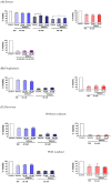
Silibinin reduced reactive oxygen species (ROS) levels and oxidative damage when co-administered with a combination of isoniazid (INH) and pyrazinamide (PZA). Positive controls were treated with the oxidising agent tert-butyl hydroperoxide (TBHP) 200 μM for 2 h. To avoid excessive hepatocyte death, the concentrations of INH and PZA were limited to 40 mM and 10 mM, respectively, when treated in combination (I/P 40/10) over 24 h. (A) 50 μM silibinin reduced intracellular ROS levels (t-test, p = 0.0466). (B) Silibinin decreased carbonylation levels, a marker of oxidative damage in proteins, at 25 μM (one-way ANOVA, p = 0.0015) and 50 μM (one-way ANOVA, p = 0.0023). (C) Silibinin reduced lipid peroxidation levels as measured by the thiobarbituric acid reactive substances (TBARS) assay at 25 μM (one-way ANOVA, p < 0.0001) and 50 μM (one-way ANOVA, p = 0.0007). (D) Silibinin’s reduction of ROS levels at 50 μM was independent of DNA oxidative damage reduction as visually assessed, and as measured quantitatively by tail moment and olive moments. Administration of silibinin alone did not trigger DNA fragmentation. Data represent mean ± S.E.M. of at least two replicates. * p < 0.05, ** p < 0.01, *** p < 0.001 vs. vehicle control co-administered with I/P 40/10.
Figure 3
Figure 3
Silibinin reduced reactive oxygen species (ROS) levels and oxidative damage when co-administered with a combination of isoniazid (INH) and pyrazinamide (PZA). Positive controls were treated with the oxidising agent tert-butyl hydroperoxide (TBHP) 200 μM for 2 h. To avoid excessive hepatocyte death, the concentrations of INH and PZA were limited to 40 mM and 10 mM, respectively, when treated in combination (I/P 40/10) over 24 h. (A) 50 μM silibinin reduced intracellular ROS levels (t-test, p = 0.0466). (B) Silibinin decreased carbonylation levels, a marker of oxidative damage in proteins, at 25 μM (one-way ANOVA, p = 0.0015) and 50 μM (one-way ANOVA, p = 0.0023). (C) Silibinin reduced lipid peroxidation levels as measured by the thiobarbituric acid reactive substances (TBARS) assay at 25 μM (one-way ANOVA, p < 0.0001) and 50 μM (one-way ANOVA, p = 0.0007). (D) Silibinin’s reduction of ROS levels at 50 μM was independent of DNA oxidative damage reduction as visually assessed, and as measured quantitatively by tail moment and olive moments. Administration of silibinin alone did not trigger DNA fragmentation. Data represent mean ± S.E.M. of at least two replicates. * p < 0.05, ** p < 0.01, *** p < 0.001 vs. vehicle control co-administered with I/P 40/10.
Figure 4
Figure 4
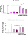
Silibinin reduced apoptosis when co-administered with a combination of INH and PZA by maintaining mitochondrial membrane potential. Various concentrations of silibinin were co-administered with I/P 40/10 over 18 h. (A) The administration of I/P 40/10 significantly increased caspase-3 activity (one-way ANOVA, p < 0.0001). The co-administration of silibinin with I/P 40/10 reduced the activity of caspase-3 when silibinin was administered at 25 μM (one-way ANOVA, p < 0.0001) and 50 μM (one-way ANOVA, p < 0.0001), suggesting that there was a reduction in apoptotic activity. The positive control was treated with camptothecin (CPT) 5 μM for 24 h. (B) The administration of I/P 40/10 negatively affected LO2 cells’ membrane potential (one-way ANOVA, p < 0.0001). Silibinin of 50 μM reduced the percentage of cells whose membrane potential was negatively affected by I/P 40/10 (one-way ANOVA, p = 0.0234). Positive control was treated with the oxidising agent TBHP 200 μM for 1 h. Data represent mean ± S.E.M. of three replicates. * p < 0.05, *** p < 0.001 vs. vehicle control co-administered with I/P 40/10, ^^^ p < 0.001 vs. respective vehicle controls.
Figure 5
Figure 5
Silibinin induced expression of proteins in the nuclear factor (erythroid-derived 2)-like 2 (Nrf2)–antioxidant response element (ARE) pathway and restored protein expression. Vehicle control was treated with 0.05% v/v dimethyl sulfoxide (DMSO). ‘−’ denotes conditions without toxicant cotreatment; ‘+’ denotes conditions with toxicant cotreatment. (A) In LO2, silibinin’s reduction of ROS levels when co-administered with I/P 40/10 was independent of heme oxygenase-1 (HO-1) protein restoration. The administration of I/P 40/10 significantly reduced HO-1 levels without silibinin (one-way ANOVA, p = 0.0150), or with silibinin at 25 μM (one-way ANOVA, p = 0.0051) and 50 μM (one-way ANOVA, p = 0.0022). Positive controls were treated with the Nrf2–ARE inducer trans-cinnamaldehyde (CA) 50 μM for 24 h. Data represent mean ± S.E.M. of three replicates. * p < 0.05, ** p < 0.01 vs. negative control. (B) In transforming growth factor-α transgenic mouse hepatocytes (TAMH), 50 μM silibinin alone induced sulfiredoxin 1 (Srxn1) expression (one-way ANOVA, p = 0.0237), but the co-administration of silibinin with 40 mM INH did not restore Srxn1 expression to pre-suppression levels (one-way ANOVA, p = 0.0551). In contrast, 50 μM silibinin restored HO-1 expression (one-way ANOVA, p = 0.0333), but did not induce HO-1 when was administered alone (one-way ANOVA, p = 0.0564). This effect did not extend to glutamate-cysteine ligase catalytic subunit (Gclc) and NAD(P)H quinone dehydrogenase 1 (NQO1) restoration. Positive controls were treated with the Nrf2–ARE inducer sulphoraphane (SU) 10 μM for 24 h. Data represent mean ± S.E.M. of two replicates. * p < 0.05 vs. vehicle control, ^ p < 0.05 vs. vehicle control co-administered with hepatotoxic regimen.

Similar articles

Cited by

References

    1. Global Tuberculosis Report 2019. World Health Organization; Geneva, Switzerland: 2019.
    1. Kwon Y.S., Jeong B.H., Koh W.J. Tuberculosis: Clinical trials and new drug regimens. Curr. Opin. Pulm. Med. 2014;20:280–286. doi: 10.1097/MCP.0000000000000045. - DOI - PubMed
    1. Cano-Paniagua A., Amariles P., Angulo N., Restrepo-Garay M. Epidemiology of drug-induced liver injury in a University Hospital from Colombia: Updated RUCAM being used for prospective causality assessment. Ann. Hepatol. 2019;18:501–507. doi: 10.1016/j.aohep.2018.11.008. - DOI - PubMed
    1. Abbara A., Chitty S., Roe J.K., Ghani R., Collin S.M., Ritchie A., Kon O.M., Dzvova J., Davidson H., Edwards T.E., et al. Drug-induced liver injury from antituberculous treatment: A retrospective study from a large TB centre in the UK. BMC Infect. Dis. 2017;17:231. doi: 10.1186/s12879-017-2330-z. - DOI - PMC - PubMed
    1. Ramappa V., Aithal G.P. Hepatotoxicity Related to Anti-tuberculosis Drugs: Mechanisms and Management. J. Clin. Exp. Hepatol. 2013;3:37–49. doi: 10.1016/j.jceh.2012.12.001. - DOI - PMC - PubMed
  NODES
admin 34
Note 2
twitter 2