Category:Cells
basic structural and functional unit of all organisms | |||||
| Upload media | |||||
| Instance of |
| ||||
|---|---|---|---|---|---|
| Subclass of |
| ||||
| Part of |
| ||||
| Has part(s) |
| ||||
| Different from | |||||
| |||||
Subcategories
This category has the following 7 subcategories, out of 7 total.
Media in category "Cells"
The following 133 files are in this category, out of 133 total.
-
12985 2005 Article 81 Fig2 HTML.jpg 1,200 × 824; 104 KB
-
2024-08-11 Kasteel Wijchen binnen 24.jpg 6,240 × 3,512; 11.81 MB
-
208031 EPFL David Suter Sox2.jpg 1,304 × 734; 52 KB
-
A Cell Post Plasmolysis.jpg 960 × 720; 94 KB
-
Bernard-S.-Marasa-etal-2010-WI38.png 615 × 372; 194 KB
-
Biofilm di stafilococcus aureus in un catetere.png 1,092 × 416; 624 KB
-
Blood Anemia.jpg 1,280 × 720; 187 KB
-
Camillo Golgi, Golgi cell Type I. Wellcome L0002017.jpg 1,184 × 1,522; 814 KB
-
Cardiac Stem Cell Differentiation.png 512 × 444; 64 KB
-
CardiacMuscle - longtitudinal.jpg 640 × 480; 188 KB
-
Cell metabolism and color.jpg 3,888 × 2,592; 1.21 MB
-
Cell mosaic.png 1,106 × 1,100; 1.17 MB
-
Cell tower with sun set.jpg 1,080 × 1,080; 49 KB
-
CellAdhesion.jpg 1,344 × 1,024; 203 KB
-
Cells mosaic.png 1,470 × 1,060; 2.26 MB
-
Cells on plane.png 1,496 × 614; 1.8 MB
-
Cells Selectively Absorb Short Nanotubes (5881057302).jpg 504 × 202; 112 KB
-
Cells?.jpg 5,184 × 3,456; 6.91 MB
-
Cellular spread of Sendai virus.jpg 3,000 × 2,250; 393 KB
-
Cellular Universe.jpg 818 × 672; 74 KB
-
Cellular Uptake NPs.jpg 1,536 × 1,103; 2.11 MB
-
Chlorella with yellow indicative of chlorophyll.jpg 396 × 400; 14 KB
-
Connexion of neuves.jpg 1,376 × 1,032; 126 KB
-
CSIRO ScienceImage 1478 Cells.jpg 2,657 × 1,751; 5.59 MB
-
Cuboidal Epithelium Section.jpg 640 × 480; 338 KB
-
DAPI-stained nuclei of HeLa cells.jpg 12,788 × 12,788; 16.97 MB
-
Day 298 - West Midlands Police - Custody CCTV Cameras (8121509733).jpg 1,516 × 1,080; 599 KB
-
Ddt24102.jpg 661 × 335; 87 KB
-
Descubrimiento de las células.png 708 × 1,177; 633 KB
-
Differentiation of Stem Cells Into Neurons.jpg 1,884 × 835; 845 KB
-
Educatina 3.jpg 828 × 442; 37 KB
-
Embolized Uterus (44507244114).jpg 1,350 × 1,124; 646 KB
-
Embrione del piede di topo - sezione.png 819 × 1,225; 1.53 MB
-
Erythrocytes (red blood cells) Rouleaux stacking.gif 440 × 440; 3.3 MB
-
Eukaryotic cells.jpg 1,122 × 793; 98 KB
-
Fiber ends Cx50-1.jpg 3,432 × 3,840; 1.47 MB
-
Fibroblastos 100x a.jpg 2,048 × 1,536; 257 KB
-
FISH imaging of myc RNA in RPE.pdf 987 × 987; 25 KB
-
Flower petal cells image 2.jpg 3,072 × 4,096; 5.6 MB
-
Flower petal cells.jpg 3,072 × 4,096; 4.75 MB
-
Fractals in cells of a flower.jpg 640 × 480; 84 KB
-
Gas vesicle TEM.pdf 487 × 562; 659 KB
-
Glomerulus Diameter Measurement.jpg 501 × 168; 24 KB
-
Grapes.png 1,024 × 1,024; 662 KB
-
Halobacterial Gas Vesicles.pdf 487 × 779; 758 KB
-
HEK cell transfection.jpg 680 × 512; 184 KB
-
HEK293FTinvitro.jpg 311 × 313; 29 KB
-
HeLa Cell (5940515305).jpg 800 × 484; 175 KB
-
HeLa Cultured Cancer Cells Dying Without Glucose.jpg 2,835 × 1,807; 2.12 MB
-
Hepatocyte Culture.tif 894 × 653; 1.47 MB
-
Hifa generativa bifurcada.jpg 1,378 × 1,034; 316 KB
-
How Cells Make Friends - panoramio.jpg 2,177 × 2,856; 441 KB
-
Human Cheek Cells.jpg 640 × 426; 125 KB
-
Isochrysis galbana 1200x VID 20220319 190422.ogg 46 s, 1,920 × 1,080; 4.57 MB
-
Isochrysis galbana all stages VID 20220319 185958.ogg 2 min 36 s, 1,920 × 1,080; 14.99 MB
-
Isochrysis galbana all stages VID 20220319 190656.ogg 1 min 54 s, 1,920 × 1,080; 11.09 MB
-
Isochrysis galbana VID 20220319 190939.ogg 3 min 28 s, 1,920 × 1,080; 20.06 MB
-
Isochrysis galbana VID 20220319 205646.ogg 10 min 52 s, 1,920 × 1,080; 62.61 MB
-
Isochrysis galbana VID 20220319 211125.ogg 16 min 44 s, 1,920 × 1,080; 94.35 MB
-
Isochrysis galbana.ogg 1 min 27 s, 1,920 × 1,080; 9.91 MB
-
Juvenile Granulosa Cell Tumor, Ovary (25394681457).jpg 2,455 × 2,191; 2.75 MB
-
LEF Expression in Human Lung Tissue Lysates.jpg 650 × 700; 132 KB
-
Lung cells.jpg 640 × 480; 74 KB
-
Metaphase3.jpg 3,024 × 4,032; 1.49 MB
-
Metaphase5.jpg 626 × 470; 77 KB
-
Micro1.jpg 960 × 1,280; 94 KB
-
MicroElectrode Array Stretching Simulating und Recording Equipment.jpg 3,024 × 4,032; 642 KB
-
Microscopic image of stem cells, Hues 9 stained with DAPI.png 1,360 × 1,024; 1.9 MB
-
Mouse - handwriting on back.jpg 8,141 × 5,788; 4.65 MB
-
Mucosal Leiomyoma of the Sigmoid Colon (44711063484).jpg 1,996 × 1,834; 943 KB
-
Muestra de cultivos microbianos.jpg 6,000 × 4,000; 10.57 MB
-
Neubauer Haemocytomer.jpg 4,000 × 3,000; 6.22 MB
-
Neurology Introduction.ogv 5 min 0 s, 1,280 × 720; 79.8 MB
-
Neutral red uptake assay. Rats embrionic fibroblasts.jpg 2,048 × 1,536; 381 KB
-
Normal and multipolar mitosis.tif 1,024 × 1,024; 3.14 MB
-
Normal Diarthrodial Joint (28498602677).jpg 2,054 × 2,303; 1.37 MB
-
Onion Root.jpg 4,096 × 3,286; 12 MB
-
Papillary carcinoma of the thyroid, FNA, Pap stain. (40700342535).jpg 2,201 × 1,953; 1.46 MB
-
Papillary Thyroid Carcinoma, FNA (41592403851).jpg 2,244 × 2,020; 1.36 MB
-
Papillary Thyroid Carcinoma, FNA, Giemsa stain (40700344115).jpg 2,458 × 1,891; 1.54 MB
-
Partial Hydatidiform Mole (32978625038).jpg 2,323 × 2,723; 2.48 MB
-
Pilar (Trichilemmal) Cyst of the Foot (48327336537).jpg 2,555 × 2,566; 2.3 MB
-
Placenta Percreta (30040338617).jpg 2,079 × 3,777; 1.98 MB
-
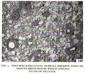
Pneumonia forming around bronchioles.png 350 × 310; 259 KB
-
Pone.0008930.g004.jpg 662 × 346; 196 KB
-
Pseudomembranous Colitis (45642113041).jpg 3,043 × 1,089; 1.5 MB
-
Purple cells.jpg 3,000 × 3,000; 631 KB
-
Red Fluorescence Microscopy.jpg 3,024 × 4,032; 359 KB
-
Reticular Cells.jpg 400 × 308; 46 KB
-
Schleiden; cellular tissue of plants Wellcome M0010608.jpg 2,550 × 4,159; 4 MB
-
SeV organ infection specificity.png 221 × 269; 37 KB
-
Skeletonema costatum single chain.jpg 1,023 × 912; 183 KB
-
Skin cells.JPG 382 × 383; 29 KB
-
Small monastic cells building.jpg 4,476 × 2,983; 1.93 MB
-
SmeT and SmeDEF transcription start sites.jpg 1,225 × 664; 232 KB
-
SmeT model.jpg 995 × 481; 62 KB
-
Sneaky Mice.tif 1,388 × 1,040; 4.14 MB
-
Speckled HEp-2 cells, immunofluorescence (16099409590).jpg 1,257 × 1,257; 940 KB
-
Standart cell.jpg 5,184 × 3,456; 2.83 MB
-
Stem cells, day 1 after passage.tiff 1,392 × 1,040; 4.14 MB
-
Stem cells, day 3 after passage.jpg 1,388 × 1,040; 1.11 MB
-
Stem cells, day 3 after passage.tiff 1,392 × 1,040; 4.14 MB
-
Stem cells, Hues 9 p 38, day 1 after passage.jpg 1,384 × 1,036; 830 KB
-
Stem cells, Hues 9 p 38, day 2 after passage.jpg 1,384 × 1,036; 1.01 MB
-
Stem cells, Hues 9, day 2 after passage.tiff 1,392 × 1,040; 4.14 MB
-
Tem Cell.png 1,024 × 1,024; 1.43 MB
-
TEORIA CELULAR.pdf 1,241 × 1,754, 4 pages; 185 KB
-
Teoría celular.png 720 × 1,180; 460 KB
-
Tetraselmis suecica 2.ogg 5 min 58 s, 1,920 × 1,080; 34.47 MB
-
Tetraselmis suecica all 3 stages 2.jpg 6,000 × 8,000; 5.06 MB
-
Tetraselmis suecica all 3 stages 3.jpg 6,000 × 8,000; 5.05 MB
-
Tetraselmis suecica all 3 stages 4.jpg 6,000 × 8,000; 5.2 MB
-
Tetraselmis suecica all 3 stages 5.jpg 6,000 × 8,000; 5.66 MB
-
Tetraselmis suecica all 3 stages 6.jpg 6,000 × 8,000; 5.09 MB
-
Tetraselmis suecica all 3 stages 7.jpg 6,000 × 8,000; 5.06 MB
-
Tetraselmis suecica all 3 stages 8.ogg 5 min 8 s, 1,920 × 1,080; 29.7 MB
-
Tetraselmis suecica all 3 stages 9.jpg 6,000 × 8,000; 4.98 MB
-
Tetraselmis suecica in a vegetative non-motile stage. 01.ogg 1 min 6 s, 1,920 × 1,080; 6.45 MB
-
Tetraselmis suecica in a vegetative non-motile stage. 02.ogg 1 min 3 s, 1,920 × 1,080; 6.23 MB
-
Tetraselmis suecica in a vegetative non-motile stage. 03.jpg 6,000 × 8,000; 4.67 MB
-
The cell and protoplasm (1940) (20399395028).jpg 2,190 × 2,924; 1.65 MB
-
Tipos-de-transporte-celular.jpg 1,517 × 1,270; 485 KB
-
Tumor Cells.png 1,920 × 1,080; 4.61 MB
-
Агрегаты сперматозоидов во флуоресцентом микроскопе.JPG 1,017 × 896; 57 KB


