Category:Lateral sulcus
fold of the brain (primary motor cortex) separating the frontal and parietal lobes superiorly from the temporal lobe inferiorly. | |||||
| Upload media | |||||
| Instance of |
| ||||
|---|---|---|---|---|---|
| Subclass of |
| ||||
| Named after | |||||
| |||||
Lateral sulcus (Lateral fissure, Sylvian fissure) .
Media in category "Lateral sulcus"
The following 60 files are in this category, out of 60 total.
-
A human head dissected; "In memoriam". Wellcome L0074866.jpg 4,817 × 5,611; 4.43 MB
-
Acquedotto di Silvio.svg 516 × 406; 58 KB
-
Blausen 0103 Brain Sensory&Motor ar.png 1,600 × 960; 5.87 MB
-
Blausen 0103 Brain Sensory&Motor.png 1,600 × 960; 993 KB
-
Brodmann areas inside of lateral sulcus close up.png 772 × 637; 211 KB
-
Brodmann areas inside of lateral sulcus.png 792 × 839; 1.91 MB
-
Brodmann areas of human lateral frontal cortex.png 1,891 × 889; 2.31 MB
-
Cambridge Natural History Mammalia Fig 273.png 532 × 374; 33 KB
-
-
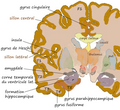
Coronal hippocampe.png 543 × 499; 298 KB
-
Cunningham cerebral sulci.png 1,252 × 790; 2.84 MB
-
Five sulci measured.png 1,660 × 1,215; 2.36 MB
-
FrontalCapts.png 2,309 × 911; 927 KB
-
FrontalCaptsLateral.png 1,252 × 829; 558 KB
-
Girolamo Fabrici d'Acquapendente Tabulae Picae 1600.png 772 × 1,145; 1.32 MB
-
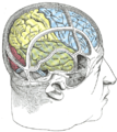
Gray1197.png 444 × 500; 47 KB
-
Gray1198.png 446 × 350; 24 KB
-
Gray658.png 361 × 450; 40 KB
-
Gray724.png 600 × 588; 73 KB
-
Gray725 lateral sulcus ramus posterior.png 255 × 600; 62 KB
-
Gray726 lateral sulcus.svg 992 × 573; 143 KB
-
Gray727 lateral fissure.svg 1,025 × 598; 19 KB
-
Gray739.png 500 × 379; 186 KB
-
Human and chimp brain.png 1,010 × 1,346; 836 KB
-
Human brain frontal (coronal) section description 2.JPG 702 × 487; 43 KB
-
Human brain frontal (coronal) section description2.JPG 702 × 487; 42 KB
-
Human brain lateral view description.JPG 701 × 487; 49 KB
-
Human temporal lobe areas.png 1,793 × 1,513; 1.63 MB
-
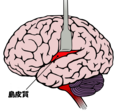
Insula cortex ja.png 700 × 645; 128 KB
-
J. Voort Kamp in Caspar Bartholin Institutiones Anatomicae.png 781 × 457; 316 KB
-
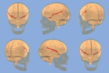
Lateral sulcus.gif 250 × 250; 1.9 MB
-
Lateral sulcus.ogv 10 s, 608 × 608; 1.59 MB
-
Lateral sulcus.png 324 × 209; 27 KB
-
Lateral sulcus2.png 896 × 566; 157 KB
-
Lobes.png 909 × 671; 382 KB
-
LobesCaptsLateral.png 1,201 × 710; 441 KB
-
Operculum.png 700 × 405; 59 KB
-
Operculum1.jpg 1,065 × 796; 146 KB
-
ParietCapts lateral.png 1,139 × 758; 407 KB
-
ParietCapts.png 2,337 × 878; 819 KB
-
PretermSurfaces HiRes es.png 876 × 190; 158 KB
-
PretermSurfaces HiRes ja.png 876 × 190; 146 KB
-
PretermSurfaces HiRes.png 876 × 190; 165 KB
-
PZSL1907Page0814.png 2,063 × 3,201; 6.1 MB
-
Regeczy780.jpg 1,308 × 1,167; 158 KB
-
Sillons vue externe.png 888 × 492; 361 KB
-
Slide2HAN.JPG 960 × 720; 96 KB
-
Slide2KLI.JPG 960 × 720; 87 KB
-
Slide2STE.JPG 960 × 720; 106 KB
-
Slide3HAN.JPG 960 × 720; 117 KB
-
Slide4HAN.JPG 960 × 720; 112 KB
-
Sobo 1909 645.png 1,227 × 750; 2.64 MB
-
Sobo 1911 644.png 2,496 × 1,692; 12.1 MB
-
Squamous Suture and Sylvian Fissure 1.png 1,345 × 1,051; 1.02 MB
-
Squamous Suture and Sylvian Fissure 2.png 1,345 × 1,022; 920 KB
-
Squamous Suture and Sylvian Fissure 3.png 1,345 × 1,071; 1.84 MB
-
Superficial anatomy of the inferior parietal lobule (IPL).png 454 × 390; 349 KB
-
TempCaptsLateral.png 947 × 684; 383 KB
-
Temporaal kwab.png 816 × 592; 130 KB
-
Tonotopic maps in human auditory cortex.jpg 1,177 × 1,280; 273 KB


