File:Abortus.PNG
Abortus.PNG (199 × 151 pixels, file size: 10 KB, MIME type: image/png)
Captions
Captions
Summary
edit| DescriptionAbortus.PNG |
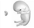
English: Two drawings of an abortus alongside each other. On the left is an embryo at 4 weeks after fertilization (6 weeks gestational age, crown-rump length is about 0.2 inches). On the right is a fetus 8 weeks after fertilization (10 weeks gestational age, crown-rump length is about 1.25 inches). |
| Date | |
| Source | Own work by uploader using images already uploaded to Wikimedia Commons |
| Author | Ferrylodge |
Uses other images already uploaded here and here.
Info regarding image of fetus on the right
editThis is a drawing of a human fetus at 10 weeks' gestational age (i.e. 8 weeks after fertilization). A color version of this image is available at Wikimedia here.
See larger version at 3D Pregnancy archive copy at the Wayback Machine. A rotatable 3D version of this photo is available here archive copy at the Wayback Machine, and a sketch is available here archive copy at the Wayback Machine.
The company behind 3DPregnancy.com is Tribal Internet Projects, a Dutch-based publisher of family websites. 3Dpregnancy.com was launched in 2007.[1] archive copy at the Wayback Machine
This particular picture was drawn by Melchior Meijer who is a 3D artist. He and 3Dpregnancy.com used various resources to produce the illustration, including books, DVD's and websites to verify how a fetus looks at this stage of development.
Some of the resources relied upon to create this image were as follows:
A Child is Born (A book by Lennart Nilsson)
In the Womb (DVD)
In the Womb (Book)
Kidshealth.org (Website)
When comparing this image to other images, it should be kept in mind that this stage of development is often referenced using different numerical descriptions. This is an approximate drawing of a fetus eight weeks after fertilization, i.e. at the beginning of the ninth week after fertilization. This is equivalent to a gestational age of about ten weeks, i.e. at the beginning of the eleventh week of gestational age. This drawing can be compared to other online images of a fetus at approximately the same stage of development, including the following images:
I. Drawing and movie of fetus at eight weeks and two days after fertilization, from the Endowment for Human Development;
II. Motion-picture archive copy at the Wayback Machine 4D ultrasound of fetus at eight weeks and two days after fertilization, from the Endowment for Human Development;
III. Photograph of fetus during ninth week after fertilization, from Thomas W. Sadler, Langman's Medical Embryology, page 90 (2006) via Google Books;
IV. Photograph archive copy at the Wayback Machine with detailed annotations at 8 weeks after fertilization, from online course in embryology for medicine students developed by the universities of Fribourg, Lausanne and Bern (Switzerland) with the support of the Swiss Virtual Campus;
V. Drawing of fetus at ten weeks’ gestational age, from KidsHealth.org which has a medical review board;
VI. Drawing of fetus at ten weeks' gestational age, from Michigan Department of Community Health;
VII. Drawing archive copy at the Wayback Machine of fetus at ten weeks' gestational age, from A.D.A.M. via About.com.
VIII. Photo of intact fetus removed from 44 year-old female who was diagnosed with carcinoma in situ of cervix (early stage cancer of womb). Abortion was deemed inevitable for future health of the woman. This fetus is at 10 weeks gestation (i.e. from LMP), instead of 10 weeks from fertilisation.
Note that by the fetal stage, the tail is gone. See Mayo Clinic website. An atrophied embryonic tail bud remains, but typically there is no tail.[2] archive copy at the Wayback Machine Additionally, note that a human fetus does not have gills. See Stanley J. Ulijaszek, Francis E. Johnston, M. A. Preece The Cambridge Encyclopedia of Human Growth and Development, pages 161-162 (1998). Also see James S. Trefil The Nature of Science, page 309 (2003). A fetus 8 weeks after fertilization is typically about 1.25 inches crown to rump. This donor-approved image is in black and white, although the originally-uploaded version is in a pinkish color. According to the pro-choice organization "Life and Liberty for Women" archive copy at the Wayback Machine, the color of a fetus after removal from the uterus (e.g. after an abortion) depends upon the method of removal. Gray skin will result from “laminaria through an intra-amniotic injection." On the other hand, "If the procedure was done while the fetus was alive, its skin would be the pinkish color....", as in the drawing that was originally uploaded.
The owners of this image donated it to Wikimedia. The original version that the owners donated had a watermark which has subsequently been removed according to Wikimedia policy. See here. The present image (without watermark) was donated by Wouter Vergeer <wouter.vergeer@tribal.nl> on Wednesday, August 29, 2007 on behalf of 3DPregnancy.com. Mr. Vergeer stated in an email to Ferrylodge on that date:
| “ | For now we are only willing to share these four 3D pictures under the CC-SY-BA license. Lets first see how this goes before we decide to share more or, perhaps in the future, all our works....I noticed you changed the picture and removed the CC-SY-BA notification. Which is fine by us. We just added them to make sure everybody saw they were released under CC-SY-BA license and nobody would delete them. We did notice, however that the quality of the images has decreased
quite a lot due to the cropping. I enclosed new versions of the images at better quality with this email. Would you be so kind to upload these? Or should we do this? The company behind 3DPregnancy.com is Tribal Internet Projects. We are Dutch based publisher of family websites. We are working on a corporate website containing more company information. If you have any more questions, please let me know. And thank you for your involvement. With kind regards / Met vriendelijke groet, Wouter Vergeer Managing Director Tribal Internet Projects BV Larenweg 24 5234 KA 's-Hertogenbosch The Netherlands Toll Free form U.S. and Canada: 1-888-766-5577 Tel.: +31 (0)73 6158113 (NEW NUMBER) Fax.: +31 (0)73 6124756 Internet: www.tribal.nl E-mail: wouter.vergeer@tribal.nl |
” |
This image is now licensed under a "free" license.
The color image has been converted to black and white, in response to some objections here. The staff of 3D Pregnancy was notified, and they replied (at Mon, 7 Jan 2008 10:55:23):
| “ | Thank you for notifying me. The black and white version as currently online, are fine! Thnx for the change. With kind regards, Wouter. | ” |
Licensing
edit- You are free:
- to share – to copy, distribute and transmit the work
- to remix – to adapt the work
- Under the following conditions:
- attribution – You must give appropriate credit, provide a link to the license, and indicate if changes were made. You may do so in any reasonable manner, but not in any way that suggests the licensor endorses you or your use.
- share alike – If you remix, transform, or build upon the material, you must distribute your contributions under the same or compatible license as the original.

|
Permission is granted to copy, distribute and/or modify this document under the terms of the GNU Free Documentation License, Version 1.2 or any later version published by the Free Software Foundation; with no Invariant Sections, no Front-Cover Texts, and no Back-Cover Texts. A copy of the license is included in the section entitled GNU Free Documentation License.http://www.gnu.org/copyleft/fdl.htmlGFDLGNU Free Documentation Licensetruetrue |
File history
Click on a date/time to view the file as it appeared at that time.
| Date/Time | Thumbnail | Dimensions | User | Comment | |
|---|---|---|---|---|---|
| current | 16:26, 4 March 2009 |  | 199 × 151 (10 KB) | Ferrylodge (talk | contribs) | Add scale. |
| 15:57, 21 February 2009 |  | 154 × 128 (9 KB) | Ferrylodge (talk | contribs) | {{Information |Description={{en|1=Two drawings of an abortus alongside each other. On the left is an embryo at 4 weeks after fertilization (6 weeks gestational age). On the right is a fetus 8 weeks after fertilization (10 weeks gestational age).}} |Sour |
You cannot overwrite this file.
File usage on Commons
The following page uses this file:
File usage on other wikis
The following other wikis use this file:
- Usage on en.wikipedia.org
- Usage on hi.wikipedia.org
- Usage on ka.wikipedia.org
- Usage on ms.wikipedia.org