Category:Biomechanics
study of the structure and function of the mechanical aspects of biological systems | |||||
| Upload media | |||||
| Instance of | |||||
|---|---|---|---|---|---|
| Subclass of | |||||
| |||||
English: Biomechanics is the application of mechanical principles to living organisms.
Subcategories
This category has the following 20 subcategories, out of 20 total.
A
- Animal flight mechanics (6 F)
- Animations of biomechanics (14 F)
B
- Biomechanical testing (1 F)
C
- Cellular mechanotransduction (59 F)
- Ciliary movement (23 F)
D
- De motu animalium (15 F)
E
F
G
I
- Injury biomechanics (19 F)
M
N
- Necrobotics (3 F)
S
T
V
W
Media in category "Biomechanics"
The following 163 files are in this category, out of 163 total.
-
1931 Smithsonian miscellaneous collections Snodgrass 1929 p082 Fig. 44.jpg 1,424 × 888; 117 KB
-
21-S-C4-JK-455e-biomech-push-up-front.png 1,554 × 2,132; 186 KB
-
22-S-C5-C3-JK-biomechanics-deltoid.png 2,745 × 2,815; 287 KB
-
22-S-C5-C3-JK-biomechanics-sauropod.png 2,454 × 1,926; 278 KB
-
3-Element Windkessel Model.svg 646 × 478; 12 KB
-
3036810 13239 2010 27 Fig1 HTML.png 512 × 339; 52 KB
-
A cell-based framework for modeling cardiac mechanics - 10237 2022 1660 MOESM1 ESM.webm 4.5 s, 7,680 × 4,320; 6.97 MB
-
A Psychophysics Experiment on the Control of Reaching Movements.png 1,370 × 923; 2.26 MB
-
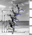
Akrobati, Lokální těžiště a hmotnosti 2.png 762 × 936; 535 KB
-
Akrobati, Lokální těžiště a hmotnosti 3 výpočet sw Mathcad.png 598 × 382; 121 KB
-
Akrobati, Lokální těžiště a hmotnosti.png 609 × 664; 409 KB
-
At biomechatronics, there are many ankles (3462800815).jpg 1,600 × 1,200; 507 KB
-
B09685P005 (1).jpg 2,496 × 1,664; 1.59 MB
-
B09717P006.jpg 1,728 × 1,152; 862 KB
-
Baletka, En pointe, Biomechanika, Silový rozbor, Metoda řezu.png 1,112 × 952; 229 KB
-
Beakon.jpg 473 × 268; 15 KB
-
Bewiss2überblick1.jpg 3,510 × 2,551; 1.13 MB
-
Biomechanics-mechanics-relation.png 786 × 308; 59 KB
-
Biomechanics-sumo-anatomy-muscles.png 1,295 × 703; 812 KB
-
Biomechinjury1.svg 1,152 × 864; 12 KB
-
Body Weight Support Treadmill Training.jpg 1,319 × 1,351; 199 KB
-
Boxing Heavy Bag Analysis.webm 24 s, 1,920 × 1,080; 124.82 MB
-
The brake pedal force capability of adult females (IA brakepedalforcec557radl).pdf 947 × 1,270, 32 pages; 1.06 MB
-
Broken fixed arm.jpg 826 × 370; 68 KB
-
Cardiacoutput4.jpg 670 × 331; 56 KB
-
Cardiacoutput7.jpg 500 × 498; 32 KB
-
Centroid human.png 652 × 598; 67 KB
-
Clamped beam solved example, book Biomechanika 1 by Karel Frydrýšek.png 833 × 593; 749 KB
-
Comparaison blessures de chat par nombre d'étages.JPG 272 × 336; 14 KB
-
Cyclogram Gastev TSIT.jpg 735 × 494; 81 KB
-
Differential of body.png 1,186 × 511; 1.33 MB
-
EB1911 Chiroptera Fig. 18.jpg 600 × 269; 56 KB
-
EMG - SIMI.jpg 1,333 × 1,000; 249 KB
-
Esquema rigidez de los órganos.gif 787 × 401; 43 KB
-
External Fixator Prospon (Cooperation VSB - Technical University and MEDIN).png 1,697 × 848; 1,006 KB
-
External-External-Fixator-Pelvis-Acetabulum.png 753 × 690; 162 KB
-
Fall-down.png 895 × 495; 545 KB
-
Femur-fractura-nail-artificial-bone.png 1,440 × 1,440; 1.73 MB
-
FigOppositeAsymmetry.pdf 162 × 493; 9 KB
-
Force Plate-Mounted Stairs by AMTI.jpg 3,008 × 2,000; 1.56 MB
-
Giovanni Borelli - lim joints (De Motu Animalium).jpg 446 × 548; 95 KB
-
Graphical description of the Fick principle..jpg 500 × 210; 21 KB
-
H-C Wikepedia.pdf 1,239 × 1,752; 303 KB
-
Hill muscle model.svg 231 × 403; 2 KB
-
History Centroid Man.png 565 × 872; 354 KB
-
Homo footprints.jpg 1,552 × 928; 153 KB
-
IBV Fachada.jpg 1,639 × 1,131; 1.26 MB
-
Imprints from skeletal foot (8616851548).jpg 1,072 × 1,000; 203 KB
-
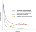
Input Impedance Windkessel models.PNG 662 × 596; 55 KB
-
Instrumented stairway.jpg 1,659 × 1,403; 602 KB
-
Inversion du pied.jpg 4,000 × 3,000; 1.74 MB
-
Jinf15.gif 336 × 325; 7 KB
-
Kiisa Nishikawa on The Company of Biologists.jpg 907 × 921; 132 KB
-
Kistler plates.jpg 1,977 × 1,469; 377 KB
-
Klouby člověka, mechanika, biomechanika.png 952 × 2,124; 2.52 MB
-
Kontrollschleifen der Motorik.jpg 2,360 × 2,453; 600 KB
-
Kontrollschleifen der Motorik.svg 600 × 350; 2 KB
-
Laboratorio Biomecánica.jpg 979 × 1,306; 669 KB
-
Laetoli Footprints Site A (1).jpg 685 × 342; 296 KB
-
Laetoli Footprints Site A (2).jpg 249 × 652; 215 KB
-
Laetoli footprints.png 1,511 × 1,885; 1.46 MB
-
Lower limb amputation levels.jpg 635 × 335; 49 KB
-
Mantis shrimp muscle.png 442 × 610; 98 KB
-
Medusa- Hydraulic Propulsion.jpg 7,002 × 5,100; 2.95 MB
-
MomentumHead.svg 496 × 383; 8 KB
-
OpenSim (Leg).jpg 1,152 × 847; 91 KB
-
Ortopedie, Biomechanika, operace, .png 1,015 × 980; 621 KB
-
Pas pronateur-supinateur-normal.png 400 × 297; 40 KB
-
PatrickLFamerL83.jpg 1,337 × 1,939; 205 KB
-
Pisada prono-supino-normal.svg 400 × 297; 89 KB
-
Plethysmography.jpg 400 × 320; 55 KB
-
Pneumatically Energized and Actuated Robotic Leg (PEARL).jpg 600 × 800; 71 KB
-
Proce.jpg 472 × 257; 49 KB
-
Prosthetic foot.jpg 6,000 × 4,000; 15.47 MB
-
Pseudoelastic response (stress vs stretch ratio).png 489 × 389; 13 KB
-
Pulse contour analysis..jpg 501 × 587; 45 KB
-
Representative Kinesiology Images.jpg 500 × 687; 333 KB
-
Rick Hansen.jpg 512 × 772; 61 KB
-
Rigid bodies.jpg 426 × 492; 40 KB
-
Spongiosabälkchen des proximalen Femurs korrespondierend zu den Spannungstrajectorien.png 1,596 × 1,586; 3.02 MB
-
Stabiloplatforma-sport.JPG 768 × 1,024; 333 KB
-
Strukturierung Mechanik.gif 720 × 512; 24 KB
-
Tendon-to-bone attachment with Williams singularity.png 1,074 × 402; 186 KB
-
The Hill-type model.jpg 493 × 217; 19 KB
-
The Muybridge Medal of the ISB.png 200 × 202; 63 KB
-
Tibia-CT-Biomechanics-part1.png 990 × 814; 362 KB
-
Tibia-CT-Biomechanics-part2.png 1,119 × 701; 392 KB
-
Two figures in motion (8615747543).jpg 2,976 × 3,244; 1.02 MB
-
UBC-BME-INJ-20-001.png 767 × 426; 24 KB
-
UBC-BME-INJ-20-010.png 772 × 534; 77 KB
-
UBC-BME-INJ-20-012.png 586 × 628; 184 KB
-
UBC-BME-INJ-20-014.png 1,294 × 668; 77 KB
-
UBC-BME-INJ-20-018-1.png 1,385 × 535; 91 KB
-
UBC-BME-INJ-20-018-2.png 705 × 277; 45 KB
-
UBC-BME-INJ-20-018-3.png 705 × 277; 48 KB
-
UBC-BME-INJ-20-018-4.png 705 × 277; 45 KB
-
UBC-BME-INJ-20-018-5.png 705 × 277; 48 KB
-
UBC-BME-INJ-20-019-1.png 862 × 560; 52 KB
-
UBC-BME-INJ-20-019-2.png 824 × 632; 59 KB
-
UBC-BME-INJ-20-019-3.png 914 × 582; 49 KB
-
UBC-BME-INJ-20-020.png 881 × 517; 87 KB
-
UBC-BME-INJ-20-021.png 982 × 680; 62 KB
-
UBC-BME-INJ-20-024-1.png 1,162 × 890; 68 KB
-
UBC-BME-INJ-20-024-2.png 747 × 556; 21 KB
-
UBC-BME-INJ-20-025-1.png 771 × 703; 73 KB
-
UBC-BME-INJ-20-027.png 717 × 326; 32 KB
-
UBC-BME-INJ-20-030.png 1,515 × 714; 171 KB
-
UBC-BME-KNM-20-002.png 1,112 × 1,208; 186 KB
-
UBC-BME-KNM-20-003.png 1,056 × 1,258; 269 KB
-
UBC-BME-KNM-20-005-1.png 538 × 1,082; 124 KB
-
UBC-BME-KNM-20-005-2.png 928 × 514; 162 KB
-
UBC-BME-KNM-20-005.png 1,162 × 340; 70 KB
-
UBC-BME-KNM-20-006.png 1,142 × 598; 117 KB
-
UBC-BME-KNM-20-007-1.png 954 × 818; 112 KB
-
UBC-BME-KNM-20-007-2.png 952 × 830; 130 KB
-
UBC-BME-KNM-20-008.png 1,424 × 1,080; 351 KB
-
UBC-BME-KNM-20-009.png 1,150 × 720; 121 KB
-
UBC-BME-KNM-20-010.png 1,454 × 692; 123 KB
-
UBC-BME-KNM-20-011.png 1,400 × 956; 167 KB
-
UBC-BME-KNM-20-013.png 1,426 × 700; 163 KB
-
UBC-BME-KNM-20-014.png 1,448 × 408; 121 KB
-
UBC-BME-KNM-20-015.png 904 × 480; 78 KB
-
UBC-BME-KNT-20-001.png 1,002 × 1,208; 317 KB
-
UBC-BME-KNT-20-002-1.png 1,062 × 608; 62 KB
-
UBC-BME-KNT-20-002-2.png 1,182 × 520; 303 KB
-
UBC-BME-KNT-20-003.png 378 × 502; 55 KB
-
UBC-BME-KNT-20-004.png 652 × 622; 141 KB
-
UBC-BME-KNT-20-005.png 710 × 558; 151 KB
-
UBC-BME-KNT-20-006-2.png 772 × 396; 178 KB
-
UBC-BME-KNT-20-008.png 756 × 588; 114 KB
-
UBC-BME-KNT-20-011-1.png 492 × 474; 42 KB
-
UBC-BME-KNT-20-011-2.png 532 × 510; 25 KB
-
UBC-BME-KNT-20-012.png 2,024 × 784; 379 KB
-
UBC-BME-KNT-20-013.png 438 × 520; 61 KB
-
UBC-BME-KNT-20-014-2.png 710 × 812; 85 KB
-
UBC-BME-KNT-20-015-01.png 776 × 824; 66 KB
-
UBC-BME-KNT-20-015-04.png 686 × 662; 49 KB
-
UBC-BME-STA-20-001.png 284 × 444; 49 KB
-
UBC-BME-STA-20-002-3.png 730 × 666; 156 KB
-
UBC-BME-STA-20-003-1.png 506 × 310; 73 KB
-
UBC-BME-STA-20-003.png 846 × 520; 154 KB
-
UBC-BME-STA-20-004-1.png 430 × 458; 47 KB
-
UBC-BME-STA-20-004-2.png 670 × 674; 121 KB
-
UBC-BME-STA-20-004-3.png 372 × 342; 28 KB
-
UBC-BME-STA-20-004-4.png 360 × 386; 30 KB
-
UBC-BME-STA-20-005.png 660 × 480; 121 KB
-
UBC-BME-STA-20-006.png 1,096 × 364; 48 KB
-
UBC-BME-STA-20-007-1.png 1,600 × 866; 72 KB
-
UBC-BME-STA-20-007-2.png 1,228 × 896; 88 KB
-
UBC-BME-STA-20-010.png 1,258 × 698; 555 KB
-
UBC-BME-STA-20-011.png 1,272 × 626; 191 KB
-
Ulm Helmholtzstraße 14 Institut für Unfallchirurgische Forschung und Biomechanik 2019 03 03.jpg 6,035 × 3,477; 12.74 MB
-
V09984P039.jpg 1,955 × 1,728; 1.55 MB
-
Van Mow Miami 2014B.jpg 391 × 509; 163 KB
-
Vodouš Tringa totanus silový rozbor.png 1,450 × 980; 884 KB


