Category:Uterus
Deutsch: Die Gebärmutter – lat. Uterus, griech. μέτρα (metra) oder ὑστέρα (hystera) – ist ein weibliches Geschlechtsorgan.
English: The uterus (Latin word for womb) is a major female hormone-responsive reproductive sex organ of most mammals, including humans.
major female hormone-responsive reproductive sex organ of most mammals including humans | |||||
| Upload media | |||||
| Instance of |
| ||||
|---|---|---|---|---|---|
| Subclass of |
| ||||
| Part of | |||||
| Facet of | |||||
| Has use | |||||
| |||||
Subcategories
This category has the following 11 subcategories, out of 11 total.
- Anatomy of uterus (30 F)
*
A
- Uterus in art (45 F)
- Artificial womb (5 F)
C
E
H
- Hystrometer (1 F)
U
- Videos about uterus (5 F)
Media in category "Uterus"
The following 110 files are in this category, out of 110 total.
-
Ampulla tube uterinae, the ampulla of the fallopian tube.jpg 1,400 × 512; 834 KB
-
Berlin, Museum europäischer Kulturen MEK, Votivgabe NIK 0470.jpg 2,144 × 3,216; 6.02 MB
-
Cervical canal mucus.png 645 × 518; 419 KB
-
Chart showing growth of uterus and vagina. Wellcome L0002176EB.jpg 1,177 × 1,502; 644 KB
-
De-Gebärmutter.ogg 1.5 s; 14 KB
-
De-Uterus.ogg 1.4 s; 13 KB
-
Developmental dynamics of the ovarian ligaments correlate with ovary morphogenesis.jpg 6,551 × 4,488; 3.57 MB
-
Die inneren Geschlechtstheile einer 35 Jahre alten Frau.jpg 1,145 × 888; 1,007 KB
-
Embryology during the fourth month of gestation. Canal of Nuck.png 1,073 × 598; 725 KB
-
Embryology during the third month of gestation. Canal of Nuck.png 3,000 × 1,974; 1.25 MB
-
En-us-uterus.ogg 1.2 s; 13 KB
-
Endometrium of rabbit uterus at 14 h-3days of pseudopregnancy.png 1,365 × 599; 1.24 MB
-
Expression and distribution of MECA-79 in the uterus cropped.png 2,949 × 1,221; 6.04 MB
-
Expression and distribution of MECA-79 in the uterus.png 2,992 × 3,313; 2.28 MB
-
Fehlbildungen des menschlichen Uterus 1.png 1,600 × 1,000; 48 KB
-
Female rabbit.png 1,157 × 939; 1.65 MB
-
Female reproductive tract in mammals 1.png 2,100 × 1,348; 78 KB
-
Fmi 1491274 1.jpg 470 × 259; 13 KB
-
Fr-utérus.ogg 1.2 s; 15 KB
-
Gebärmutter mit Embryo im Längsschnitt - Uterus longitudinal section with embryo.jpg 1,961 × 1,735; 525 KB
-
Growing Up (1928) 17.png 882 × 1,155; 674 KB
-
Growing Up (1928) 7.png 744 × 820; 1.1 MB
-
Healthcare professional educating on pelvic floor.jpg 3,390 × 2,323; 1.6 MB
-
Histopathological changes of uterine tissues for all groups.png 839 × 928; 1.01 MB
-
Hunterian Museum Specimen.jpg 6,000 × 4,000; 13.94 MB
-
Immuno-localization of PCNA after treatment with estradiol (a-b).png 829 × 568; 847 KB
-
Kirkes' handbook of physiology (1907) (14769687492).jpg 1,091 × 918; 435 KB
-
Les voies génitales femelles chez les mammifères.png 2,122 × 1,348; 105 KB
-
Malformación uterina.png 1,600 × 1,000; 94 KB
-
Nl-uterus.ogg 1.2 s; 15 KB
-
Number of endometrial glands in the cross-section of the uterus of a rat.jpg 1,344 × 1,124; 2.17 MB
-
Ovary and uterus.jpg 1,959 × 632; 1.5 MB
-
PullingUterusOver1.jpg 3,003 × 2,733; 897 KB
-
PushingUterusOver.jpg 3,023 × 2,433; 802 KB
-
Rabbit uterus at 18 days of pseudopregnancy.png 1,158 × 1,155; 2.4 MB
-
Rabbit uterus at 3-7 days of pseudopregnancy.png 1,019 × 795; 1.31 MB
-
Rabbit uterus at different stages of pseudopregnancy.png 1,379 × 906; 1.71 MB
-
Rabbit uterus during pseudopregnancy showing, telocytes.png 1,376 × 679; 766 KB
-
Reproductive system - Uterine cycle -- Smart-Servier.png 1,311 × 1,059; 258 KB
-
Retroverted and Retroflexed Uterus 1.png 2,500 × 1,000; 196 KB
-
The expansion and relocation of the Müllerian duct leave the ovary fully encapsulated.jpg 6,435 × 2,459; 2.51 MB
-
Uterine histological structure of a rat.jpg 1,100 × 882; 1.36 MB
-
Uterine malformation.png 1,600 × 1,000; 97 KB
-
Uterine tissue of female rats treated with (4 mgkg AgNps) H&E stain.jpg 1,200 × 921; 186 KB
-
Utero arcuato 1.png 820 × 480; 27 KB
-
Uterosacral ligaments connected to uterus.jpg 660 × 588; 80 KB
-
Uterus arcuatus 1.png 820 × 480; 28 KB
-
Uterus bicornis - Zoologie 1.png 790 × 730; 35 KB
-
Uterus bicornis 1.png 650 × 500; 23 KB
-
Uterus didelphys 1.png 400 × 340; 9 KB
-
Uterus of a rat, Mallory stain.jpg 4,608 × 3,072; 7.32 MB
-
Uterus of female rats treated with (2 mgkg AgNps) H&E stain.jpg 1,200 × 825; 180 KB
-
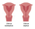
Uterus septus 1.png 600 × 500; 22 KB
-
Uterus unicornis 1.png 620 × 380; 18 KB
-
Uterus, Berengarius, 1523 Wellcome M0001523.jpg 3,841 × 2,893; 2.34 MB
-
UTERUSCAVITYPICTUREFROMVA.tif 300 × 240; 309 KB
-
Vagina septa 1.png 650 × 500; 25 KB
-
Weibliche Geschlechtsgänge bei Säugetieren 1.png 2,233 × 1,348; 174 KB
-
Женские половые органы.png 1,080 × 1,080; 433 KB
