File:Metastasis sites for common cancers.svg

Original file (SVG file, nominally 680 × 1,090 pixels, file size: 1.64 MB)
Captions
Captions
Contents
Summary
edit| DescriptionMetastasis sites for common cancers.svg |
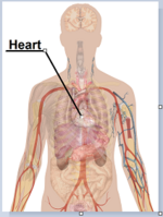
English: Main sites of metastases for common cancer types. Primary cancers are denoted by "...cancer" (except for skin melanoma) and their main metastasis sites are denoted by "...metastases".
List of included informationThe included cancer types are the ones causing most death as per data from the US in 2008.[1]
Not included information of major importance
References
|
| Date | |
| Source | All imaged used are in the Public Domain |
| Author | Mikael Häggström |
| Other versions |
[edit]
|
Licensing
edit| This file is made available under the Creative Commons CC0 1.0 Universal Public Domain Dedication. | |
| The person who associated a work with this deed has dedicated the work to the public domain by waiving all of their rights to the work worldwide under copyright law, including all related and neighboring rights, to the extent allowed by law. You can copy, modify, distribute and perform the work, even for commercial purposes, all without asking permission.
http://creativecommons.org/publicdomain/zero/1.0/deed.enCC0Creative Commons Zero, Public Domain Dedicationfalsefalse |
Human body diagramseditMain article at: Human body diagrams Template location:Template:Human body diagrams How to derive an imageeditDerive directly from raster image with organseditThe raster (.png format) images below have most commonly used organs already included, and text and lines can be added in almost any graphics editor. This is the easiest method, but does not leave any room for customizing what organs are shown. Adding text and lines: Derive "from scratch"editBy this method, body diagrams can be derived by pasting organs into one of the "plain" body images shown below. This method requires a graphics editor that can handle transparent images, in order to avoid white squares around the organs when pasting onto the body image. Pictures of organs are found on the project's main page. These were originally adapted to fit the male shadow/silhouette.
Organs:
Derive by vector templateeditThe Vector templates below can be used to derive images with, for example, Inkscape. This is the method with the greatest potential. See Human body diagrams/Inkscape tutorial for a basic description in how to do this.
Examples of derived worksedit
Licensingedit
|
File history
Click on a date/time to view the file as it appeared at that time.
| Date/Time | Thumbnail | Dimensions | User | Comment | |
|---|---|---|---|---|---|
| current | 10:53, 19 October 2018 |  | 680 × 1,090 (1.64 MB) | Jmarchn (talk | contribs) | Draw now with 3 layers (draw, arrows and text) |
| 07:17, 17 June 2011 |  | 680 × 1,090 (1.65 MB) | Mikael Häggström (talk | contribs) | Retry | |
| 05:02, 23 May 2011 |  | 680 × 1,090 (1.65 MB) | Mikael Häggström (talk | contribs) | Bunny-jumping, because it won't accept my latest edit. It's always the previous version that shows correct | |
| 05:00, 23 May 2011 |  | 621 × 767 (1.18 MB) | Mikael Häggström (talk | contribs) | see above | |
| 04:56, 23 May 2011 |  | 680 × 1,090 (1.65 MB) | Mikael Häggström (talk | contribs) | Now it's to previous one that shows correct! Reverting. | |
| 04:55, 23 May 2011 |  | 680 × 1,090 (1.65 MB) | Mikael Häggström (talk | contribs) | No response, so redid | |
| 04:54, 23 May 2011 |  | 680 × 1,090 (1.65 MB) | Mikael Häggström (talk | contribs) | Made pancreas arrows blue to distinguish from the red of lung cancer | |
| 16:12, 22 May 2011 |  | 680 × 1,090 (1.65 MB) | Mikael Häggström (talk | contribs) | darker green | |
| 15:57, 22 May 2011 |  | 680 × 1,090 (1.65 MB) | Mikael Häggström (talk | contribs) | Ups, forgot to write out colorectal cancer | |
| 15:55, 22 May 2011 |  | 680 × 1,090 (1.65 MB) | Mikael Häggström (talk | contribs) | {{Information |Description ={{en|1=f}} |Source ={{own}} |Author =Mikael Häggström |Date =f |Permission = |other_versions = }} |
You cannot overwrite this file.
File usage on Commons
The following 9 pages use this file:
- File:Metastasis sites for common cancers-IT.png
- File:Metastasis sites for common cancers-IT.svg
- File:Metastasis sites for common cancers-IT Inkscape.png
- File:Metastasis sites for common cancers-IT Workaround.svg
- File:Metastasis sites for common cancers-IT librsvg.png
- File:Metastasis sites for common cancers-IT rendersvg.png
- File:Metastasis sites for common cancers-ca.svg
- File:Metastasis sites for common cancers.svg
- Template:Other versions/Metastasis sites for common cancers
File usage on other wikis
The following other wikis use this file:
- Usage on ar.wikipedia.org
- Usage on en.wikipedia.org
- Usage on eu.wikipedia.org
- Usage on fa.wikipedia.org
- Usage on ha.wikipedia.org
- Usage on hy.wikipedia.org
- Usage on sr.wikipedia.org
- Usage on tl.wikipedia.org
- Usage on zh.wikipedia.org
Metadata
This file contains additional information such as Exif metadata which may have been added by the digital camera, scanner, or software program used to create or digitize it. If the file has been modified from its original state, some details such as the timestamp may not fully reflect those of the original file. The timestamp is only as accurate as the clock in the camera, and it may be completely wrong.
| Width | 680 |
|---|---|
| Height | 1090 |


