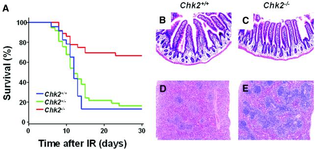
Fig. 1. Reduced radiosensitivity of Chk2–/– mice and Chk2–/– splenocytes in vivo. (A) Kaplan–Meier survival curve of age-matched 8–16-week-old Chk2+/+ (n = 23), Chk2+/– (n = 37) and Chk2–/– (n = 36) mice after exposure to 8 Gy of X-rays. Data are combined from two separate experiments. (B and C) Normal appearance of intestine in Chk2+/+ (B) and Chk2–/– (C) mice 1 day after IR. (D and E) Atrophy of white pulp of the spleen and reduction of splenocyte number in Chk2+/+ mice (D), but not in Chk2–/– mice (E), 8 days after exposure to IR.

An official website of the United States government
Here's how you know
Official websites use .gov
A
.gov website belongs to an official
government organization in the United States.
Secure .gov websites use HTTPS
A lock (
) or https:// means you've safely
connected to the .gov website. Share sensitive
information only on official, secure websites.
