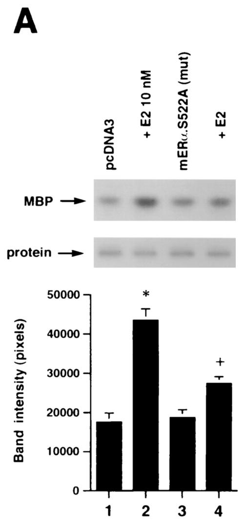
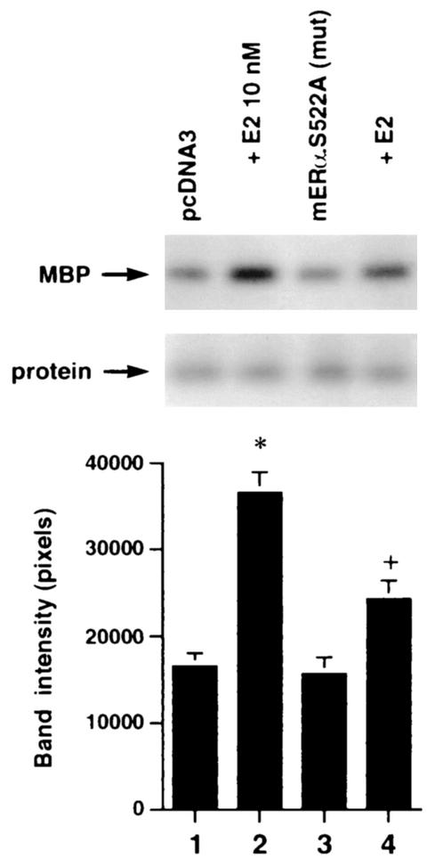
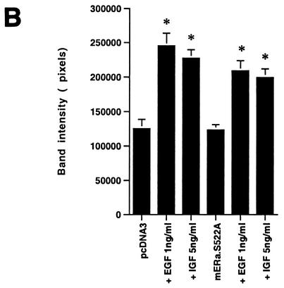
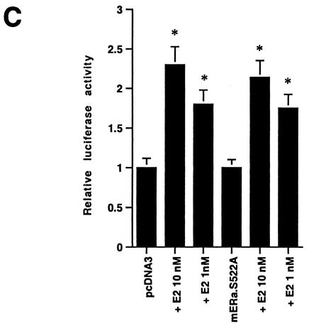
FIG. 5.
(A) Expression of ERα S522A inhibits E2 activation of ERK. MCF-7 (left) or ZR-75-1 (right) breast cancer cells (which express endogenous ER) were transfected transiently to express either pcDNA3 (control) or S522A mutant ERα. The cells were then exposed to 10 nM E2 for 8 min, after which they were lysed, and immunoprecipitated ERK was assayed for activity by using MBP as a substrate. Precipitated ERK protein is shown in the lower gels, and the bar graphs each reflect three experiments combined. *, P < 0.05 for pcDNA3 in the absence versus the presence of E2; +, P < 0.05 for comparison of E2 treatments of pcDNA3-expressing versus ERα S522A-expressing cells. (B) ERα S522A does not impair EGF or IGF-1 activation of ERK. Data from three experiments are combined. *, P < 0.05 for pcDNA3-transfected or ERα S522A-expressing MCF-7 cells in the absence versus the presence of EGF or IGF-1. (C) E2 comparably activates an ERE-luciferase reporter in untransfected MCF-7 cells and MCF-7 cells transfected to express ERα S522A. Bar graph shows results for three experiments combined. *, P < 0.05 for pcDNA3- or ERα S522A-transfected MCF-7 cells without versus with E2.


