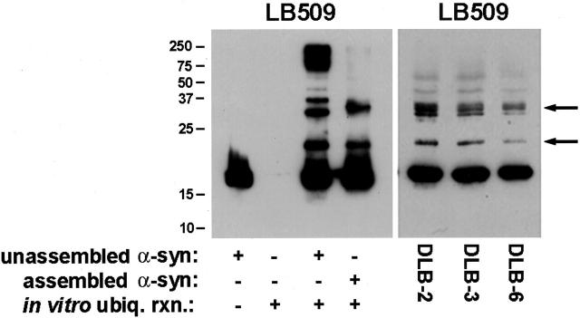
Figure 5.
In vitro ubiquitination of monomeric and fibrillar α-syn. Left: Unassembled or fibrillar α-syn (0.5 mg/ml) was subjected to in vitro ubiquitination as described in Materials and Methods. Equal volumes of unassembled α-syn, ubiquitination reaction without α-syn, ubiquitination reaction with unassembled α-syn, and ubiquitination reaction with fibrillar α-syn were loaded in separate lanes of 15% SDS-polyacrylamide gels that were transferred electrophoretically onto nitrocellulose and analyzed by Western blotting with LB509. Right: Western blot analysis (using LB509) of the SDS-soluble fraction of cingulate cortex from three DLB cases. Arrows indicate ubiquitinated forms of α-syn. The mobility of molecular mass markers (kd) is depicted to the left of the panel.

