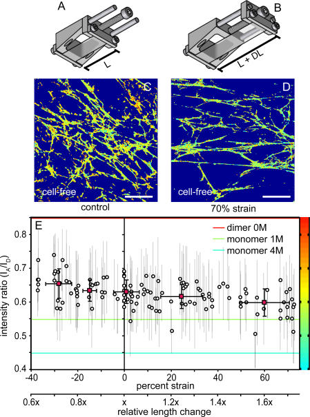
Figure 5. Fn Conformation after Application of Exogenous Strain.
A schematic of the strain device is shown in the relaxed configuration with length L before (A) and length L + DL after (B) application of strain. PDMS sheets were covalently modified with Fn-u as described in Materials and Methods, and fibroblast cells were cultured for 24 h in the presence of amine/cys Fn-DA and excess Fn-u. Cells were extracted in mild detergent. Color-coded I A/I D ratiometric images are shown for a field of view without application of stretch (C) and after application of 70% elongation strain with 28% transverse compression (D). Region of interest analysis on individual fibrils was used to determine the impact of elongation on I A/I D on a per fibril basis (circles, mean ± standard deviation), and binned averages were calculated for fibrils between −37% and −20%, −20% and −10%, −10% and 10%, 10% and 40%, and 40% and 73% strain (red squares, mean ± standard deviations) (E). Abscissa is also plotted as relative length change. Solution values for dimeric Fn-DA in 0 M GdnHCl and monomeric Fn-DA in 1 and 4 M GdnHCl are shown as horizontal red, green, and blue lines, respectively. Scale bars = 50 μm.

