Figure 5.


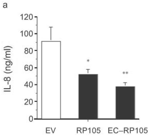
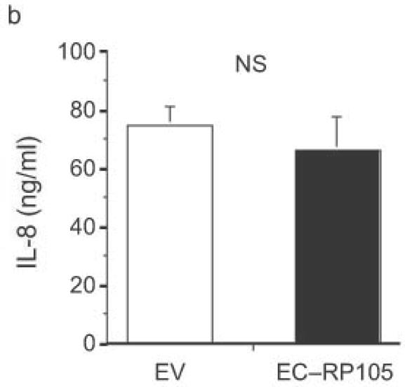
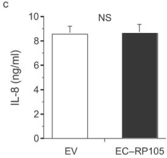
The extracellular domain of RP105 is sufficient to effect suppression of TLR4 signaling. (a) HEK293 cells stably expressing CD14 and TLR4 were transiently transfected with MD-2 and MD-1, along with EV, RP105 or the extracellular domain of RP105 (EC-RP105), as indicated. Cells were subsequently stimulated with purified E. coli K235 LPS (10 ng/ml). Means +/− SE of triplicate cultures in a single experiment are depicted, representative of an experimental n = 4 *p < 0.02, **p < 0.002, compared with RP105-deficient cells. (b, c) HEK293 cells stably expressing CD14 and TLR2 were transiently transfected with MD-2 and MD-1, along with EV or EC-RP105, as indicated. Cells were subsequently stimulated with (b) Zymosan A (10 μg/ml) or (c) IL-1β (100 ng/ml) as noted. Means +/− SE of replicate cultures (n =9) are depicted. NS, not significant.
