Abstract
Melanoma chondroitin sulfate proteoglycan (MCSP) is an early cell surface melanoma progression marker implicated in stimulating tumor cell proliferation, migration, and invasion. Focal adhesion kinase (FAK) plays a pivotal role in integrating growth factor and adhesion-related signaling pathways, facilitating cell spreading and migration. Extracellular signal–regulated kinase (ERK) 1 and 2, implicated in tumor growth and survival, has also been linked to clinical melanoma progression. We have cloned the MCSP core protein and expressed it in the MCSP-negative melanoma cell line WM1552C. Expression of MCSP enhances integrin-mediated cell spreading, FAK phosphorylation, and activation of ERK1/2. MCSP transfectants exhibit extensive MCSP-rich microspikes on adherent cells, where it also colocalizes with α4 integrin. Enhanced activation of FAK and ERK1/2 by MCSP appears to involve independent mechanisms because inhibition of FAK activation had no effect on ERK1/2 phosphorylation. These results indicate that MCSP may facilitate primary melanoma progression by enhancing the activation of key signaling pathways important for tumor invasion and growth.
Keywords: melanoma chondroitin sulfate proteoglycan; FAK; integrin; ERK1/2; cell spreading
Introduction
Metastatic melanoma is one of the fastest rising skin cancers (Houghton and Polsky, 2002; Geller and Annas, 2003). In certain patient groups, primary melanomas progress to malignancy via discrete but overlapping stages including dysplasia, radial growth phase (RGP), invasive vertical growth phase (VGP), and metastasis. Alterations in cell–cell and cell–ECM interactions are also associated with these stages of tumor progression. Changes in the expression or function of adhesion molecules such as integrins, Mel-CAM/MUC18, CD44, ICAM-1, cadherins, and cell surface proteoglycans (PGs) have all been documented in the progression of primary melanomas (Bogenrieder and Herlyn, 2002; Li et al., 2002).
Melanoma chondroitin sulfate proteoglycan (MCSP) is uniformly and abundantly expressed in most human melanoma lesions (Ferrone et al., 1988) and has been implicated in tumor invasion (Iida et al., 2001). MCSP expression is an ominous prognostic factor in acral letiginous melanoma (Kageshita et al., 1993) and in nonmelanoma tumors such as infantile acute myeloid leukemia (Hilden et al., 1997). The protein can be expressed with or without covalently attached chondroitin sulfate glycosaminoglycan, and is therefore considered a “part-time” cell surface PG. Chondroitin sulfate modification of the core protein has been linked to its ability to bind the heparin-binding domain of fibronectin (FN; Iida et al., 1992, 1996). MCSP acts as a coreceptor for α4β1 integrin to modulate cell adhesion and spreading by mechanisms dependent on the small Rho family GTPase Cdc42 and the adaptor protein p130cas (p130 crk-associated substrate; Eisenmann et al., 1999). MCSP is also associated with the transmembrane matrix metalloproteinase MT3-MMP on VGP melanoma cells, and in vitro facilitates invasion into type I collagen gels as well as degradation of denatured type I collagen (Iida et al., 2001).
Our findings for MCSP function are complemented by analyses of NG2, the rat homologue of MCSP, which appears to be important for cell proliferation and migration (Burg et al., 1998; Chekenya et al., 1999; Nishiyama, 2001). NG2 interacts with a variety of ECM components, including FN, collagens type II, V, and VI, laminin, and tenascin to alter cellular morphology and proliferation (Stallcup, 2002; Majumdar et al., 2003). Collectively, the available results support a role for MCSP/NG2 as signal-transducing molecules that initiate or modify intracellular signal cascades important for cell adhesion, motility, and invasion.
Integrins are a family of heterodimeric adhesion receptors that mediate both cell–ECM and cell–cell adhesion. Integrins initiate multiple cellular signals that profoundly influence shape, proliferation, differentiation, invasion, metastasis, apoptosis, and anoikis (Dedhar, 1999; Giancotti and Ruoslahti, 1999; Juliano, 2002). Altered expression of a number of integrins has been linked to the progression of malignant melanoma, and increased expression of α4β1 integrin is associated with transformation from RGP to VGP (Johnson, 1999; Hood and Cheresh, 2002). α4β1 integrin binds to the CS1-binding domain of FN (Wayner et al., 1989; Iida et al., 1992) and to the vascular endothelial cell adhesion molecule VCAM (Mould et al., 1994). Soluble antagonists of α4β1 integrin inhibit melanoma metastasis in animal models, emphasizing the importance of this receptor in melanoma biology (Danen et al., 1998).
FAK is a major nonreceptor tyrosine kinase activated after integrin-mediated adhesion to ECM proteins such as FN. Among other things, FAK serves to integrate signaling pathways between growth factor receptors and integrins (Sieg et al., 2000) and is implicated in facilitating cell survival and regulating cell spreading, migration, and invasion (Schlaepfer and Hunter, 1996; Hauck et al., 2002). Interaction between FAK and the cytoplasmic tail of β1 integrins results in autophosphorylation of FAK tyrosine 397 (FAK pY397) that can lead to stimulation of a cell-signaling cascade that ultimately activates the Ras/MAPK/extracellular signal–regulated kinase (ERK) pathway in some cells (Guan, 1997; Sieg et al., 2000). Increased expression or constitutive activation of FAK correlates with increased invasion and metastasis in many malignancies, including melanoma, emphasizing a potential link between FAK activation and tumor progression (Kahana et al., 2002; Hecker and Gladson, 2003).
Integrin-mediated adhesion also impacts on growth factor–stimulated activation of the ERK/MAPK pathway, a major effector of tumor cell migration, growth, and survival (Aplin and Juliano, 1999; Howe et al., 2002). Integrin-mediated activation of ERK/MAPK can occur through FAK-dependent and independent mechanisms, although the exact means of activation are unknown (Lin et al., 1997; Schlaepfer and Hunter, 1997; Barberis et al., 2000). Constitutive activation of ERK 1 and 2 is associated with more advanced melanoma tumors, where it promotes anchorage-independent growth and survival (Mansour et al., 1994; Conner et al., 2003; Satyamoorthy et al., 2003).
In this report, we have cloned and expressed MCSP in an RGP human melanoma cell line that lacks endogenous MCSP. Cells transfected with MCSP exhibited integrin-mediated cell spreading and markedly enhanced phosphorylation of FAK compared with mock transfectants. MCSP-expressing cells also exhibited a higher basal level of ERK1/2 phosphorylation, which was further increased by engaging α4β1 integrin. Enhanced ERK1/2 phosphorylation appeared to be independent of FAK phosphorylation, as overexpression of a dominant-negative focal adhesion–related nonkinase (FRNK) completely inhibited phosphorylation of endogenous FAK, but had no effect on ERK1/2 phosphorylation. MCSP also functions as a coreceptor for α4β1 integrin, and confocal analysis showed extensive colocalization between MCSP and the α4 integrin subunit. Therefore, elevated MCSP expression in early tumors may facilitate melanoma progression by enhancing the activation of FAK and ERK1/2 (as well as other signaling pathways) associated with tumor progression.
Results
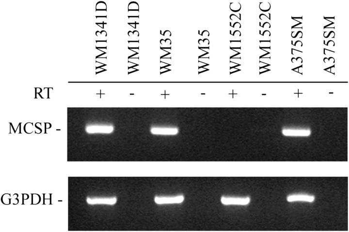
MCSP participates in cell–ECM interactions and is expressed on most human melanoma cell lines (Ferrone et al., 1988). Initially, human melanoma cell lines grown from tumors at various stages of progression (Satyamoorthy et al., 1997; Eisenmann et al., 1999) were screened for MCSP expression. Of the fifteen cell lines tested, only one, WM1552C, which was isolated from a patient with an RGP tumor, failed to express detectable levels of MCSP mRNA (Fig. 1). By contrast, another RGP cell line, WM35, expressed easily detectable MCSP mRNA, as did the invasive VGP cell line WM1341D and the metastatic cell line A375SM (Fig. 1). Surface expression of MCSP (or absence in WM1552C cells) was verified by flow cytometry using mAb 9.2.27 (unpublished data), which recognizes the core protein of MCSP.
Figure 1.
Expression of MCSP mRNA in melanoma cell lines by RT-PCR. Poly(A)+ RNA was isolated from each of the cell lines indicated and equal amounts were reverse transcribed (± reverse transcriptase), followed by PCR amplification with primers specific for MCSP or glyceraldehyde-3-phosphate dehydrogenase (G3PDH), as a control.
Cloning full-length human MCSP
Full-length human MCSP cDNA was generated by RT-PCR amplification of mRNA isolated from A375SM cells as described in the Materials and methods. Analysis of the full-length clone revealed a number of variations in nucleotide and amino acid sequence compared with previous reports (Pluschke et al., 1996). Short sequences encompassing the regions of variance were amplified from A375SM, WM35, and WM1341D cells by RT-PCR to verify that the nucleotide variations were not cell line dependent or an artifact of cloning. We also compared the sequences to the human genome database and found that, in every case, the human genome sequence matched that found in our clone. We have concluded that our MCSP clone more accurately reflects the sequence of MCSP expressed in human melanomas and have submitted the complete sequence to GenBank (accession no. AY359468).
Of the eleven differences found in amino acid sequence (highlighted in Fig. 2), eight of these changes (H477R, Y478H, L486P, C631R, H715Q, S716G, T717A, and L942H) reflect amino acids that are identical to the residue at the corresponding position in the rat homologue NG2, based on protein sequence alignment using the Wisconsin GCG package (Nishiyama et al., 1991; Pluschke et al., 1996). The other three alterations (E1208R, P1405A, and P1557R) represent novel changes to the amino acid sequence not previously reported in either MCSP or NG2. Perhaps the most notable change is at residue 631, where arginine has replaced cysteine, thereby reducing the number of potential disulfide bridges in the NH2-terminal cysteine region of MCSP. This change may affect folding of the NH2-terminal portion of the protein, which includes the laminin-G domains. Other structural features of the MCSP ectodomain, including the putative chondroitin sulfate attachment sites and newly defined CSPG repeats, appear to be unaffected by these changes.
Figure 2.
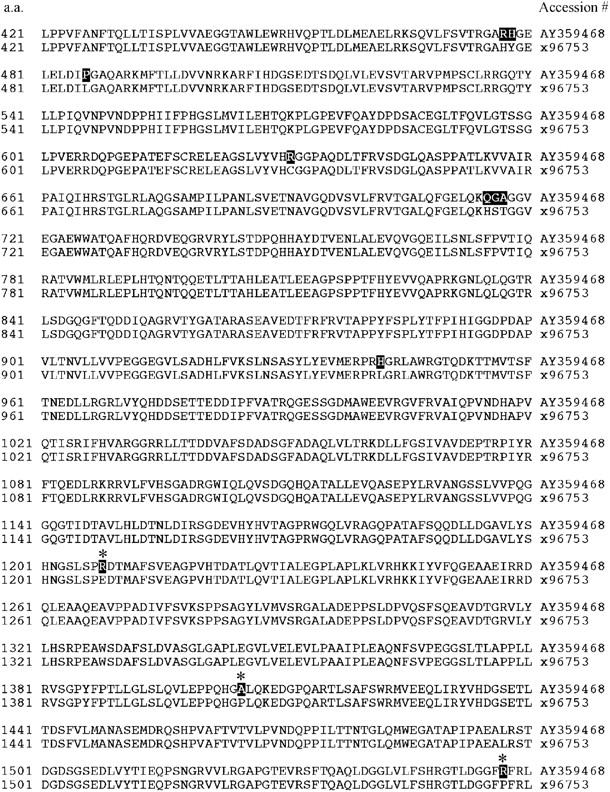
Partial amino acid sequence alignment of the MCSP cDNA generated from A375SM melanoma cells and the published sequence. Highlighted sequences represent revisions of the original sequence (Pluschke et al., 1996). The new and original sequence data are available from GenBank/EMBL/DDBJ under accession no. AY359468 and X96753, respectively. Asterisks represent novel amino acid changes not previously reported for MCSP or NG2.
Transfected MCSP induces adhesion and spreading of melanoma cells
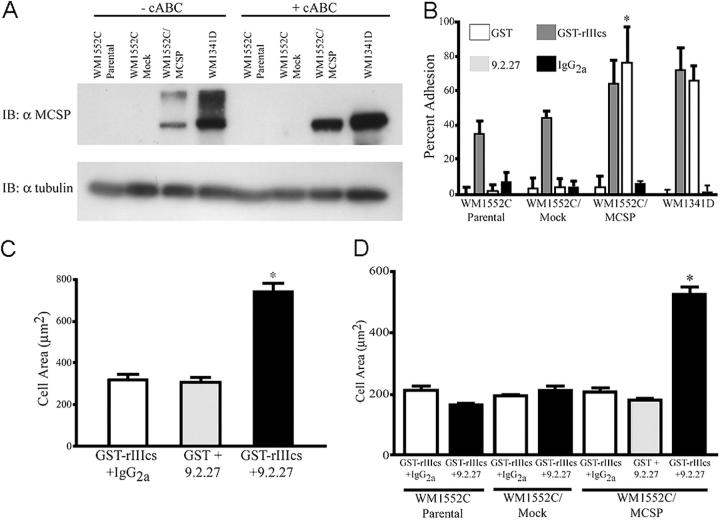
WM1552C cells transfected with cDNA encoding the MCSP core protein were generated as outlined in the Materials and methods, resulting in a stable population of cells that was >95% positive for MCSP. Flow cytometric analysis of MCSP expression on transfected cells revealed levels similar to those observed on cells expressing endogenous MCSP (unpublished data). Immunoblotting of total cell extracts was used to determine if the MCSP core protein was also expressed as a chondroitin sulfate PG on transfected cells (Fig. 3 A). Two immunoreactive bands at ∼400 and 250 kD were detected in extracts of WM1552C/MCSP cells, which is similar to what was observed in WM1341D cells that express endogenous MCSP. The larger mol wt species represents core protein modified with chondroitin sulfate glycosaminoglycan, as evidenced by sensitivity to treatment with chondroitinase ABC (Fig. 3 A, cABC). The level of α4β1 integrin on the surface of WM1552C cells was unaffected by expression of MCSP when evaluated by flow cytometry (unpublished data).
Figure 3.
MCSP supports adhesion and induces spreading of WM1552C/MCSP-transfected cells. (A) WM1552C cells were stably transfected with MCSP (WM1552C/MCSP) and expression verified by immunoblot. Whole-cell lysates from cells incubated ±0.5 U/ml cABC were fractionated by SDS-PAGE and probed with anti-MCSP core protein mAb 9.2.27. (B) Cells were plated in 96-well plates on 3 μg/ml GST, 3 μg/ml GST-rIIIcs, 1 μg/ml mAb 9.2.27, or 1 μg/ml IgG2a, allowed to adhere for 30 min at 37°C, and the number of adherent cells was determined by formazan absorbance. Data shown represent the mean of triplicate wells, ± SD. (C) WM1341D- and (D) WM1552C-transfected cells were serum starved overnight and plated on chimeric substrata using the same concentrations as in B. Plates were incubated at 37°C for 1 h, washed, fixed, and stained as described in the Materials and methods. Cell areas of a random 50 cells/well from triplicate wells were quantified by tracing the cell border using NIH Image software. *, P < 0.001 by two-tailed t test.
We have previously used chimeric substrates that can selectively bind α4β1 integrin (GST-rIIIcs, a minimal FN fragment containing the CS1 integrin-binding sequence) and MCSP (using antibody against the extracellular portion of the MCSP core protein) as a model for ligands (Eisenmann et al., 1999). We have also shown that metastatic cells adherent on CS1 spread when the MCSP core protein was also engaged (Iida et al., 1995; Eisenmann et al., 1999). Wells coated with a recombinant GST fusion protein containing the CS1 α4β1 integrin-binding domain of FN (GST-rIIIcs) promoted high levels of adhesion of both WM1552C and WM1341D cells (Fig. 3 B). Wells coated with the anti-MCSP mAb 9.2.27 also supported high adhesion levels of both WM1341D cells and WM1552C/MCSP transfectants (but not parental or mock WM1552C cells; Fig. 3 B). MCSP-expressing cells did not significantly spread on surfaces coated only with integrin-binding (GST-rIIIcs/IgG2a) or PG-binding (GST/mAb 9.2.27) substrates; however, chimeric GST-rIIIcs/mAb 9.2.27 (integrin/PG-binding) surfaces promoted extensive cell spreading (Fig. 3, C and D).
MCSP expression enhances phosphorylation of FAK and ERK1/2
Because FAK is a key member of integrin-mediated signaling pathways and initial cell spreading is regulated partly through FAK activity in many cells (Guan, 1997; Schlaepfer et al., 1999), we tested whether MCSP could induce FAK activation. WM1341D cells were serum starved overnight and were then plated on the various surfaces as indicated (Fig. 4). Engaging integrin alone on surfaces coated with GST-rIIIcs/IgG2a caused a modest increase in the level of FAK pY397 that peaked at 30 min after plating (Fig. 4 A). By contrast, plating the WM1341D cells onto integrin/PG-binding substrata resulted in enhanced levels of FAK pY397 much greater than those observed in cells adherent only via α4β1 integrin. The kinetics of phosphorylation at FAK Y397 were similar on both substrates (Fig. 4 A). Plating cells on surfaces coated with GST/mAb 9.2.27, used to stimulate MCSP alone, did not result in increased FAK pY397 (Fig. 4 B), indicating that MCSP does not directly stimulate FAK phosphorylation.
Figure 4.
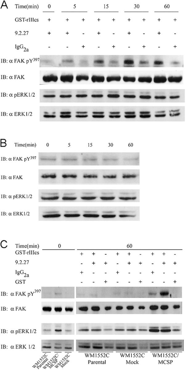
MCSP stimulates FAK Y 397 and ERK1/2 phosphorylation in melanoma cells. Cells were serum starved overnight and plated on various chimeric substrata using the same concentrations described in Fig. 3 B. (A) WM1341D cells were allowed to adhere to the specified substrata at 37°C for the indicated times, lysed with SDS sample buffer, and immunoblotted with total and phosphospecific FAK and ERK1/2 antibodies as indicated. (B) WM1341D cells were allowed to adhere to plates coated with mAb 9.2.27 and GST for the indicated times at 37°C, lysed in SDS sample buffer, and lysates were evaluated by immunoblotting as in A. (C) WM1552C parental and transfectant cells were plated on the indicated substrata and allowed to adhere for 1 h at 37°C. Cells were lysed in SDS sample buffer and analyzed for levels of FAK and ERK1/2 phosphorylation as in A.
The ERK/MAPK pathway has been implicated in cell spreading and is one of the downstream effectors of activated FAK (Guan, 1997; Schlaepfer et al., 1999). We reprobed the blots to determine if there is a relationship between FAK pY397 and ERK1/2 phosphorylation in these cells. Phosphorylated ERK (pERK) 1/2 was easily detected in the VGP WM1341D cells (Fig. 4 A); it did not appreciably vary in these cells during the time course of the assay (0–60 min). The level of pERK1/2 was equal in cells plated on either the integrin- or the integrin/PG-binding substrates (Fig. 4 A), in contrast to what was observed with FAK phosphorylation. Engaging MCSP alone also had no effect on the level of pERK1/2 in these cells (Fig. 4 B). ERK1/2 is constitutively activated in suspended WM1341D cells (zero time point) and was not further phosphorylated after cell adhesion (Fig. 4, A and B). Therefore, FAK activation induced by adhesion of WM1341D cells does not appear to have an effect on the level of ERK phosphorylation.
As was observed for the WM1341D cells, WM1552C/MCSP transfectants exhibited low levels of FAK pY397 when plated onto the integrin-binding surfaces for 60 min (Fig. 4 C). However, the WM1552C/MCSP transfectants (but not the parental or mock-transfected cells) showed a robust level of FAK pY397 when plated onto the integrin/PG-binding surfaces (Fig. 4 C), similar to what was observed in the WM1341D cells that express endogenous MCSP (Fig. 4 A).
The WM1552C cells differ from the WM1341D cells with respect to regulating the levels of ERK1/2 phosphorylation. WM1552C/MCSP cells in suspension maintained easily detectable levels of pERK1/2, whereas parental and mock transfectants exhibited very low levels of pERK1/2 under suspension conditions (Fig. 4 C, zero time point). Plating of cells onto the chimeric substrate for 1 h at 37°C resulted in greater levels of ERK1/2 stimulation within the MCSP transfectants (Fig. 4 C), which may be due, in part, to the higher baseline level observed in suspended cells. Elevated levels of pERK1/2 were not observed in cells plated on surfaces that bind only MCSP (Fig. 4 C; GST/mAb 9.2.27), demonstrating that increased levels of pERK1/2 in cells plated on the chimeric substrate were related to engagement of integrin.
MCSP enhances FAK activation in cells adherent to FN-related ligands
Next, we wanted to determine if MCSP expression activated signal transduction pathways when cells contacted FN-derived ligands, as FN contains multiple cell adhesion sites that bind to a number of integrins and cell surface PGs. We used a low coating concentration (0.5 μg/ml) of a recombinant FN fragment (GST-FN51) that contains both the heparin-binding domain and multiple CS1 sites for binding both PG and α4β1 integrin, respectively. WM1552C/MCSP cells contacting this fragment exhibit elevated levels of FAK pY397 within 30 min of plating when compared with mock counterparts (Fig. 5 A). MCSP transfectants also showed enhanced FAK phosphorylation when plated on low coating concentrations of intact FN (Fig. 5 B); however, this enhanced FAK phosphorylation was not evident when higher coating concentrations (5.0 and 10.0 μg/ml) of either GST-FN51 or FN were used (unpublished data). mAb 9.2.27 (but not mAb 149.53) could inhibit the enhanced FAK phosphorylation when used as a soluble antagonist (Fig. 5 C). The epitope for mAb 149.53 has been mapped to core protein residues 1846–1857 (unpublished data), whereas that recognized by mAb 9.2.27 appears to be located toward the NH2-terminal end of the core protein (unpublished data). These results suggest that a specific domain(s) within the extracellular portion of the MCSP core protein is important for promoting FAK phosphorylation in melanoma cells. The levels of pERK1/2 were less affected by adhesion to the GST-FN51 fragment or to intact FN than to the chimeric substrate (Fig. 5). Furthermore, antibodies against the MCSP core protein that inhibited enhanced FAK phosphorylation had no inhibitory effect on pERK1/2 levels in MCSP-expressing cells (Fig. 5 C). These results further indicate that the activation of FAK and ERK1/2 is regulated by distinct mechanisms in WM1552C/MCSP cells.
Figure 5.
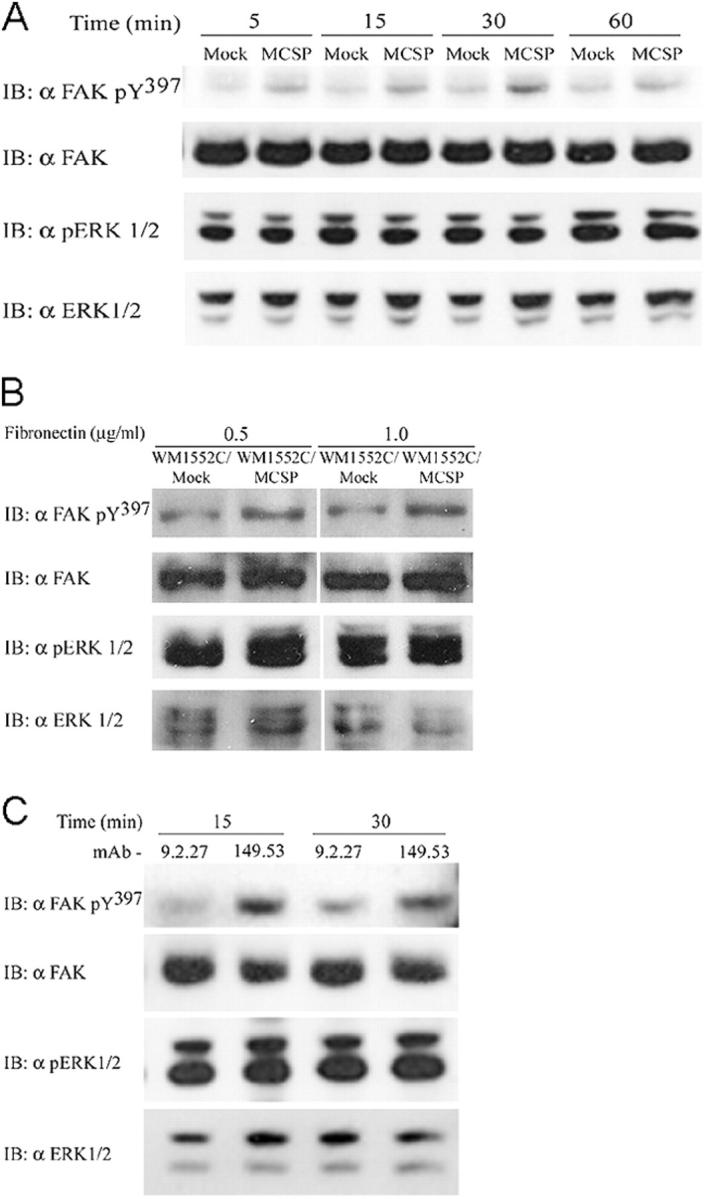
Plating on GST-FN51 or FN promotes FAK Y 39 7 phosphorylation. WM1552C/Mock and WM1552C/MCSP cells were plated at 37°C in 35-mm Petri dishes coated with 0.5 μg/ml GST-FN51 for the indicated times (A), or with FN for 1 h (B) in SIFM. The cells were lysed by addition of SDS sample buffer and the lysates were evaluated for FAK and ERK1/2 phosphorylation/expression by immunoblotting as indicated. White lines indicate that intervening lanes have been spliced out. (C) Serum-starved WM1552C/MCSP cells were released and pretreated with 5 μg/ml of either anti-MCSP mAb 9.2.27 or mAb 149.53 for 30 min at 37°C. Cells were plated on FN51-coated dishes (0.5 μg/ml) for the indicated times at 37°C, lysed in SDS sample buffer, and analyzed by immunoblotting with the indicated antibodies.
MCSP and α4 integrin colocalize on adherent melanoma cells
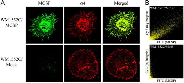
Cells were plated onto surfaces coated with GST-FN51 and were allowed to adhere and spread for 30 min at RT. After fixation, both MCSP and α4 integrin were detected on cell surfaces using MCSP core protein or α4 integrin-specific antibodies, followed by secondary antibodies labeled with Cy3 (α4 integrin) or FITC (MCSP). The distribution of these receptors was then evaluated by confocal microscopy (Fig. 6). Under these conditions, both MCSP and mock-transfected WM1552C cells adhere and spread extensively on the GST-FN51 fragment. However, the MCSP-expressing cells develop extensive microspike-type structures, consistent with an involvement of Cdc42 in MCSP-related signaling pathways. MCSP was distributed at the tips of these microspikes and at the basal surface of adherent cells in a perinuclear fashion (Fig. 6 A). The distribution of α4 integrin was similar to MCSP, also localizing at microspikes and in a perinuclear fashion on the basal surface of adherent cells (Fig. 6 A). Mock-transfected cells also spread extensively; however, the morphology of the cells was quite distinct from that of the MCSP-expressing cells, with broader lamellae that contained a circumferential staining pattern for α4 integrin (Fig. 6 A). These lamellae also lacked the extensive microspike formations characteristic of MCSP-expressing cells.
Figure 6.
MCSP and α4 integrin colocalize upon engagement on GST-FN51. (A) WM1552C/Mock and MCSP cells were plated on coverslips coated with 5 μg/ml GST-FN51 for 1 h at 37°C in SIFM, and were then fixed. Cells were double stained with polyclonal anti-MCSP antibody (FITC) and anti-α4 integrin mAb (Cy3), followed by the appropriate secondary antibody as indicated. The colocalization of these receptors upon engagement is evident in the merged images (yellow). (B) Z sections from the adherent boundary of the cell were analyzed for the colocalization of MCSP (FITC) and α4 integrin (Cy3) using the Fluoview™ software colocalization processor. Pixel pairs demonstrating staining for both receptors plot at a 45° angle on the scattergram (WM1552C/MCSP cells), whereas pixels staining with only one fluorophore fall along the corresponding axis (WM1552C/Mock cells).
Adherent cells were next scanned in XYZ coordinates, the Z sections corresponding to the substrate proximal region of adherent cells extracted and scatter diagrams generated using the Fluoview™ software colocalization processor as outlined in the Materials and methods. Images containing pixel pairs that overlap perfectly are described by a straight line that transects the scatter diagram at a 45° slope (Fig. 6 B). Processing the data from the MCSP transfectants in this way reveals a high degree of colocalization of α4 integrin and MCSP core protein in the adherent cells. In contrast, a scatter diagram for cells that express only α4 integrin shows only linear distribution of pixels along the y axis, dependent on the relative intensity of α4 integrin staining (Fig. 6 B).
Impact of FRNK expression on FAK and ERK1/2 phosphorylation
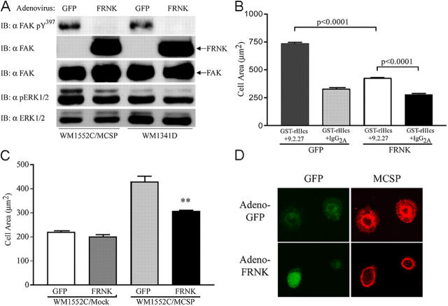
Alternative splicing of FAK leads to expression of a truncated protein that contains the focal adhesion–_targeting domain, but lacks the catalytic and src-binding domains. This product, FRNK, represents a naturally expressed COOH-terminal region of FAK that acts as an inhibitor of FAK function. We used an adenoviral construct to overexpress FRNK in melanoma cells expressing endogenous MCSP and our transfectant cells (Fig. 7). Overnight infection with the FRNK/GFP adenovirus resulted in robust overexpression of FRNK in both WM1552C/MCSP and WM1341D cells (Fig. 7 A). Overexpression of FRNK led to complete inhibition of FAK Y397 phosphorylation in cells adherent on chimeric substrata. By contrast, levels of pERK1/2 in either adherent cell population were unaffected (Fig. 7 A).
Figure 7.
Overexpression of dominant-negative FAK (FRNK) inhibits FAK phosphorylation and MCSP-mediated melanoma cell spreading. (A) Cells were infected in SIFM with an adenoviral FRNK/GFP construct or an adenoviral construct expressing only GFP at 37°C overnight, released, and allowed to adhere on plates coated with GST-rIIIcs/mAb 9.2.27 for 1 h at 37°C. Cell lysates were analyzed for tyrosine phosphorylation of FAK Y397 by immunoblotting. (B) WM1341D melanoma cells were infected as in A, allowed to spread for 1 h at 37°C in wells coated with GST-rIIIcs/mAb 9.2.27 or GST-rIIIcs/IgG2a, and the cell areas were measured as described in the Materials and methods. (C) WM1552C/Mock and MCSP-transfected cells were infected overnight as in A. Cells were plated in wells coated with GST-rIIIcs/9.2.27 for 1 h at 37°C, and the adherent cells were measured as described in the Materials and methods. **, P < 0.001 by two-tailed t test. (D) WM1552C/MCSP cells infected with either adeno-GFP or adeno-FRNK were plated on coverslips coated with 5 μg/ml GST-FN51, fixed, and stained for MCSP. Representative images from confocal analysis are shown for GFP (green) and MCSP staining (red).
Overexpression of FRNK in either the VGP WM1341D cells (Figs. 7 B) or WM1552C/MCSP transfectants (Fig. 7 C) significantly inhibited cell spreading by 60–75% when compared with control cells. MCSP distribution was also dramatically affected by overexpression of FRNK in these cells (Fig. 7 D). Distribution was more uniform in mock-infected WM1552C/MCSP cells fully adherent and spread on GST-FN51–coated surfaces, with MCSP present on the cell body and the extensive array of microspikes (Fig. 7 D, Adeno-GFP). In contrast, FRNK expression reduced the spreading of WM1552C/MCSP cells adherent on GST-FN51 and also caused MCSP to be distributed in a circumferential manner with reduced microspike formation and extension (Fig. 7 D, Adeno-FRNK).
Discussion
MCSP (and the rat homologue NG2) are cell surface PGs implicated in the adhesion, migration, and invasion of tumor cells. MCSP is expressed on the vast majority of melanoma lesions and melanoma cell lines (Ferrone et al., 1988), suggesting that it is an important contributing factor to the progression of primary tumors. By using defined ligands for either α4β1 integrin or the MCSP core protein, we have shown that simultaneous engagement of both α4β1 integrin and MCSP is required to stimulate cell spreading, suggesting that MCSP acts as a coreceptor for integrin (Iida et al., 1992, 1995). The core protein of NG2 can directly bind certain components of the ECM as well as certain growth factors (e.g., PDGF and bFGF; Burg et al., 1998; Goretzki et al., 1999). Several cell-signaling molecules have been implicated in MCSP (NG2)-induced spreading, including GTPases (e.g., Cdc42, Rac1) and the adaptor protein p130cas (Eisenmann et al., 1999; Majumdar et al., 2003).
We have cloned human MCSP from A375SM melanoma cells and have determined that there are several differences in amino acid sequence that distinguish our clone from previous reports (Pluschke et al., 1996). We considered that these changes might be the results of tumor-specific mutations in the core protein; however, we have verified our sequence in multiple human melanoma cell lines, and the amino acid sequence we reported also equates to what is found in the human genome database. The biological/structure significance of these changes remains to be determined; however, they are mostly localized to the central rodlike domain of the core protein that contains a newly predicted structural domain known as the CSPG repeat (Staub et al., 2002). One noteworthy change is in residue 631, in which a previously reported cysteine in MCSP is replaced by arginine where it might affect previously predicted disulfide bridge formation within the core protein (Pluschke et al., 1996).
Melanoma cells expressing endogenous MCSP or stably transfected with MCSP failed to spread or exhibit high levels of FAK pY397 when plated on surfaces coated with a minimal integrin-binding ligand. By contrast, chimeric substrates that also engage MCSP promoted robust spreading and enhanced FAK phosphorylation. MCSP appeared to enhance the extent, rather than the rate, of FAK phosphorylation, based on a comparison of the kinetics of FAK activation in cells adherent on the GST-rIIIcs fragment versus the chimeric substrate. Because plating cells onto a surface coated only with anti-MCSP mAb had no detectable effect on FAK phosphorylation, we conclude that MCSP works as coreceptor to enhance α4β1 integrin-mediated FAK activation in these cells.
Although the chimeric substrate model is useful to isolate the relative contributions of MCSP and α4β1 integrin, it was also necessary to determine if cells responded in a similar manner on ECM-related ligands that are more relevant in vivo. MCSP-expressing cells exhibited enhanced levels of FAK pY397 when adhering to surfaces coated with either GST-FN51 or intact FN. MCSP-specific antibodies that recognize distinct determinants on the extracellular portion of MCSP had a differential effect on FAK phosphorylation stimulated by adhesion to GST-FN51, indicating that a specific domain(s) within the extracellular portion of the core protein is important for enhancing integrin-stimulated FAK phosphorylation. Importantly, it was necessary to use a low coating concentration of GST-FN51 or FN to observe the effects of MCSP on enhancing levels of FAK pY397. MCSP-expressing cells in contact with higher concentrations of these ligands did not exhibit significant elevation in the level of FAK pY397 compared with mock transfectants. These results suggest that one important function of MCSP may be to amplify signals from the ECM that are normally transmitted by integrins (and possibly other receptors).
MCSP-expressing cells adherent on GST-FN51 demonstrated a striking morphology relative to mock-transfected controls. The MCSP-expressing cells exhibited extensive microspike formation, which has been observed previously in reports examining subcellular distribution of MCSP (Garrigues et al., 1986). Confocal analysis of MCSP-transfected cells demonstrated that both MCSP and α4 integrin are localized to the basal surface of adherent cells and on microspikes that form in response to GST-FN51 ligand. Although colocalization of MCSP and α4 integrin may enhance the efficiency of receptor cooperation, caution must be exercised to avoid overinterpretation of these data. We have previously shown that anti-MCSP antibody–coated beads can stimulate cells to undergo cell spreading when GST-rIIIcs is coated onto plates, indicating that complete colocalization of the two receptors may not be required to stimulate cell spreading (Iida et al., 1995).
The present work has also shown that, although the WM1552C/Mock cells adhered and spread on high concentrations of GST-FN51 fragment, the morphology was quite different than that exhibited by MCSP-expressing cells. Although these cells failed to spread on GST-rIIIcs, the GST-FN51 fragment contains multiple cellular recognition sites, several of which bind α4β1 integrin (Iida et al., 1992; Sharma et al., 1999). Multiple integrin recognition sites within GST-FN51 may act as “synergy sites” to stimulate cell spreading more efficiently than GST-rIIIcs alone. These results clearly show that the signals triggered by MCSP cause distinctive changes in the organization of the cytoskeleton that are not triggered by α4β1 integrin alone.
Overexpressing dominant-negative FRNK in both WM1341D cells and WM1552C transfectants inhibited FAK activation and cell spreading. FRNK also had a striking effect on the subcellular distribution of MCSP in adherent cells, causing it to localize in a striking circumferential pattern. Although small GTPases such as Cdc42 are associated with actin-mediated formation of leading edges in spreading/migrating cells, FAK can facilitate cell spreading in part by inhibiting the activation of RhoA (Burridge and Chrzanowska-Wodnicka, 1996; Wakatsuki et al., 2003). Melanoma cells overexpressing FRNK still form microspikes, but lack the ability to extend their leading edges to the same extent as mock-infected cells, consistent with a model in which FRNK interferes with the inhibition of the RhoA/myosin pathway in these cells (Wakatsuki et al., 2003). It is quite possible that actin-mediated polymerization, which would likely be intact in the presence of FRNK, is important for driving MCSP to the leading edge of adherent cells that are attempting to spread.
MCSP also enhances the activation of the ERK/MAPK pathway in WM1552C cells, as detected by an enhanced level of phosphorylated ERK1/2 within these cells. WM1552C/MCSP transfectants show increased levels of pERK1/2 in suspension, and plating onto either the chimeric or integrin-bound substrates led to further stimulation of pERK1/2. By contrast, cells adherent to surfaces coated with only anti-MCSP mAb 9.2.27 had low levels of pERK1/2 that were not elevated over those detected in suspended cells, consistent with the importance of integrins in promoting adhesion-induced elevations of ERK/MAPK phosphorylation (Lin et al., 1997; Barberis et al., 2000; Danen and Yamada, 2001).
MCSP also appeared to enhance FAK and ERK1/2 phosphorylation by different mechanisms. Simultaneous engagement of both MCSP and α4β1 integrin was required to obtain maximal FAK phosphorylation, whereas enhanced levels of pERK1/2 within these cells were not affected by chimeric vs. integrin-only binding substrates. MCSP-induced increases in the level of FAK pY397 were completely inhibited by overexpression of FRNK, whereas FRNK had no detectable effect on the level of pERK1/2 within these cells. Furthermore, WM1552C/MCSP cells exhibited elevated levels of pERK1/2 even when in suspension, where there was little if any activation of FAK. Finally, although soluble mAb 9.2.27 inhibited FAK activation in cells contacting GST-FN51, it had no effect on the level of pERK1/2 in these cells. Although links between FAK activation and stimulation of the RAS/MAPK pathway have been reported (Schlaepfer et al., 1999), this finding is consistent with the many reports showing that integrin-mediated activation of MAPK can also occur via FAK-independent mechanisms (Lin et al., 1997; Barberis et al., 2000; Danen and Yamada, 2001). Although the mechanisms by which MCSP enhanced the level of ERK1/2 phosphorylation under these conditions is not yet known, the data suggest that it is by mechanisms distinct from MCSP-enhanced activation of FAK.
In contrast to the WM1552C/MCSP cells, the level of pERK1/2 within the WM1341D cells was not increased when these cells were plated onto surfaces coated with integrin-binding ligands. We attribute this to the high level of pERK1/2 already present in WM1341D cells even when suspended, as we have previously shown (Neudauer and McCarthy, 2003). Constitutive activation of the ERK/MAPK pathway is associated with more advanced melanomas (Satyamoorthy et al., 2003; Smalley, 2003). The mechanisms leading to constitutive activation of this pathway are multifaceted and can include the expression of autocrine growth factors or activating mutations within the upstream B-Raf kinase, the latter of which have been associated with the majority of human melanomas and melanoma cell lines. Our findings suggest that expression of MCSP can be an additional contributing factor to the sustained elevation of ERK/MAPK signaling within melanoma cells. Sustained activation of the ERK/MAPK pathway has implications in the growth and invasion of melanoma cells, suggesting that MCSP expression may contribute to the progression of primary tumors.
Although MCSP is highly expressed in both primary and metastatic lesions (Ferrone et al., 1988), its function in melanoma progression is not well understood. Elevated levels of activated FAK are found in many tumors, including melanoma, and are linked to increased growth, survival, and invasion (Kahana et al., 2002; Hecker and Gladson, 2003). Constitutive activation of the ERK/MAPK pathway is also associated with malignant progression of melanomas. The current results suggest that sustained activation of these pathways in melanoma may be related in part to overexpression of MCSP. Our data are consistent with a model in which MCSP serves to amplify signals from the extracellular environment that are initiated by other cell surface receptors. In the case of FAK activation, which has been linked in some cells to integration of both growth factor receptor and integrin-related pathways, MCSP may function to reduce the requirement of ligands for these receptors (Sieg et al., 2000). The results would be to give MCSP-expressing cells a selective advantage in the dynamic and competitive microenvironment of a progressing tumor, consistent with observations documenting the sustained expression of MCSP in a high portion of early- to late-stage melanomas. A more complete understanding of mechanisms associated with MCSP function could provide new therapeutic _targets in the treatment of melanoma patients with advanced primary tumors or malignant disease.
Materials and methods
Cell culture
WM35, WM1552C, and WM1341D human melanoma cells were provided by Dr. Meenhard Herlyn (Wistar Institute, Philadelphia, PA). Cells were maintained in 4:1 MCDB 153/Leibovitz's L-15 medium supplemented with 5 μg/ml insulin and 2% FBS. A375SM human melanoma cells were provided by Dr. Isaiah J. Fidler (M.D. Anderson Hospital Cancer Center, Houston, TX) and cultured in MEM supplemented with 10% FBS, MEM vitamin solution, 50 μg/ml gentamicin, and 1 mM sodium pyruvate.
Antibodies and reagents
Anti-MCSP mAbs 9.2.27 and 149.53 were developed and characterized as described previously (Morgan et al., 1981; Giacomini et al., 1983). Other antibodies were purchased from indicated companies: anti-tubulin from Oncogene Research Products, anti-FAK pY397 phosphospecific antibody from Biosource International, Inc., anti-FAK from Upstate Biotechnology, anti-phospho p44/42 MAPK (pERK1/2) and anti-p44/42 MAPK (ERK1/2) from Cell Signaling Technology, Inc., anti-α4 integrin from CHEMICON International, normal mouse IgG2a and goat anti–mouse IgG FC from ICN Pharmaceuticals, and FITC donkey anti–rabbit and Cy3-donkey anti–mouse secondary antibodies from Jackson ImmunoResearch Laboratories. cABC was purchased from Sigma-Aldrich.
Preparation of pAb against MCSP
Purified MCSP, isolated from placental tissue, was donated by Dr. Robert C. Spiro (Orquest Inc., Mountain View, CA). pAb against MCSP was generated by immunizing a 6–8-wk-old female New Zealand White rabbit with keyhole limpet hemocyanin MCSP protein according to a standard protocol. The Ig fraction was separated on DEAE-agarose as described previously (Iida et al., 1992). Specificity of the antibody was determined by immunoprecipitation and immunoblot analysis of A375SM melanoma cells, a metastatic cell line previously shown to express high levels of MCSP (Iida et al., 1992, 1995).
Recombinant FN fragments and human FN
The recombinant GST fusion proteins of FN fragment rIIIcs (GST-rIIIcs) and FN fragment 51 (GST-FN51) were purified as described previously (Eisenmann et al., 1999). Human plasma FN was purified as a by-product of factor VIII production by sequential ion exchange and gelatin affinity chromatography as described previously (McCarthy et al., 1988).
MCSP mRNA expression in melanoma cell lines
mRNA was isolated from human melanoma cell lines using the Oligotex Direct mRNA Kit (QIAGEN) and quantified with the Ribogreen® RNA Quantitation Kit (Molecular Probes, Inc.). Full-length cDNA was generated with the SuperScript™ Preamplification System (Invitrogen). PCR amplification of a 450-bp fragment of MCSP was performed using primers 5′ MCSP middle and 3′ MCSP middle (described below).
Generation of full-length MCSP construct
Full-length MCSP cDNA was reverse transcribed from mRNA isolated from A375SM cells as described above. The MCSP cDNA was amplified by PCR with the eLONGase® enzyme mix (Invitrogen). Two halves spanning a unique BamHI site in the MCSP sequence were amplified, one encompassing bases 1–3511 (primer set A) and the second bases 3061–7011 (primer set B). Primer set A 5′-CTAGAATTCGATGCAGTCCGGCCGCGGCCCCCCACTTC-3′ 5′-CAGCTGTGACGTGGTAGTGGACCTCATCC-3′ (3′ MCSP middle, 5′ MCSP EcoRI) and primer set B 5′-CAGACCATCAGCCGGATCTTCCATGTG-3′ (5′ MCSP middle) 5′-GCAGTCTAGATGCCTGTCCCTGGCCCGATC-3′ (3′ MCSP XbaI) PCR fragments were digested with the appropriate enzymes (BamHI and EcoRI for primer set A, BamHI and XbaI for primer set B) and were ligated into vector pcDNA 3.1(+) (CLONTECH Laboratories, Inc.). The resulting full-length constructs were verified by sequencing. Sequence for the full-length MCSP is available at GenBank/EMBL/DDBJ (accession no. AY359468).
Generation of stable transfectants
WM1552C cells were transfected with either vector alone (mock) or vector containing full-length MCSP using FuGENE™ 6 (Roche). Transfected cells were selected and maintained by culturing in the presence of 0.25 mg/ml G418. Cells expressing MCSP were further selected by staining with mAb 9.2.27 followed with Cy3 donkey anti–mouse secondary antibody, and the Cy3-positive cells were selected and collected on a FACSVantage™ cytometer (Becton Dickinson). Positive cells were collected and cultured in growth medium supplemented with 0.25 mg/ml G418.
SDS-PAGE and immunoblotting
For detection of MCSP core protein and PG expression, cells were resuspended in serum- and insulin-free medium (SIFM) at 106 cells/ml and were incubated at 37°C for 20 min with or without 0.5 U cABC/ml. Cells were lysed in SDS sample buffer and proteins were fractionated with 6% SDS-PAGE. For the FAK phosphorylation assay, 35-mm Petri dishes were coated with 0.5 ml of either GST or GST-rIIIcs (3 μg/ml) and purified goat anti–mouse IgG Fc antibodies (final dilution 1:500). After overnight incubation at 37°C, plates were blocked for 1 h with 0.3% BSA (wt/vol) in PBS and were then incubated with 1 μg/ml mAb 9.2.27 or IgG2a (isotype control) for 2 h at 37°C. Cells were starved overnight, released with 5 mM EDTA in PBS, resuspended in SIFM at 106 cells/ml, and incubated at 37°C for 30 min with or without 0.5 U cABC. 3 × 10⁵ cells were then added to each dish and incubated at 37°C for indicated times. 4× SDS sample buffer was added to each plate and the lysate was passed through a 27-gauge needle to shear DNA. Immunoblotting was performed using standard techniques.
Cell adhesion and spreading assays
Triplicate wells in Immulon-I microtiter plates (Fisher Scientific) were coated overnight with 100 μl of 3 μg/ml GST, GST-rIIIcs, and/or 1:500 goat anti–mouse IgG Fc antibodies in PBS. Plates were blocked for 1 h at 37°C with 150 μl SIFM containing 0.3% BSA and 20 mM Hepes (adhesion medium). Antibody-coated wells were incubated for 2 h at 37°C with 1 μg/ml of either IgG2a or mAb 9.2.27, and were then washed with adhesion medium to remove unbound substrate. Suspended cells (100 μl) at 105/ml in adhesion medium were added to each well, the plates were incubated at 37°C for 30 min, and the wells were gently washed to remove nonadherent cells. The number of adherent cells was determined by formazan absorbance using the Celltiter 96® Aqueous Non-radioactive Cell Proliferation Assay (Promega). Data are presented as the percentage of total input cells ± SD, based on a standard curve. For spreading, adherent cells were photographed at 400× and cell areas were measured with NIH Image v1.62. Data shown are the average area of 50 cells/well from triplicate wells, ± SEM.
Confocal microscopy and colocalization scatterplots
Cells were plated on coverslips coated with 5.0 μg/ml FN51 for 1 h at 37°C in SIFM, washed twice with PBS, fixed with 4% PFA for 30 min at RT, and blocked with 1% donkey serum. For dual staining, cells were incubated simultaneously with both primary antibodies for 1 h at RT followed by washing with PBS. Appropriate secondary antibodies were added and the coverslips were incubated for 1 h at 37°C. Coverslips were washed four times in PBS and mounted using Gel/Mount™ mounting media (Biomeda). A laser-scanning system (FV-500; Olympus) with a 60× plan-apochromatic oil objective was used to image the samples. Z sections corresponding to the region of the cell in closest proximity to the substrate were extracted from image stacks. Pixels having the same location in the two images were paired (P1, P2) for analysis and plotted in a scatter diagram defined by grayscale values derived from the source image using the Fluoview™ software colocalization processor (Olympus).
Adenoviral constructs
A replication-defective adenovirus encoding human FRNK (amplified from human lung fibroblast CCL-20; American Type Culture Collection) was used to overexpress this protein in melanoma cells. FRNK cDNA was subcloned into the BamHI and ClaI sites of pBSKs vector. The FRNK insert was then subcloned into the MIGR1-HCMV plasmid containing a separate promoter and coding region for GFP. The MIGR1-HCMV expression cassette containing FRNK/GFP was excised and subcloned into pAxCAwt vector (TaKaRa) and linearized according to the manufacturer's instructions. The linearized pAxCAwt + FRNK was introduced into HEK293 cells, and adenovirus-FRNK/GFP was amplified from cell extracts and was purified by CsCl gradient centrifugation. An adenovirus expressing GFP alone was used as a control. The multiplicity of viral infection was determined by viral dilution in HEK293 cells. GFP expression was routinely monitored by flow cytometry to estimate the level of infection of melanoma cells, which was 95% or greater.
Acknowledgments
The authors would like to thank Dr. Ralph A. Reisfeld (Scripps Research Institute, La Jolla, CA) for providing mAb 9.2.27. The authors are indebted to Dr. Meenhard Herlyn for his critical suggestions that led to these experiments and to Dr. Eva Turley for providing feedback and constructive input during the preparation of this manuscript.
This work was supported by National Institutes of Health grants RO1 CA82295 (to J.B. McCarthy) and P01 CA89480, and FV-500-NCI Shared Instrument grant 1S10RR16851.
J. Yang and M.A. Price contributed equally to this paper.
Abbreviations used in this paper: cABC, chondroitinase ABC; ERK, extracellular signal–regulated kinase; FN, fibronectin; FRNK, focal adhesion–related nonkinase; MCSP, melanoma chondroitin sulfate proteoglycan; pERK, phosphorylated ERK; PG, proteoglycan; RGP, radial growth phase; SIFM, serum- and insulin-free medium; VGP, vertical growth phase.
References
- Aplin, A.E., and R.L. Juliano. 1999. Integrin and cytoskeletal regulation of growth factor signaling to the MAP kinase pathway. J. Cell Sci. 112:695–706. [DOI] [PubMed] [Google Scholar]
- Barberis, L., K.K. Wary, G. Fiucci, F. Liu, E. Hirsch, M. Brancaccio, F. Altruda, G. Tarone, and F.G. Giancotti. 2000. Distinct roles of the adaptor protein Shc and focal adhesion kinase in integrin signaling to ERK. J. Biol. Chem. 275:36532–36540. [DOI] [PubMed] [Google Scholar]
- Bogenrieder, T., and M. Herlyn. 2002. Cell-surface proteolysis, growth factor activation and intercellular communication in the progression of melanoma. Crit. Rev. Oncol. Hematol. 44:1–15. [DOI] [PMC free article] [PubMed] [Google Scholar]
- Burg, M.A., K.A. Grako, and W.B. Stallcup. 1998. Expression of the NG2 proteoglycan enhances the growth and metastatic properties of melanoma cells. J. Cell. Physiol. 177:299–312. [DOI] [PubMed] [Google Scholar]
- Burridge, K., and M. Chrzanowska-Wodnicka. 1996. Focal adhesions, contractility, and signaling. Annu. Rev. Cell Dev. Biol. 12:463–518. [DOI] [PubMed] [Google Scholar]
- Chekenya, M., H.K. Rooprai, D. Davies, J.M. Levine, A.M. Butt, and G.J. Pilkington. 1999. The NG2 chondroitin sulfate proteoglycan: role in malignant progression of human brain tumours. Int. J. Dev. Neurosci. 17:421–435. [DOI] [PubMed] [Google Scholar]
- Conner, S.R., G. Scott, and A.E. Aplin. 2003. Adhesion-dependent activation of the ERK1/2 cascade is by-passed in melanoma cells. J. Biol. Chem. 278:34548–34554. [DOI] [PubMed] [Google Scholar]
- Danen, E.H., and K.M. Yamada. 2001. Fibronectin, integrins, and growth control. J. Cell. Physiol. 189:1–13. [DOI] [PubMed] [Google Scholar]
- Danen, E.H., C. Marcinkiewicz, I.M. Cornelissen, A.A. van Kraats, J.A. Pachter, D.J. Ruiter, S. Niewiarowski, and G.N. van Muijen. 1998. The disintegrin eristostatin interferes with integrin α4β1 function and with experimental metastasis of human melanoma cells. Exp. Cell Res. 238:188–196. [DOI] [PubMed] [Google Scholar]
- Dedhar, S. 1999. Integrins and signal transduction. Curr. Opin. Hematol. 6:37–43. [DOI] [PubMed] [Google Scholar]
- Eisenmann, K.M., J.B. McCarthy, M.A. Simpson, P.J. Keely, J.L. Guan, K. Tachibana, L. Lim, E. Manser, L.T. Furcht, and J. Iida. 1999. Melanoma chondroitin sulphate proteoglycan regulates cell spreading through Cdc42, Ack-1 and p130cas. Nat. Cell Biol. 1:507–513. [DOI] [PubMed] [Google Scholar]
- Ferrone, S., M. Temponi, D. Gargiulo, G.A. Scassellati, R. Cavaliere, and P.G. Natali. 1988. Selection and utilization of monoclonal antibody defined melanoma associated antigens for immunoscintigraphy in patients with melanoma. Radiolabeled Monoclonal Antibodies for Imaging and Therapy. Vol. 152. S.C. Srivastava, editor. Plenum Publishing Corp., New York/London. 55–73.
- Garrigues, H.J., M.W. Lark, S. Lara, I. Hellstrom, K.E. Hellstrom, and T.N. Wight. 1986. The melanoma proteoglycan: restricted expression on microspikes, a specific microdomain of the cell surface. J. Cell Biol. 103:1699–1710. [DOI] [PMC free article] [PubMed] [Google Scholar]
- Geller, A.C., and G.D. Annas. 2003. Epidemiology of melanoma and nonmelanoma skin cancer. Semin. Oncol. Nurs. 19:2–11. [DOI] [PubMed] [Google Scholar]
- Giacomini, P., A.K. Ng, R.R. Kantor, P.G. Natali, and S. Ferrone. 1983. Double determinant immunoassay to measure a human high-molecular-weight melanoma-associated antigen. Cancer Res. 43:3586–3590. [PubMed] [Google Scholar]
- Giancotti, F.G., and E. Ruoslahti. 1999. Integrin signaling. Science. 285:1028–1032. [DOI] [PubMed] [Google Scholar]
- Goretzki, L., M.A. Burg, K.A. Grako, and W.B. Stallcup. 1999. High-affinity binding of basic fibroblast growth factor and platelet-derived growth factor-AA to the core protein of the NG2 proteoglycan. J. Biol. Chem. 274:16831–16837. [DOI] [PubMed] [Google Scholar]
- Guan, J.L. 1997. Role of focal adhesion kinase in integrin signaling. Int. J. Biochem. Cell Biol. 29:1085–1096. [DOI] [PubMed] [Google Scholar]
- Hauck, C.R., D.A. Hsia, and D.D. Schlaepfer. 2002. The focal adhesion kinase—a regulator of cell migration and invasion. IUBMB Life. 53:115–119. [DOI] [PubMed] [Google Scholar]
- Hecker, T.P., and C.L. Gladson. 2003. Focal adhesion kinase in cancer. Front. Biosci. 8:s705–s714. [DOI] [PubMed] [Google Scholar]
- Hilden, J.M., F.O. Smith, J.L. Frestedt, R. McGlennen, W.B. Howells, P.H. Sorensen, D.C. Arthur, W.G. Woods, J. Buckley, I.D. Bernstein, and J.H. Kersey. 1997. MLL gene rearrangement, cytogenetic 11q23 abnormalities, and expression of the NG2 molecule in infant acute myeloid leukemia. Blood. 89:3801–3805. [PubMed] [Google Scholar]
- Hood, J.D., and D.A. Cheresh. 2002. Role of integrins in cell invasion and migration. Nat. Rev. Cancer. 2:91–100. [DOI] [PubMed] [Google Scholar]
- Houghton, A.N., and D. Polsky. 2002. Focus on melanoma. Cancer Cell. 2:275–278. [DOI] [PubMed] [Google Scholar]
- Howe, A.K., A.E. Aplin, and R.L. Juliano. 2002. Anchorage-dependent ERK signaling—mechanisms and consequences. Curr. Opin. Genet. Dev. 12:30–35. [DOI] [PubMed] [Google Scholar]
- Iida, J., A.P. Skubitz, L.T. Furcht, E.A. Wayner, and J.B. McCarthy. 1992. Coordinate role for cell surface chondroitin sulfate proteoglycan and α4β1 integrin in mediating melanoma cell adhesion to fibronectin. J. Cell Biol. 118:431–444. [DOI] [PMC free article] [PubMed] [Google Scholar]
- Iida, J., A.M. Meijne, R.C. Spiro, E. Roos, L.T. Furcht, and J.B. McCarthy. 1995. Spreading and focal contact formation of human melanoma cells in response to the stimulation of both melanoma-associated proteoglycan (NG2) and α4β1 integrin. Cancer Res. 55:2177–2185. [PubMed] [Google Scholar]
- Iida, J., A.M. Meijne, J.R. Knutson, L.T. Furcht, and J.B. McCarthy. 1996. Cell surface chondroitin sulfate proteoglycans in tumor cell adhesion, motility and invasion. Semin. Cancer Biol. 7:155–162. [DOI] [PubMed] [Google Scholar]
- Iida, J., D. Pei, T. Kang, M.A. Simpson, M. Herlyn, L.T. Furcht, and J.B. McCarthy. 2001. Melanoma chondroitin sulfate proteoglycan regulates matrix metalloproteinase-dependent human melanoma invasion into type I collagen. J. Biol. Chem. 276:18786–18794. [DOI] [PubMed] [Google Scholar]
- Johnson, J.P. 1999. Cell adhesion molecules in the development and progression of malignant melanoma. Cancer Metastasis Rev. 18:345–357. [DOI] [PubMed] [Google Scholar]
- Juliano, R.L. 2002. Signal transduction by cell adhesion receptors and the cytoskeleton: functions of integrins, cadherins, selectins, and immunoglobulin-superfamily members. Annu. Rev. Pharmacol. Toxicol. 42:283–323. [DOI] [PubMed] [Google Scholar]
- Kageshita, T., N. Kuriya, T. Ono, T. Horikoshi, M. Takahashi, G.Y. Wong, and S. Ferrone. 1993. Association of high molecular weight melanoma-associated antigen expression in primary acral lentiginous melanoma lesions with poor prognosis. Cancer Res. 53:2830–2833. [PubMed] [Google Scholar]
- Kahana, O., M. Micksche, I.P. Witz, and I. Yron. 2002. The focal adhesion kinase (P125FAK) is constitutively active in human malignant melanoma. Oncogene. 21:3969–3977. [DOI] [PubMed] [Google Scholar]
- Li, G., K. Satyamoorthy, and M. Herlyn. 2002. Dynamics of cell interactions and communications during melanoma development. Crit. Rev. Oral Biol. Med. 13:62–70. [DOI] [PubMed] [Google Scholar]
- Lin, T.H., A.E. Aplin, Y. Shen, Q. Chen, M. Schaller, L. Romer, I. Aukhil, and R.L. Juliano. 1997. Integrin-mediated activation of MAP kinase is independent of FAK: evidence for dual integrin signaling pathways in fibroblasts. J. Cell Biol. 136:1385–1395. [DOI] [PMC free article] [PubMed] [Google Scholar]
- Majumdar, M., K. Vuori, and W.B. Stallcup. 2003. Engagement of the NG2 proteoglycan triggers cell spreading via rac and p130cas. Cell. Signal. 15:79–84. [DOI] [PubMed] [Google Scholar]
- Mansour, S.J., W.T. Matten, A.S. Hermann, J.M. Candia, S. Rong, K. Fukasawa, G.F. Van de Woude, and N.G. Ahn. 1994. Transformation of mammalian cells by constitutively active MAP kinase kinase. Science. 265:966–970. [DOI] [PubMed] [Google Scholar]
- McCarthy, J.B., M.K. Chelberg, D.J. Mickelson, and L.T. Furcht. 1988. Localization and chemical synthesis of fibronectin peptides with melanoma adhesion and heparin binding activities. Biochemistry. 27:1380–1388. [DOI] [PubMed] [Google Scholar]
- Morgan, A.C., Jr., D.R. Galloway, and R.A. Reisfeld. 1981. Production and characterization of monoclonal antibody to a melanoma specific glycoprotein. Hybridoma. 1:27–36. [DOI] [PubMed] [Google Scholar]
- Mould, A.P., J.A. Askari, S.E. Craig, A.N. Garratt, J. Clements, and M.J. Humphries. 1994. Integrin α4β1-mediated melanoma cell adhesion and migration on vascular cell adhesion molecule-1 (VCAM-1) and the alternatively spliced IIICS region of fibronectin. J. Biol. Chem. 269:27224–27230. [PubMed] [Google Scholar]
- Neudauer, C.L., and J.B. McCarthy. 2003. Insulin-like growth factor I-stimulated melanoma cell migration requires phosphoinositide 3-kinase but not extracellular-regulated kinase activation. Exp. Cell Res. 286:128–137. [DOI] [PubMed] [Google Scholar]
- Nishiyama, A. 2001. NG2 cells in the brain: a novel glial cell population. Hum. Cell. 14:77–82. [PubMed] [Google Scholar]
- Nishiyama, A., K.J. Dahlin, J.T. Prince, S.R. Johnstone, and W.B. Stallcup. 1991. The primary structure of NG2, a novel membrane-spanning proteoglycan. J. Cell Biol. 114:359–371. [DOI] [PMC free article] [PubMed] [Google Scholar]
- Pluschke, G., M. Vanek, A. Evans, T. Dittmar, P. Schmid, P. Itin, E.J. Filardo, and R.A. Reisfeld. 1996. Molecular cloning of a human melanoma-associated chondroitin sulfate proteoglycan. Proc. Natl. Acad. Sci. USA. 93:9710–9715. [DOI] [PMC free article] [PubMed] [Google Scholar]
- Satyamoorthy, K., E. DeJesus, A.J. Linnenbach, B. Kraj, D.L. Kornreich, S. Rendle, D.E. Elder, and M. Herlyn. 1997. Melanoma cell lines from different stages of progression and their biological and molecular analyses. Melanoma Res. 7:S35–S42. [PubMed] [Google Scholar]
- Satyamoorthy, K., G. Li, M.R. Gerrero, M.S. Brose, P. Volpe, B.L. Weber, P. Van Belle, D.E. Elder, and M. Herlyn. 2003. Constitutive mitogen-activated protein kinase activation in melanoma is mediated by both BRAF mutations and autocrine growth factor stimulation. Cancer Res. 63:756–759. [PubMed] [Google Scholar]
- Schlaepfer, D.D., and T. Hunter. 1996. Signal transduction from the extracellular matrix—a role for the focal adhesion protein-tyrosine kinase FAK. Cell Struct. Funct. 21:445–450. [DOI] [PubMed] [Google Scholar]
- Schlaepfer, D.D., and T. Hunter. 1997. Focal adhesion kinase overexpression enhances ras-dependent integrin signaling to ERK2/mitogen-activated protein kinase through interactions with and activation of c-Src. J. Biol. Chem. 272:13189–13195. [DOI] [PubMed] [Google Scholar]
- Schlaepfer, D.D., C.R. Hauck, and D.J. Sieg. 1999. Signaling through focal adhesion kinase. Prog. Biophys. Mol. Biol. 71:435–478. [DOI] [PubMed] [Google Scholar]
- Sharma, A., J.A. Askari, M.J. Humphries, E.Y. Jones, and D.I. Stuart. 1999. Crystal structure of a heparin- and integrin-binding segment of human fibronectin. EMBO J. 18:1468–1479. [DOI] [PMC free article] [PubMed] [Google Scholar]
- Sieg, D.J., C.R. Hauck, D. Ilic, C.K. Klingbeil, E. Schaefer, C.H. Damsky, and D.D. Schlaepfer. 2000. FAK integrates growth-factor and integrin signals to promote cell migration. Nat. Cell Biol. 2:249–256. [DOI] [PubMed] [Google Scholar]
- Smalley, K.S. 2003. A pivotal role for ERK in the oncogenic behaviour of malignant melanoma? Int. J. Cancer. 104:527–532. [DOI] [PubMed] [Google Scholar]
- Stallcup, W.B. 2002. The NG2 proteoglycan: past insights and future prospects. J. Neurocytol. 31:423–435. [DOI] [PubMed] [Google Scholar]
- Staub, E., B. Hinzmann, and A. Rosenthal. 2002. A novel repeat in the melanoma-associated chondroitin sulfate proteoglycan defines a new protein family. FEBS Lett. 527:114–118. [DOI] [PubMed] [Google Scholar]
- Wakatsuki, T., R.B. Wysolmerski, and E.L. Elson. 2003. Mechanics of cell spreading: role of myosin II. J. Cell Sci. 116:1617–1625. [DOI] [PubMed] [Google Scholar]
- Wayner, E.A., A. Garcia-Pardo, M.J. Humphries, J.A. McDonald, and W.G. Carter. 1989. Identification and characterization of the T lymphocyte adhesion receptor for an alternative cell attachment domain (CS-1) in plasma fibronectin. J. Cell Biol. 109:1321–1330. [DOI] [PMC free article] [PubMed] [Google Scholar]


