Abstract
Fibronectin matrix assembly is a multistep, integrin-dependent process. To investigate the role of integrin dynamics in fibronectin fibrillogenesis, we developed an antibody-chasing technique for simultaneous tracking of two integrin populations by different antibodies. We established that whereas the vitronectin receptor αvβ3 remains within focal contacts, the fibronectin receptor α5β1 translocates from focal contacts into and along extracellular matrix (ECM) contacts. This escalator-like translocation occurs relative to the focal contacts at 6.5 ± 0.7 μm/h and is independent of cell migration. It is induced by ligation of α5β1 integrins and depends on interactions with a functional actin cytoskeleton and vitronectin receptor ligation. During cell spreading, translocation of ligand-occupied α5β1 integrins away from focal contacts and along bundles of actin filaments generates ECM contacts. Tensin is a primary cytoskeletal component of these ECM contacts, and a novel dominant-negative inhibitor of tensin blocked ECM contact formation, integrin translocation, and fibronectin fibrillogenesis without affecting focal contacts. We propose that translocating α5β1 integrins induce initial fibronectin fibrillogenesis by transmitting cytoskeleton-generated tension to extracellular fibronectin molecules. Blocking this integrin translocation by a variety of treatments prevents the formation of ECM contacts and fibronectin fibrillogenesis. These studies identify a localized, directional, integrin translocation mechanism for matrix assembly.
Keywords: fibronectin, integrin, tensin, vitronectin, extracellular matrix
Introduction
Cells secrete and organize extracellular matrix (ECM), which provides structural support for cell adhesion, migration, and tissue organization, as well as external regulation of cellular functions (Hay 1991). Matrix assembly by cells can convert soluble fibronectin (FN) molecules into elaborate fibrillar matrices in a process involving integrins and the cytoskeleton. Extensive knowledge now exists about integrin functions, FN molecular requirements for fibrillogenesis, and cytoskeletal constituents of cell–matrix adhesive contacts. Nevertheless, it is not yet clear how these three components combine to generate FN fibrils by mechanisms that should be continuously dynamic and spatially coordinated. FN matrix assembly is a cell-dependent process that is triggered at specific sites on the cell surface (Peters and Mosher 1987) and depends on unfolding of the FN dimer (Hynes 1999; Schwarzbauer and Sechler 1999).
Integrins have a central role in FN matrix assembly. These ubiquitous α/β heterodimeric transmembrane receptors also mediate cell adhesion, migration, and bidirectional signal transduction through interaction with different ECM proteins (Hynes 1992). A number of integrins bind to FN but are not normally capable of initiating formation of fibrils (Zhang et al. 1993). The major receptor responsible for FN matrix assembly is the α5β1 integrin. Transfection of α5 integrin into CHO cells leads to a large increase in FN assembly (Giancotti and Ruoslahti 1990). Conversely, assembly of FN by fibroblasts can be inhibited by mAbs against α5 or β1 subunits (Akiyama et al. 1989; Fogerty et al. 1990) or by FN fragments containing the α5β1-binding domain (McDonald et al. 1987). A chimera containing the β1 integrin cytoplasmic tail functions as a dominant-negative inhibitor of FN assembly (LaFlamme et al. 1994). Nevertheless, in the absence of α5β1, αvβ3 (Wennerberg et al. 1996; Wu et al. 1996) and activated αIIbβ3 (Wu et al. 1995; Hughes et al. 1996) can also direct matrix assembly, which may account for FN matrix formation in α5 integrin–null mice (Yang et al. 1993).
Although some of the integrin interactions and cellular states important for matrix assembly have been identified, the relationship between these factors and the role of integrins is not clear. Truncation of the β1 cytoplasmic domain can block FN matrix assembly by severing the link between ligand-occupied integrins and the cytoskeleton (Wu et al. 1995). Similar inhibition of matrix assembly is observed when actin cytoskeleton is disrupted (Ali and Hynes 1977; Wu et al. 1995; Christopher et al. 1997). Cell-generated tension or contractility is another prerequisite for FN fibrillogenesis (Halliday and Tomasek 1995; Zhang et al. 1994, Zhang et al. 1997), possibly by exposing cryptic self-association sites within FN molecules (Aguirre et al. 1994; Hocking et al. 1994; Wu et al. 1995; Ingham et al. 1997; Zhong et al. 1998).
It has been proposed that integrins are the transmitters of this tension (Wu et al. 1995; Zhong et al. 1998). However, the actual mechanism by which integrins can induce fibrillogenesis is not known. Fibroblast integrins organize two types of α5β1-containing structures where transmission of cell-generated tension could occur: focal contacts (FC) and ECM contacts (Chen and Singer 1982; Chen et al. 1985; Singer et al. 1988). ECM contacts were recently renamed fibrillar adhesions (Zamir et al. 1999). FC are specialized adhesion sites that anchor stress fibers and provide cultured cells with firm substrate attachment (reviewed in Yamada and Geiger 1997). In contrast, ECM contacts bind fibrils of FN parallel to actin bundles and tensin, but they are deficient in other FC components, such as paxillin and vinculin (Hynes and Destree 1978; Chen and Singer 1982; Chen et al. 1985; Zamir et al. 1999).
We speculated that these structures are under different types of tension and that the isometric (static) tension at FC (Burridge 1981) might be translated into dynamic force for translocating α5β1 integrins out of FC to stretch and unfold bound FN. To test the hypothesis that α5β1 integrins undergo directional motion on the cell surface, we developed an antibody-chasing technique that permits comparisons of the dynamic behavior of two different integrin populations on the cell surface. We established that ligated α5β1 integrins actively translocate along stress fibers, moving from FC into and along ECM contacts containing tensin. At the same time, the vitronectin (VN) receptor αvβ3 remains resident within FC, but its ligation is necessary for α5β1 movement. Translocating FN receptors could initiate FN fibrillogenesis by transmitting cytoskeleton-generated forces to extracellular FN molecules. We propose a novel model for early FN fibril formation driven by localized α5β1 integrin translocation dependent on the cytoskeletal protein tensin.
Materials and Methods
Antibodies and Purified Proteins
Antibodies to human β1 integrins included rat mAb 9EG7 (PharMingen) and mouse mAbs 12G10 (Mould et al. 1995) and K20 (Immunotech). Function-blocking (rat mAb 16) and noninhibitory (rat mAb 11) anti-α5 antibodies were described previously (Akiyama et al. 1989; Miyamoto et al. 1995). Anti-FN mAb was from Transduction Laboratories; goat anti–human FN antibodies were directly labeled with FITC (Akiyama et al. 1989). Fab′ fragments were produced with an ImmunoPure™ Fab Preparation Kit (Pierce) and conjugated with FITC or TRITC as described (Akiyama et al. 1989). F-actin was detected by rhodamine-phalloidin (Molecular Probes). Secondary species–specific CY3- or FITC-conjugated antibodies were from Jackson ImmunoResearch Laboratories.
FN was purified as described by Miekka et al. 1982. VN was a gift from Dr. H. Tran (National Institute of Dental and Craniofacial Research [NIDCR], National Institutes of Health [NIH], Bethesda, Maryland). Collagen I (Vitrogen 100) was from Collagen Corp., and laminin 1 was from Trevigen. Purified FN was labeled with FITC or the Alexa™ 488 Protein Labeling Kit (Molecular Probes) according to the manufacturer's protocol.
Cell Culture and Substrate Coating
Primary human foreskin fibroblasts (HFF) were a gift from S. Yamada (NIDCR, NIH) and were used at passages 9–18 after plating on sterile untreated glass coverslips in DME (GIBCO BRL Life Technologies) supplemented with 10% FBS (Hyclone), 100 U/ml penicillin, and 100 μg/ml streptomycin (complete medium). When indicated, the coverslips were precoated by incubating 1 h at 37°C or overnight at 4°C with FN or VN at 10 μg/ml. For mixed substrates, 5 μg/ml FN was mixed with 5 μg/ml VN or poly-l-lysine.
Modified Media.
When cycloheximide was used to block endogenous FN synthesis, FN-free medium was used containing 1% FN-depleted FBS. Serum-free medium was used for evaluating effects of VN on integrin translocation. To block FN synthesis and secretion, HFF were passaged with trypsin-EDTA and cultured overnight in FN-free medium with 10–25 μg/ml cycloheximide. Inhibitors included cytochalasin D (Calbiochem), jasplakinolide (Molecular Probes), and phenylarsine oxide (ICN Biomedicals). Nocadazole, vinblastine, paclitaxel, and 2,3-butanedione 2-monoxime were from Sigma Chemical Co.
Antibody-Chasing and Conventional Immunofluorescence
HFF were plated on regular or precoated glass coverslips (12 mm; Carolina Biological Supply Company) at 2 × 10⁴ cells/coverslip. After culturing overnight, cells were incubated with warm medium containing 10 μg/ml primary antibody for 30 min. The specific amounts of serum, integrin ligands, or inhibitors are indicated in the text. In some experiments, a mixture of two primary antibodies was used. After two washes, the cells were incubated for different time periods (chasing periods) in medium without antibody. All incubations were at 37°C with 10% CO2. Samples taken at the end of each labeling and chasing period were fixed with 4% paraformaldehyde in PBS with 5% sucrose for 20 min without permeabilization. Primary antibodies were visualized with secondary CY3- or FITC-conjugated antibody. Stained samples were mounted in GEL/MOUNT™ (Biomeda Corp.) containing 1 mg/ml 1,4-phenylendiamine (Fluka) to reduce photobleaching.
For monovalent antibody labeling, HFF were sequentially labeled for 30 min each with 3 μg/ml FITC-tagged (first and third incubations) or 3 μg/ml TRITC-tagged (second incubation) Fab′ fragments of mAb 12G10. Each incubation was followed by three washes with warm medium. After each Fab′ incubation, samples were fixed and mounted as above. Conventional immunofluorescence was performed as described (LaFlamme et al. 1994).
Immunofluorescence Time-Lapse, Cell Motility, and Image Processing
For time-lapse recording, HFF were cultured in 50-mm glass microwell dishes (MatTek Corp.) in a 10% CO2 atmosphere at 37°C. Video immunofluorescence images of cells labeled with fluorochrome-tagged antibodies or FN were obtained using a Nikon Diaphot inverted microscope with Zeiss 63×/1.4 objective using a Princeton Instruments cooled CCD camera (RTE/CCD-1300; Rooper Scientific) and MetaMorph 3.5 software (Universal Imaging Corp.). Cells were then fixed and stained with antibodies as indicated.
Cell movements were monitored using a Zeiss inverted microscope with a 10× phase-contrast objective. Video images were collected with a Hamamatsu model 2400 CCD camera at 30-min intervals, digitized, and converted to Quick Time movies using MetaMorph 3.5 software; positions of nuclei were tracked to quantitate cell motility.
Immunofluorescence images from fixed cells were obtained with a Zeiss Axiophot microscope equipped with a Photometrix CH 350 cooled CCD camera. Digital images and image overlays were obtained using MetaMorph 3.5 software. The velocity of β1 integrin translocation was measured with the same software on image overlays obtained from samples chased for 30 min and 1 h by calculating distances between β3 and β1 integrin signals divided by the chasing time.
Coimmunoprecipitation and Western Blotting
Overnight cultures of HFF were solubilized on ice in RIPA buffer (150 mM NaCl, 2 mM EDTA, 1% sodium deoxycholate, 0.1% SDS, 1% Triton X-100, 10% glycerol, 50 mM Hepes, pH 7.5) containing protease inhibitors (Complete™; Boehringer Mannheim). Homogenates were centrifuged at 20,000 g for 15 min at 4°C. Immunoprecipitates using anti–β1 integrin antibodies and GammaBind™ Plus Sepharose™ (Amersham Pharmacia Biotech) were resolved on 4–12% gradient gels (Novex). After electrotransfer to nitrocellulose membranes (Novex), the filters were blocked (5% non-fat dry milk in T-TBS: 150 mM NaCl, 50 mM Tris HCl, 0.1% Tween 20, pH 7.4) for 1 h. Immunoblots were visualized using the ECL system and Hyperfilm X-ray film (Amersham Pharmacia Biotech).
Expression Plasmids and Transfection
The tensin fragment comprising residues 659 to 762 containing the actin homology 2 region (residues 674 to 706) (Chuang et al. 1995) was PCR amplified from pBSTensin using the following primers: 5′-ACGGCTAAGCTTCTGCCCAACGGCCCGGCTAGCTACAACGGGGCTGAG-3′ and 5′-GTCACGTCTAGACTACGTCCCCACGGCCGGGAAGG-ACTGTGAGCGGTG-3′. The PCR products were digested with HindIII and XbaI and cloned into a green fluorescent protein (GFP) expression plasmid based on pGZ21δxZ. The sequence was verified by DNA sequencing. Transfections were performed by electroporation as described (LaFlamme et al. 1994).
Results
Ligand-occupied β1 Integrins Translocate Centripetally on the Cell Surface in ECM Contacts
Nonoccupied integrins are diffusely distributed in the plasma membrane, but after activation and occupancy they localize into two distinct structures, FC and ECM contacts. After detachment of cells from substrates, both structures are destroyed. To determine the sequence of formation and the possible relationship between these two types of adhesive structures, we characterized the distribution and dynamics of ligand-occupied β1 integrins on the surface of spreading HFF. We used the cation and ligand-induced binding site (CLIBS) type of mAbs, which recognize extracellular epitopes expressed after ligand occupation of mouse and human (mAb 9EG7 and mAb 12G10) β1 integrin receptors. The differentiation of ECM from FC was on the basis of their morphology (axial ratio >7; Zamir et al. 1999) and presence of their characteristic fibrillar FN component.
30 min after plating, cells organized FC, but there were no detectable ECM contacts (Fig. 1 A, 0.5 h). Over the subsequent 1.5 h, the FC became larger. In addition, fibrillar structures projecting from the FC towards the cell center also appeared and lengthened over time (Fig. 1 A, 2 h). After 5 h of spreading, cells had acquired a polarized morphology, and they contained well developed β1 integrin–positive ECM contacts (Fig. 1 A, 5 h). These results establish that FC are the first structures formed during cell spreading, and they suggest that ECM contacts, which formed later, might be organized through centripetal translocation of ligand-occupied β1 integrins from FC.
Figure 1.
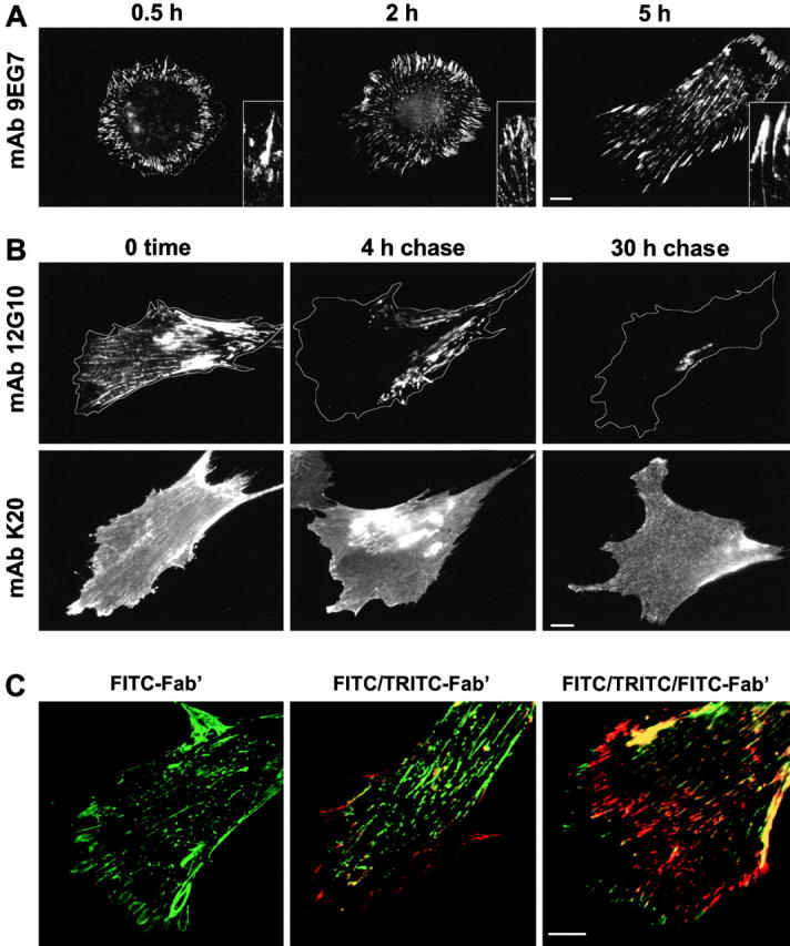
Cell surface dynamics of β1 integrins examined by conventional immunofluorescence (A) and an antibody-chasing technique (B and C). (A) HFF were allowed to spread on coverslips in complete medium for the indicated periods of time, then fixed and stained with anti–β1 integrin antibody (mAb 9EG7). (B) Overnight cultures of HFF in complete medium were labeled in vivo for 30 min with mAb 12G10 or mAb K20. Unbound antibodies were rinsed away, and labeled cells were fixed at 0 time, or chased for 4 or 30 h, then fixed and stained with secondary antibody without permeabilization. Cell borders were traced from the corresponding phase-contrast images. (C) HFF were labeled in succession for 30 min each with FITC-conjugated mAb 12G10 Fab′ fragments (FITC-Fab′), followed by rhodamine-conjugated 12G10 Fab′ (FITC/TRITC-Fab′), then again with FITC-12G10 Fab′ (FITC/TRITC/FITC-Fab′). The last two panels are merged digital FITC and rhodamine images. Bars, 10 μm.
Such integrin dynamics might occur continuously on the surface of spread cells, but conventional immunofluorescence techniques only permit evaluation of antigen distribution at the time of fixation and are not adequate to probe for exchanges of antigens between established structures. To test our hypothesis concerning direct β1 integrin translocation and to compare the dynamic behavior of integrin populations on the cell surface of spread cells, we designed antibody-chasing experiments in which living cells were labeled with nonperturbing anti-integrin antibodies (see Materials and Methods). Labeled cells were incubated in culture medium for different time periods (chasing time), which allowed normal cell surface dynamics to continue and redistribute the antibody-tagged integrins. At the end of labeling and after each chasing period, samples were fixed and stained with species-specific fluorochrome-conjugated secondary antibodies. Cells were not permeabilized to eliminate the possibility of false-positive results due to endocytosed primary antibody. Such endocytosis was detected to occur during labeling, but the endocytosed antibodies were located centrally, distant from the sites of dynamics examined in this study (data not shown). In subsequent studies, a second or third round of labeling could be performed using antibodies labeled directly with different fluorochromes.
Thus, by comparing the distribution patterns of the labeled integrins immediately after antibody incubation (0 time) with those obtained after different chasing periods, we were able to follow the redistribution of labeled integrin populations. We also compared results using these cation and ligand-induced binding site antibodies with those using mAb K20, a noninhibitory antibody that binds to all β1 integrins.
As expected, immediately after labeling, ligand-occupied β1 integrins were localized to both focal and ECM contacts (Fig. 1 B, mAb 12G10, 0 time). However, after 4 h of chasing, the labeled population of β1 integrins was found only within ECM contacts (Fig. 1 B, 4 h chase). Some staining of these fibrillar structures could be detected even after 30 h of chasing, with no FC staining (Fig. 1 B, 30 h chase). Similar results were obtained by using mAb 9EG7 (data not shown), supporting the hypothesis of continuous translocation of ligand-occupied β1 integrins from the FC to (and possibly within) ECM contacts. In contrast, mAb K20-labeled β1 integrins showed strong diffuse staining that persisted throughout all periods of chasing (Fig. 1 B, mAb K20), consistent with free diffusion of nonactivated β1 integrins on the cell surface.
Because in vivo antibody labeling might induce integrin dimerization and resultant artifacts, we prepared FITC- or rhodamine-conjugated Fab′ fragments from mAb 12G10. Sequential incubation with these fragments revealed the same centripetal transition of labeled integrins (Fig. 1 C). Since this transition was observed with monomeric antibody tags, the distinctive redistribution of ligand-occupied integrins was not due to clustering induced by bivalent IgG molecules. Moreover, we observed the appearance of new ligand-occupied integrins in FC at the leading edge of cells (Fig. 1 C, FITC/TRITC-Fab′), which moved as a red-labeled wave towards the cell body (Fig. 1 C, FITC/TRITC/FITC-Fab′). The preservation of rhodamine staining also indicates that the disappearance of the signal from FC was not due to simple loss of antibody from these structures but, instead, translocation of labeled integrins away from FC. This wave-like pattern of staining, with an initial appearance at the leading cell edge followed by translocation towards the cell body, indicates the presence of a persistent and directed flow of ligand-occupied β1 integrins on the surface of cultured fibroblasts. Similar behavior of activated β1 integrins was observed in Swiss 3T3 cells and primary mouse fibroblasts (data not shown), indicating that this phenomenon is typical in fibroblastic cells.
α5β1 Is the Translocating Integrin Heterodimer
The β1 integrin subunit can pair with at least 10 different α subunits to form heterodimeric receptors that display differing ligand specificities. To define the ligand specificity of the translocating β1 heterodimers, the ability of different integrin ligands to trigger integrin withdrawal from FC was compared using antibody-chasing experiments (Fig. 2 A). Human fibroblasts grown in FN-free medium were treated with cycloheximide in order to inhibit the synthesis and secretion of endogenous ECM proteins. To ensure better spreading, cells were plated on VN-coated coverslips. Under these conditions, the only ECM protein present as a substrate and in the medium was VN. After initial incubation of cells with mAb 9EG7 together with different ligands, ligand-occupied β1 integrins showed localization to FC regardless of the type of the ligand added (Fig. 2 A, 0 time). However, after 1 h of chasing, the labeled β1 integrins demonstrated clear ligand-dependent differences in distribution. The presence of VN alone was not capable of inducing redistribution of the β1 signal from FC (Fig. 2 A, compare 0 time with 1 h chase). This result allowed us to evaluate the effect of the other integrin ligands. Collagen- and laminin-treated cells showed irregularly shaped β1-positive aggregates concentrated at the cell body (Fig. 2 A, 1 h chase, COL and LN). These aggregates were located predominantly on the dorsal cell surface and most likely represented β1 receptors occupied with collagen and laminin. Importantly, however, FC were still well labeled, indicating that laminin and collagen ligands are not capable of inducing β1 integrin withdrawal from FC. Only FN was effective, indicating that fibril-associated translocation from FC into and within ECM contacts is a distinctive feature of certain β1-containing integrin receptor(s) for FN (Fig. 2 A, 1 h chase, FN).
Figure 2.
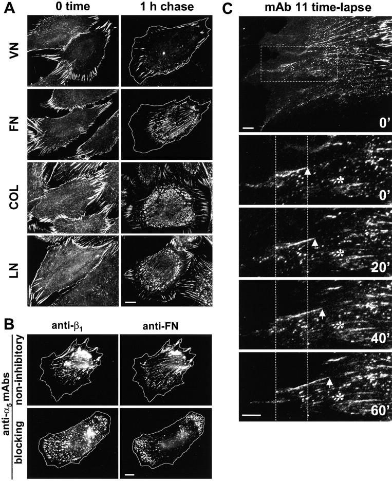
Identification of the translocating integrin heterodimer. (A) HFF were cultured overnight on VN-coated coverslips in FN-free medium with 25 μg/ml cycloheximide, and then labeled for 30 min in vivo with anti–β1 integrin antibody mAb 12G10. Cells were fixed either without chasing (0 time) or after a 1-h chase. Antibody labeling and chasing were performed in the presence of 50 μg/ml of the following ECM proteins: VN, FN, collagen I (COL), or laminin 1 (LN). Note that only FN was able to induce translocation of the β1 integrin signal out of FC. Bar, 10 μm. (B) HFF were cultured in complete medium containing 50 μg/ml of anti-α5 antibodies mAb 11 (noninhibitory) or mAb 16 (function-blocking). The cells were assayed by a 1-h chase of β1 integrins with mAb 12G10 (anti-β1), fixed, and double-stained with anti-FN. Cell contours were traced from corresponding phase-contrast images. Bar, 10 μm. (C) HFF were cultured overnight in complete medium on glass coverslips, then labeled with 10 μg/ml Alexa 488–tagged anti–α5 integrin antibody mAb 11 for 20 min. Samples were rinsed and analyzed by time-lapse microscopy at 20-min intervals as described in Materials and Methods. The top panel shows the first image, and the dashed line outlines the region shown enlarged in the lower four panels. Arrows indicate α5 integrins extending in a fibrillar structure from FC. Asterisks indicate a nonmotile aggregate relative to the fixed fiduciary lines. Bar, 5 μm.
Because α5β1 is a specific receptor for FN and the most abundant β1 integrin heterodimer on the surface of HFF (our unpublished results), we investigated its role in the integrin translocation process. α5β1 integrin function was selectively disrupted using an antifunctional antibody. Treatment of HFF with control (noninhibitory anti-α5 mAb 11) did not affect β1 integrin translocation into ECM contacts (Fig. 2 B, anti-β1) containing characteristic FN fibrils (Fig. 2 B, anti-FN). In contrast, cells grown in the presence of function-blocking anti-α5 mAb 16 did not organize ECM contacts. FN remained in small aggregates confined to the lamellae (Fig. 2 B, anti-FN). Labeled β1 integrins showed a punctate, nonfibrillar distribution over the whole cell surface, and positive staining of FC remained even after 1 h of chasing (Fig. 2 B, anti-β1). Cell morphology was not affected; this antibody was previously found to stimulate rather than to inhibit cell migration, perhaps due to the loss of ECM contacts (Akiyama et al. 1989). This blocking of ligand-occupied β1 integrin translocation out of FC by antibody inhibition of α5 subunit function strongly indicates that α5β1 is the major translocating heterodimer.
Consistent with this conclusion, Alexa-tagged anti-α5 mAb 11 bound to cell surface α5β1 integrins underwent a similar pattern of translocation as the anti-β1 12G10 mAb. In vivo immunofluorescence time-lapse recording (Fig. 2 C) showed that the labeled α5 integrins were initially localized within FC (Fig. 2 C, 0′), and later they extended toward the cell center as an elongating fibrillar structure (Fig. 2 C, 20′, 40′, and 60′).
Occupied α5β1 Heterodimers in Fibrillar Structures Translocate Relative to FC
Because β3 integrins (in contrast to β1) remained strictly confined within FC during the 1-h antibody-chasing period (see below), we used β3 integrin labeling of these structures as a reference point to determine the velocity of translocation of ligand-occupied β1 integrins. HFF were incubated in vivo with anti-β1 and anti-β3 antibodies simultaneously, and the two antibodies were chased for 30 min or 1 h. Immediately after antibody incubation, FITC staining for β1 integrins completely overlapped CY3 staining for β3 integrins in FC, whereas the ECM contacts were only β1 integrin–positive (Fig. 3 A, 0 time, yellow and green staining, respectively). Initial separation between green and red signals was observed after 30 min of chasing (Fig. 3 A, 0.5 h chase), and the separation appeared complete by 1 h (Fig. 3 A, 1 h chase). The withdrawal of FITC label from FC clearly indicated that activated β1 integrins translocate relative to the FC and in a direction opposite to migration of the cell (e.g., note the direction of lamellipodial extension on Fig. 3 A, phase-contrast–merged). The average rate of this translocation, measured as distance from the red to the green signal/chasing time, was 6.5 ± 0.7 μm/h. The rate of translocation did not depend on the type of substrate used for coating. It also did not appear to correlate with the rate of cell motility. Although the cells on VN-coated surfaces migrated significantly faster than the cells on FN (P < 0.0001), the translocation rates of β1 integrins under the same conditions were not different statistically (P = 0.15) (Fig. 3 B).
Figure 3.
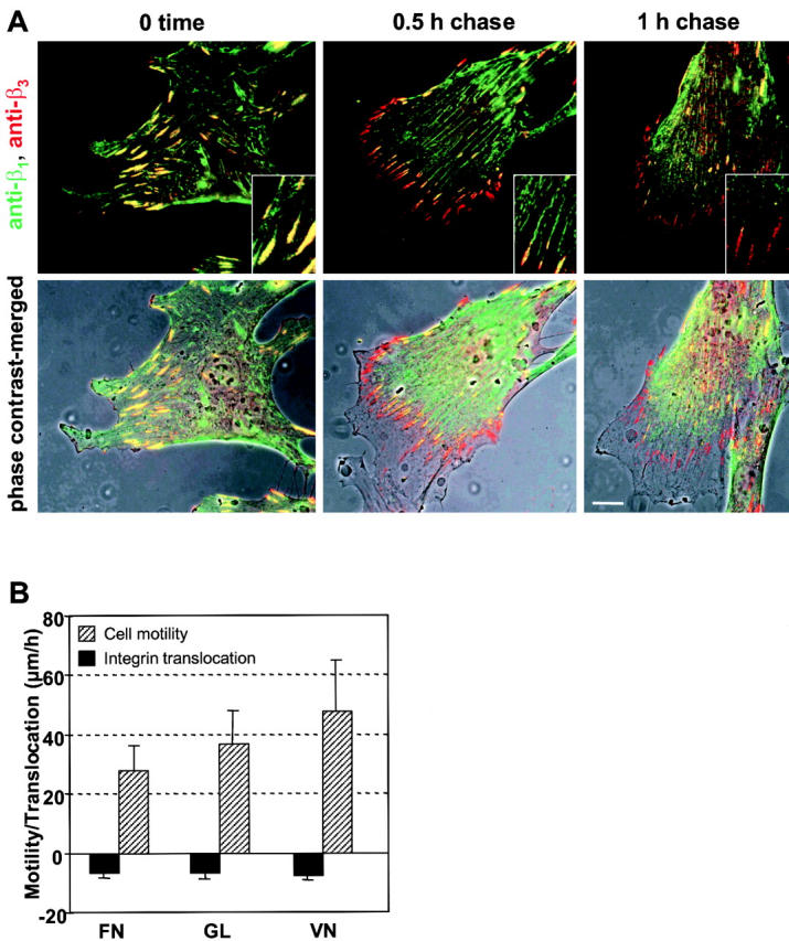
Comparisons of β1 and β3 integrin dynamics. (A) Overnight cultures of HFF on glass coverslips in complete medium were double-labeled in vivo with anti-β1 (rat mAb 9EG7) and anti-β3 (mouse mAb AP3) antibodies for 30 min. After rinsing, the cells were fixed without chasing (0 time) or after chasing for 0.5 or 1 h. Fixed, nonpermeabilized cells were stained with a mixture of FITC-conjugated anti–rat and CY3-conjugated anti–mouse goat IgG for visualization of β1 and β3 integrins, respectively. FITC and CY3 immunofluorescence images were overlaid and merged with the corresponding phase-contrast images. Bar, 10 μm. (B) HFF were plated on glass coverslips (GL), or coverslips coated with 10 μg/ml FN or VN and cultured for 16 h in complete medium. Cell motility was recorded by time-lapse video microscopy for 6 h. Corresponding immunofluorescence samples were prepared as described in A and used to measure the rate of separation between β1 and β3 integrin signals as described in Materials and Methods. Data were pooled from three independent experiments, each one measuring at least 10 cells. Error bars indicate SD.
FN Ligation and Interaction with an Intact Actin Cytoskeleton Are Required for α5β1 Integrin Translocation
Ligand-mediated occupancy and receptor clustering are necessary for activation of certain integrin functions, e.g., stimulation of actin cytoskeletal assembly in adhesion complexes (Miyamoto et al. 1995). To investigate the role of FN ligation in integrin translocation, HFF were grown overnight in the absence of FN. Synthesis and secretion of endogenous FN were blocked with cycloheximide during cell growth and antibody labeling. FN was not detectable by immunostaining and Western blotting after this treatment.
ECM contacts were absent under these conditions, but unexpectedly, both mAb 9EG7 (Fig. 4 A, 0 time) and mAb 12G10 (data not shown) continued to stain FC, detecting a subclass of β1 integrins in the putative active conformation in the absence of FN. β3 integrins were also localized within FC, resulting in yellow FC staining in merged red and green images. After 1 h of chasing, the signals for the two integrins became slightly separated, with β1 integrins concentrated in the half of FC oriented toward the cell center (Fig. 4 A, 1 h chase, no ligand). Extended chasing periods without FN showed gradual fading of the β1 signal without any translocation towards the cell center. Addition of FN induced normal translocation (Fig. 4 A, FN). After a 30-min chase in the presence of FN, β1 integrins became localized in fibrillar structures extending from FC (Fig. 4 A, 1 h chase inset). Prolonging the chase to 1 h resulted in complete separation of labeled β1 integrins from β3 in FC (Fig. 4 A, 1 h chase). Taken together, these results indicate that β1 integrins in a ligand-occupied conformation can still be detected transiently within FC even in the absence of FN, but that ligation by FN is necessary for preservation of the clustered, ligand-occupied state, and most importantly, for surface translocation of integrins away from FC.
Figure 4.
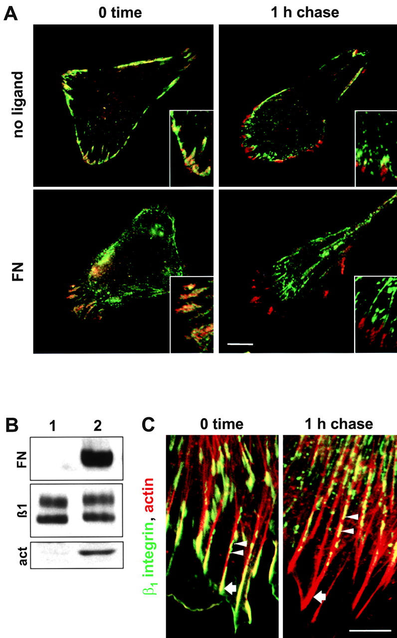
Dependence of integrin translocation on receptor ligation and interactions with actin cytoskeleton. (A) HFF were cultured as described in Fig. 2 A and then double-labeled in vivo for 30 min with anti-β1 (mAb 9EG7) and anti–β3 integrin (mAb AP3) antibodies (0 time), or chased for 1 h. Labeling and chasing were performed either with no ligand or in the presence of 100 μg/ml human plasma FN. Insets show enlargements; the bottom right panel inset was from an additional sample chased for only 30 min. Bar, 10 μm. (B) Activated β1 integrins were immunoprecipitated with mAb 9EG7 from HFF grown overnight in FN-free medium with 10 μg/ml cycloheximide without (lane 1) or with 100 μg/ml human plasma FN (lane 2). Immunoprecipitates were immunoblotted for coprecipitating FN, β1 integrin (β1), and actin (act). (C) Nontreated HFF grown in complete medium were labeled in vivo for 30 min with mAb 9EG7, fixed, and double-stained with FITC-conjugated anti–rat secondary antibody and rhodamine-phalloidin to visualize F-actin (0 time), or chased for 1 h before fixation and staining with the same mixture. Arrows indicate the position of FC, and arrowheads indicate integrin complexes along stress fibers. Bar, 5 μm.
A variety of cytoskeletal molecules organize around activated β1 integrin cytoplasmic tails in complexes that interact with actin filaments (see Yamada and Geiger 1997). To determine whether FN induces mAb 9EG7-positive β1 integrin interaction with the cytoskeleton, we compared the profiles of proteins coimmunoprecipitating with FN-ligated, translocating integrins versus nontranslocating β1 integrins. When mAb 9EG7 immunoprecipitates were prepared from cycloheximide-treated HFF grown in the absence of FN, conditions that do not favor translocation (Fig. 4 A, 1 h chase, no ligand), there was no detectable actin in the immune complexes (Fig. 4 B, lane 1). Incubation of the cells with exogenous FN, which restores translocation of β1 integrins (Fig. 4 A, 1 h chase, FN), led to the appearance of actin and FN as coimmunoprecipitating proteins (Fig. 4 B, lane 2). Furthermore, clusters of β1 integrins were observed to translocate along stress fibers when chasing was combined with staining for F-actin (Fig. 4 C). These results suggest participation of the actin cytoskeleton in integrin surface translocation.
This notion was tested further using drugs known to affect actin polymerization (Table ). Translocation was blocked by both cytochalasin D, which disrupts actin filaments, and jasplakinolide, which induces actin polymerization and stabilizes preexisting actin filaments (Bubb et al. 1994) and which promoted actin stress fibers (data not shown). These results indicate that preventing either actin polymerization or depolymerization prevents integrin redistribution, supporting the notion that an intact and functional actin cytoskeleton is required for translocation of β1 integrins.
Table 1.
Tight Association between Integrin Translocation and FN Fibril Formation but Not Cell Migration
| Conditions | Integrin translocation | Fibronectin fibrillogenesis | Velocity of cell migration (μm/h) |
|---|---|---|---|
| Control | ++ | ++ | 36.9 ± 12.0 |
| Pharmacological agents | |||
| Cycloheximide(25 μg/ml) | − | − | 5.7 ± 1.8 |
| Cycloheximide/FN(25 μg/ml) | ++ | ++ | 5.3 ± 1.6 |
| Cytochalasin D(0.4 μM) | − | − | 12.1 ± 3.9 |
| Jasplakinolide(0.1 μM) | − | − | 21.0 ± 5.6 |
| BDM (20 mM) | − | − | 14.7 ± 4.5 |
| Nocodazole(10 μM) | +++ | +++ | 9.4 ± 2.7 |
| Vinblastine(50 μM) | +++ | +++ | 12.8 ± 3.4 |
| Paclitaxel(20 μM) | ++ | ++ | 15.5 ± 6.3 |
| PAO (5–50 nM) | ++ | ++ | 6.5 ± 1.8 |
| Substrates | |||
| FN | − | − | 26.1 ± 11.1 |
| FN+VN | ++ | ++ | 20.7 ± 9.0 |
| FN+PLL | − | − | 10.3 ± 6.5 |
| VN | ++ | ++ | 47.5 ± 17.6 |
BDM, 2,3-butanedione 2-monoxime; PAO, phenylarsine oxide; and PLL, poly-l-lysine.
FN Extension Occurs on the Cell Surface in Association with Translocating α5β1 Integrins
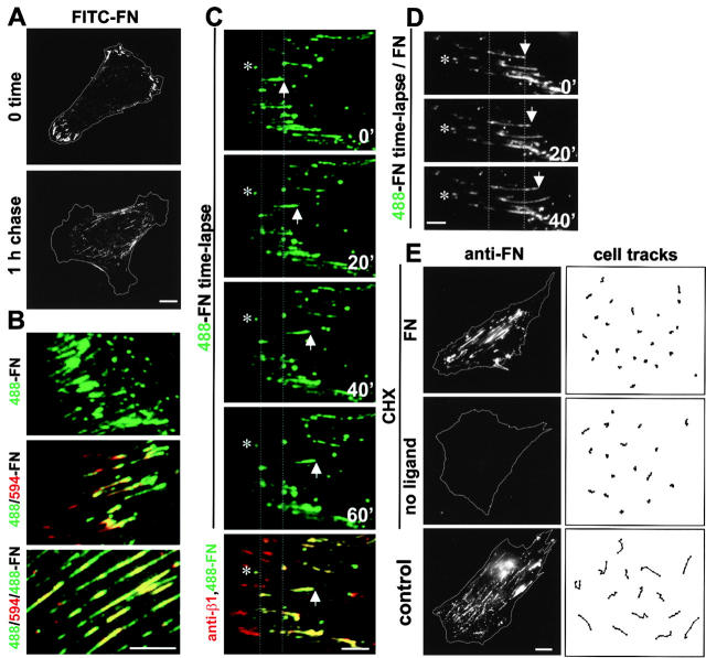
The existence of translocation of ligand-occupied α5β1 integrins from FC along ECM contacts suggested that bound FN ligand might also be translocated in a similar manner to mediate the elongation of FN fibrils (i.e., fibrillogenesis). This hypothesis was tested first by examining the dynamic behavior of FITC-labeled FN bound to the surface of cycloheximide-treated HFF. After the initial incubation, labeled FN was found in FC (Fig. 5 A, 0 time), presumably by binding to activated α5β1 integrins residing within FC under these experimental conditions (compare with Fig. 4, 0 time). A 1-h chase led to a fibril-associated withdrawal of labeled FN from FC, similar to the translocation of β1 integrins described above (Fig. 5 A, 1 h chase). Moreover, subsequent sequential incubations with FN tagged with different Alexa dyes led to segmented labeling of FN fibrils extending towards the cell center (Fig. 5 B). This pattern of formation of FN fibrils strongly resembled the wave-like pattern of surface translocation of ligand-occupied β1 integrins (compare with Fig. 1 C).
Figure 5.
Cell surface dynamics of FN. (A) HFF were treated with cycloheximide as described in Fig. 2 A and incubated in vivo for 30 min with 10 μg/ml FITC-conjugated FN. The FN label was fixed at 0 time or chased for 1 h before fixation. (B) Cycloheximide-treated HFF were labeled for 30 min in succession with 10 μg/ml Alexa 488–conjugated FN (488-FN), followed by Alexa 594–conjugated FN (488/594-FN), and finally Alexa 488–FN again (488/594/488-FN). The last two panels represent digital overlays of images obtained from the green and red channels. (C and D) HFF were cultured overnight in FN-free medium supplemented with 10 μg/ml cycloheximide, then incubated with 10 μg/ml Alexa 488–conjugated FN for 30 min. Video recording was performed in FN-free medium (C, 488-FN time-lapse) or medium containing 100 μg/ml unlabeled FN (D, 488-FN time-lapse/FN). The fibrils in D extended ∼4 μm in 40 min. At the end of the recording, samples were fixed and stained with anti–β1 integrin antibody (mAb 9EG7) and CY3-conjugated secondary antibody, and the recorded cells were examined again to determine the position of β1 integrins and labeled FN. Note the complete overlap between β1 integrin (red) and translocated labeled FN (green), resulting in yellow staining of ECM contacts in merged images, whereas FC are only β1 integrin–positive (C, bottom panel). Fiduciary lines indicate initial positions, arrows indicate tips of translocating fibril, and asterisks mark nonmotile FN aggregates. (E) Cycloheximide-treated HFF in FN-free medium were incubated with (FN) or without (no ligand) 100 μg/ml human plasma FN and recorded by time-lapse video microscopy for 6 h. Untreated cells grown in complete medium were used as controls. Representative examples of cell movements tracked at 30-min intervals over a span of 3 h are shown (cell tracks in right panels). At the end of the recording, the cells were fixed and stained with FITC-conjugated anti-FN antibodies (anti-FN). Cell borders were traced from corresponding phase-contrast images. Bars: (A and E) 10 μm; (B, C and D) 5 μm.
An alternative approach using time-lapse fluorescence microscopy revealed a stepwise translocation of FN fibrils from FC towards the cell center. Initial incubation of cycloheximide-treated cells with labeled FN and subsequent recording in FN-free medium led to formation of short translocating FN fibrils (Fig. 5 C, 488-FN time lapse), whereas incubation in the continued presence of unlabeled FN showed continuous growth and extension of the labeled fibrils (Fig. 5 D). When translocating fibrils were fixed and examined for association with ligand-occupied β1 integrins, complete colocalization of FN and β1 integrins was observed (Fig. 5 C, anti-β1,488-FN), indicating that the receptor–ligand complexes were translocating together.
It is important to note that the translocation of α5β1 integrins out of FC and the formation of ECM contacts and FN fibrils was not always continuous (Fig. 2 and Fig. 5). At any moment, ∼50% (30–70%) of the FC of a cell were active in integrin translocation. This heterogeneity, as well as the discontinuities in translocating fibrils and separation of ECM contacts from FC, suggest that the process may be switched on and off in individual FC.
The impression that α5β1 translocation does not correlate with cell migration (Fig. 3 B) was further tested under 12 more widely varying experimental or pharmacological conditions (Table ). For example, even though cell migration was reduced more than sixfold by cycloheximide (Table and Fig. 5 E), integrin translocation and FN fibrillogenesis depended solely on whether FN was present. Based on this lack of correlation under 13 different conditions, we conclude that this form of integrin translocation and early FN fibrillogenesis are independent of cell migration.
The observed fibrillar translocation of β1 integrin–labeled FN complexes, which was also detected on the surface of cells not treated with cycloheximide (data not shown), strongly suggests that the cell can redistribute and organize bound FN into fibrils in a directed manner through translocation of α5β1 integrins. However, the labeled exogenous FN could have been augmenting preexisting fibrils. To investigate initial steps of FN fibril formation, we examined freshly plated cells using immunofluorescence to follow the distribution of ligated β1 integrin as described previously by Singer et al. 1988 for the α5 subunit, but in parallel with endogenous FN. Cells plated for 30 min on VN in complete medium organized numerous small FC along the spreading edge (Fig. 6 A, anti-β1), but there was no detectable FN (Fig. 6 A, anti-FN). After 1 h, a circle of small FN-positive aggregates appeared that overlapped the inner part of FC (Fig. 6 A, 1 h). Detachment of cells from the substrate did not remove these FN circles (data not shown), indicating that the FN was secreted ventrally and firmly attached to the substrate. After an additional hour of spreading, the attached FN aggregates became reorganized into fibrils (Fig. 6 A, 2 h, anti-FN) that overlapped with fibrillar β1 integrin–positive protrusions extending from FC (Fig. 6 A, 2 h, anti-β1 and merged inset). By 5 h, all of the cells had completed the process of spreading and had a polarized shape. Numerous ECM contacts associated with FN fibrils had developed; some extended from FC (Fig. 6 A, 5 h, inset). FN fibrils were also observed away from cell bodies, indicating that FN matrix deposition had begun.
Figure 6.
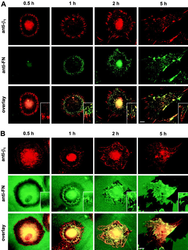
Distribution of β1 integrins and FN during cell spreading. HFF were allowed to spread in complete medium on coverslips coated with 10 μg/ml VN (A) or FN (B) for the indicated times. Cells were fixed and double-stained for β1 integrins with mAb 9EG7 (anti-β1) and FITC-conjugated anti–human FN antibody (anti-FN). Immunofluorescence images of each cell were merged by image processing. Bars, 10 μm.
A similar sequence of events was observed when HFF spread on FN precoated substrates (Fig. 6 B). As reported previously (Avnur and Geiger 1981), cells reorganized substrate-adsorbed exogenous FN into fibrils, which we found to coincide with β1 integrin–positive ECM contacts. FN began to accumulate around the inner ends of FC after 1 h (Fig. 6 B, 1 h, anti-FN). After an additional hour, ECM contacts appeared, accompanied by a characteristic scraping of FN from the coverslip surface (Fig. 6 B, 2 h). The areas cleared of FN enlarged with time of spreading (Fig. 6 B, 5 h, anti-FN). The completeness of the clearing suggests that the cells are applying considerable force to remove surface-adsorbed FN. In addition, the fact that the initial reorganization of FN into fibrils coincides topologically and temporally with the translocation of β1 integrin out of FC (Fig. 6 B, overlay) suggests that integrin translocation is the driving force for FN reorganization and fibrillogenesis.
Blocking α5β1 Integrin Translocation Blocks Early FN Fibrillogenesis
To test the link between integrin translocation and the formation of FN fibrils on the surface of HFF, we used drugs and conditions affecting cell-generated contractility and tension, actin cytoskeleton, microtubule integrity and function, and other functions. There was a complete linkage between integrin translocation and FN fibrillogenesis under 13 different experimental test conditions (Table ). For example, the myosin inhibitor 2,3-butanedione 2-monoxime inhibits contractility, prevents FN fibrillogenesis (Zhong et al. 1998), and prevents integrin translocation. Conversely, the microtubule-disrupting agents nocadazole and vinblastine, which stimulate FN matrix assembly and contractility, also slightly enhanced translocation, whereas the microtubule-stabilizing drug taxol (paclitaxel) does not affect FN fibrillogenesis (Zhang et al. 1997) and showed normal integrin translocation (Table ).
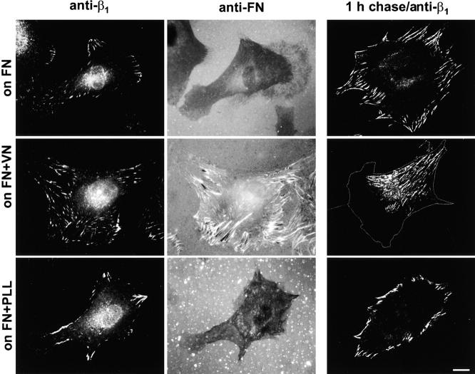
The observed constant flow of ligand-occupied α5β1 integrins from FC into the ECM contacts appeared inconsistent with the established function of FC as structures providing firm attachment and mechanical support for spread cells. This apparent contradiction could be resolved if a different integrin heterodimer residing within FC provides mechanical support during α5β1 translocation. A good candidate for this function was the VN receptor, because our antibody-chasing experiments showed that β3 integrins remained within FC during β1 integrin translocation (Fig. 3 A). Consequently, absence of VN might prevent α5β1 translocation and associated ECM contact formation and FN fibrillogenesis. To test this possibility, we plated HFF on FN or a surface coated with a mixture of FN and VN in medium lacking serum, which is the major source of VN. As predicted, in the absence of VN, cells plated for 4 h did not translocate β1 integrin from FC and were unable to reorganize FN (Fig. 7, on FN). Moreover, addition of VN to the coating substrate permitted the cells to translocate β1 integrins into ECM contacts and to reorganize FN into fibrils (Fig. 7, on FN+VN). Addition of poly-l-lysine to the coating mixture (with FN but without VN) was ineffective (Fig. 7, on FN+PLL), indicating that only specific, integrin-mediated adhesion is capable of supporting this translocation. These results together with those using a dozen other inhibitors or substrate alterations strongly support the notion that early FN fibrillogenesis and α5β1 integrin translocation are tightly linked inseparable processes and that the translocation is VN-supported and actin-dependent.
Figure 7.
Ligation of VN receptor is necessary for α5β1 integrin translocation and FN fibrillogenesis. HFF were allowed to spread for 4 h on coverslips coated with 10 μg/ml FN, a mixture of FN and VN (5 μg/ml each), or a mixture of FN and poly-l-lysine (5 μg/ml) in serum-free medium. Cells were fixed, permeabilized, and double-stained with mAb 9EG7 (anti-β1) and anti-FN antibodies, or β1 integrins were labeled in vivo for 30 min and chased for 1 h (1 h chase/anti-β1). Bar, 10 μm.
Overexpression of a Specific Tensin Domain Interferes with Integrin Translocation and FN Fibrillogenesis
Recent molecular morphometry studies have identified tensin as the major cytoskeletal protein component of ECM contacts (Zamir et al. 1999). Tensin has actin-capping, filament-binding, and cross-linking sites (Lo et al. 1994; Chuang et al. 1995), which could have roles in the formation of ECM contacts and translocation of α5β1 integrins. We searched for such functions using different recombinant tensin fragments as potential dominant-negative inhibitors of integrin translocation in transfection overexpression assays. ECM contacts and associated integrin dynamics and FN fibrillogenesis were inhibited by a domain of tensin spanning residues 659 to 762 containing the actin homology 2 region (residues 674 to 706) (Chuang et al. 1995) tagged with GFP and transiently overexpressed in HFF. Transfected cells (Fig. 8b and Fig. d) organized β1 integrin–positive FC, but they were completely devoid of ECM contacts (Fig. 8 a, compare transfected with nontransfected cell). 1 h of antibody-chasing β1 integrins showed that cells overexpressing this particular tensin fragment were unable to translocate integrins out of FC, whereas neighboring nontransfected cells readily translocated it into ECM contacts (Fig. 8c and Fig. d). FN was not present in fibrils and was detected only as diffuse staining within the area of the FC (Fig. 8e and Fig. f). There were no detectable differences in the organization of stress fibers between transfected and nontransfected cells (Fig. 8g and Fig. h). The observed alterations were proportional to the level of expression of the fusion protein as judged by the intensity of GFP signal and were absent in low-expressing cells. Overexpression of empty GFP vector or other domains of tensin did not affect integrin translocation or the formation of FN fibrils (our unpublished results). The most likely mechanism of the observed effects is a dominant-negative form of competition between the expressed fragment and endogenous tensin. These results provide a functional link between the prominent presence of tensin in ECM contacts and the integrity of these structures associated with integrin translocation and FN fibrillogenesis.
Figure 8.
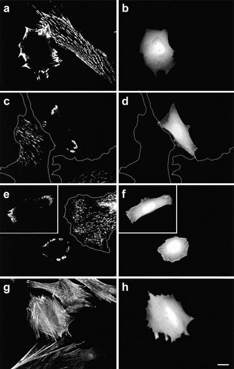
Experimental suppression of integrin translocation and FN fibrillogenesis by the tensin domain 659–762. HFF were transfected with the GFP-tagged actin homology 2 fragment of tensin and used after 36 h for in vivo labeling (a and b) and 1 h chasing of β1 integrins (c and d). Additional transfected samples were stained with anti-FN antibodies (e and f) and TRITC-phalloidin (g and h). Overexpressing cells were identified by GFP fluorescence (b, d, f, and h). Cell borders were traced from corresponding phase-contrast images. Bar, 10 μm.
Discussion
The main objective of this study was to characterize the cell surface dynamics of activated α5β1 integrins and their role in organizing the ECM. We found that ligation of α5β1 integrins by FN induced an escalator-like translocation along actin microfilament bundles. These clustered, ligand-occupied integrins moved in linear arrays out from FC towards the cell center in the form of ECM contacts bound to elongating fibrils of FN. This process may mediate initial matrix assembly by pulling a fibril of FN from the pool of FN molecules that accumulates near FC using interactions with actin cytoskeleton in a process dependent on tensin. Consistent with this concept of linked translocation-fibrillogenesis, interfering with any of the molecular components (α5β1 integrins, FN, actin, or tensin) halted both the integrin translocation process and FN fibrillogenesis, which were found to be tightly coupled under a wide variety of experimental conditions (13 different inhibitors or substrates). These findings point to a specific structure, the ECM contact, and a molecular translocation mechanism in a model proposed to explain how FN can be stretched and organized into a fibril by cellular tension via a specific local pattern of integrin movement.
We initially hypothesized that a dynamic relationship might exist between FC, which act as anchoring sites for stress fibers and are therefore under isometric tension (Burridge 1981), and ECM contacts, which coalign with FN fibrils and bundles of actin filaments (Hynes and Destree 1978; Chen and Singer 1982; Chen et al. 1985; Zamir et al. 1999; this paper). Contractile forces at FC might be translated into a mechanism for stretching FN molecules via linear translocation of FN receptors. Convincing evidence that live cells exert considerable tension on FN fibrils is provided by observing GFP-tagged FN in CHO cells (Ohashi et al. 1999). Our time-lapse data show elongation of single FN fibrils consistent with stretching. This type of tension (Zhong et al. 1998), together with putative conformational changes promoted by binding to the integrins (Sechler et al. 1996), has been suggested by others to be important for unfolding FN molecules to expose cryptic self-association sites needed for subsequent polymerization to form FN fibrils (Aguirre et al. 1994; Hocking et al. 1994; Ingham et al. 1997).
To test this hypothesis of a role for specialized integrin translocation, we developed an antibody-chasing technique, a modification of conventional immunofluorescence methods that allows simultaneous characterization of the dynamic behavior of distinct integrin populations on the cell surface. Living cells were labeled with antibodies that recognize either (a) epitopes indicating cation and ligand-induced binding sites (Bazzoni and Hemler 1998) that become exposed on integrin molecules after occupancy by a ligand, or (b) epitopes common to integrins regardless of activation or occupancy state.
Using this antibody-chasing technique and time-lapse immunofluorescence microscopy, we established that different integrin heterodimers have different dynamic behavior on the cell surface. Whereas the VN receptor αvβ3 remains resident within FC, ligated FN receptor α5β1 actively translocates from FC into and along ECM contacts on the surface of fibroblasts (Fig. 9 A). This translocation involves net displacement relative to FC with an average velocity of 6.5 ± 0.7 μm/h that is independent of cell motility. This integrin translocation is oriented along actin stress fibers, and it depends on both interaction with a functional actin cytoskeleton and ligation of VN receptors.
Figure 9.
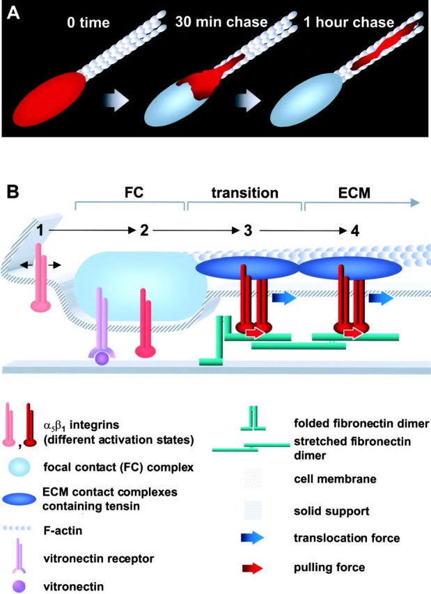
Models of integrin dynamics and FN matrix assembly. (A) Schematic representation of the translocation of labeled α5β1 integrins (red) out of FC (blue) and along actin filaments (gray) as characterized in this study. (B) Model of early FN fibrillogenesis driven by integrin translocation. (1) Nonactivated integrins are diffusely distributed on the cell surface. (2) Within FC, some α5β1 integrins can be activated even in the absence of FN (see text for details). (3) FN binding to α5β1 integrins induces formation of homogeneous clusters of this integrin; the β1 integrin cytoplasmic domains organize tensin-containing ECM contact complexes capable of translocating along actin filaments. (4) Moving ECM contact complexes pull α5β1 integrin clusters out of FC into new fibrillar ECM contacts. We propose that translocating integrins in turn pull and stretch bound FN molecules, which unfold and expose cryptic self-association sites necessary for subsequent FN polymerization. Note that components are drawn schematically and not to scale.
Tensin is thought to be involved in the interaction between integrins and the cytoskeleton, and the ECM contacts in which we observed integrin translocation were previously found to be uniquely enriched in tensin compared with other FC molecules (Zamir et al. 1999). Using a dominant-negative approach with recombinant domains from tensin, we identified a 103–amino acid fragment of tensin containing actin homology domain 2 (Chuang et al. 1995) that selectively eliminates ECM contacts, indicating that tensin is crucial for these structures. Since tensin is present in both FC and ECM contacts, it is an attractive candidate for regulating or mediating the dynamic behavior of α5β1 integrins. In fact, a similar centripetal translocation of GFP-tensin out of FC and along ECM contacts has also been observed (Zamir et al. 2000).
We conclude that transfer from FC of ligand-occupied α5β1 integrins along bundles of actin filaments during cell spreading leads to the initial formation of ECM contacts and ensures the maintenance of these structures on the surface of adherent cells. Because integrin translocation and elongation of FN fibrils are so tightly linked (including tensin dependence), we propose that α5β1 translocation is used by fibroblastic cells to promote initial FN fibrillogenesis in ECM contacts.
It is important to note that the phenomenon we report differs from general centripetal surface translocation of objects (Harris and Dunn 1972) or of ligand- or antibody-coated beads (Felsenfeld et al. 1996) with subsequent cytoskeletal strengthening (Choquet et al. 1997). Binding, anchoring, and translocation of beads can occur over the entire leading cell edge with a translocation rate of 6.6 ± 1.8 μm/min, in a process linked to cell migration and blocked selectively with low doses (5 nM) of the tyrosine phosphatase inhibitor phenylarsine oxide (Choquet et al. 1997). In contrast, the translocation we describe is confined to a narrow linear path from FC through ECM contacts with a rate of translocation that is at least 60-fold slower (0.11 ± 0.01 μm/min), involving a subset of FC. Moreover, the process is independent of cell migration and not inhibited by even 50 nM phenylarsine oxide (Table ). The behavior of α5β1 integrin–positive macroaggregates has been characterized in vivo on the surface of motile chick skeletal fibroblasts plated on laminin (Regen and Horwitz 1992). Unlike our observations, these macroaggregates remain fixed with respect to the substratum during cell movement, and most probably represent a different type of attachment.
Recently, the dynamic behavior of a GFP–β1 integrin cytoplasmic tail chimera has been characterized by Smilenov et al. 1999. FC labeled with this GFP-integrin chimera moved toward the cell center of nonmotile fibroblasts at a rate of 0.12 ± 0.08 μm/min (7.2 ± 4.8 μm/h), which is interestingly similar to the rate of activated α5β1 integrin translocation we observe (6.5 ± 0.7 μm/h). However, the two studies describe different phenomena. The translocation of activated integrins in this study was observed in motile cells moving at an average rate of 37 μm/h, whereas Smilenov et al. 1999 reported an absence of integrin tail chimera and focal contact movement in motile cells. Moreover, we measured the translocation of α5β1 integrins away from FC and along ECM contacts, i.e., movement directly compared with FC, which were relatively stationary in our motile cells. Consistent with this difference, movement of the GFP–β1 integrin chimera was reported to be accompanied by equivalent shortening of the stress fiber inserted into the focal contact, whereas we observed translocation of α5β1 integrins progressively further along a relatively stationary stress fiber inserted into a focal contact. Although the focal contact motility described by Smilenov et al. occurs exclusively in stationary cells, the translocation of α5β1 integrins out of FC that we describe occurred in both migrating and stationary fibroblasts. Nevertheless, the two types of β1 integrin movement have comparable rates, which can both be increased by treatment with nocadazole or blocked by an inhibitor of myosin contraction (2,3-butanedione 2-monoxime). These similarities suggest the intriguing possibility that the two phenomena may be powered by similar contractility and tension-dependent molecular mechanisms, even though they are otherwise distinct forms of integrin dynamics.
Our results can be incorporated into a detailed schematic model for early FN fibrillogenesis dependent on translocation of α5β1 integrins (Fig. 9 B). We propose that α5β1 heterodimers exist in at least four states (Fig. 9 B), which differ by levels of activation/occupancy and cell surface distribution. A large population of inactive or unoccupied α5β1 integrins exists evenly distributed over the entire plasma membrane of fibroblasts and can be detected by staining with general anti–β1 integrin antibodies (state 1). We suggest that only a small portion of this total β1 integrin population is recruited into FC, albeit continuously (state 2). The mechanism of this recruitment is not clear, but could either result from passive entry of inactive integrins as suggested by Brown and Juliano 1987 or by transient binding to ligand, or both mechanisms (see LaFlamme et al. 1992; Schwartz et al. 1995).
Ligation of α5β1 integrins within FC (state 3) is proposed to act as a switch initiating the formation of a new structure, the ECM contact, since adding FN switches on translocation (Fig. 4). Key steps could include integrin occupancy and clustering needed for actin cytoskeletal assembly around α5β1 tails (Yamada and Miyamoto 1995). The molecular complexes formed around cytoplasmic tails of homogeneous clusters of FN-occupied α5β1 integrins in ECM contacts are distinct from those in integrin-heterogeneous FC. First, α5β1 integrins translocate from FC and along ECM contacts, whereas αv integrins remain immobile. Second, classical FC are rich in vinculin, paxillin, and phosphotyrosine, whereas the fibrillar ECM adhesion complexes are rich in tensin and contain little or no phosphotyrosine or other FC components (Zamir et al. 1999). Third, disruption of tensin function leads to disappearance of ECM contacts without disrupting FC (this study).
Initiation of ECM contacts appeared to occur on the side of FC facing the nucleus. Interestingly, initial FN secretion/accumulation also occurs at this site during cell spreading, which most probably represents a transitional zone between FC and forming ECM contacts. The observed separation between focal and ECM contacts may result from different interactions of ECM contacts with the actin cytoskeleton (state 4). As suggested by Critchley et al. 1999, the cytoskeletal proteins that link integrins to the actin cytoskeleton may vary depending on the integrins involved. Whereas FC act as substrate-anchoring sites for stress fibers and sustain actomyosin-generated tension (Burridge and Chrzanowska-Wodnicka 1996), the complexes involved in ECM contacts are proposed to transform intracellular contractility or tension into directed movement along actin filaments. The dynamic force driving this movement might result from partially liberated isometric tension and/or actin treadmilling, and it results in centripetal surface translocation of α5β1 integrin clusters and FN. This local integrin translocation system provides a plausible mechanism for initiating FN fibrillogenesis and matrix assembly, and it identifies a novel role for tensin in these basic processes.
Acknowledgments
We thank Benjamin Geiger for valuable comments and discussions.
Footnotes
B.-Z. Katz's present address is The Hematology Institute, Tel-Aviv Medical Center, Weizman 6 Street, Tel-Aviv, Israel.
Abbreviations used in this paper: ECM, extracellular matrix; FC, focal contacts; FN, fibronectin; GFP, green fluorescent protein; HFF, human foreskin fibroblasts; VN, vitronectin.
References
- Aguirre K.M., McCormick R.J., Schwarzbauer J.E. Fibronectin self-association is mediated by complementary sites within the amino-terminal one-third of the molecule. J. Biol. Chem. 1994;269:27863–27868. [PubMed] [Google Scholar]
- Akiyama S.K., Yamada S.S., Chen W.T., Yamada K.M. Analysis of fibronectin receptor function with monoclonal antibodiesroles in cell adhesion, migration, matrix assembly, and cytoskeletal organization. J. Cell Biol. 1989;109:863–875. doi: 10.1083/jcb.109.2.863. [DOI] [PMC free article] [PubMed] [Google Scholar]
- Ali I.U., Hynes R.O. Effects of cytochalasin B and colchicine on attachment of a major surface protein of fibroblasts. Biochim. Biophys. Acta. 1977;471:16–24. doi: 10.1016/0005-2736(77)90388-1. [DOI] [PubMed] [Google Scholar]
- Avnur Z., Geiger B. The removal of extracellular fibronectin from areas of cell-substrate contact. Cell. 1981;25:121–132. doi: 10.1016/0092-8674(81)90236-1. [DOI] [PubMed] [Google Scholar]
- Bazzoni G., Hemler M.E. Are changes in integrin affinity and conformation overemphasized? Trends Biochem. Sci. 1998;23:30–34. doi: 10.1016/s0968-0004(97)01141-9. [DOI] [PubMed] [Google Scholar]
- Brown P.J., Juliano R.L. Association between fibronectin receptor and the substratumspare receptors for cell adhesion. Exp. Cell Res. 1987;171:376–388. doi: 10.1016/0014-4827(87)90170-4. [DOI] [PubMed] [Google Scholar]
- Bubb M.R., Senderowicz A.M., Sausville E.A., Duncan K.L., Korn E.D. Jasplakinolide, a cytotoxic natural product, induces actin polymerization and competitively inhibits the binding of phalloidin to F-actin. J. Biol. Chem. 1994;269:14869–14871. [PubMed] [Google Scholar]
- Burridge K. Are stress fibres contractile? Nature. 1981;294:691–692. doi: 10.1038/294691a0. [DOI] [PubMed] [Google Scholar]
- Burridge K., Chrzanowska-Wodnicka M. Focal adhesions, contractility, and signaling. Annu. Rev. Cell Dev. Biol. 1996;12:463–518. doi: 10.1146/annurev.cellbio.12.1.463. [DOI] [PubMed] [Google Scholar]
- Chen W.T., Singer S.J. Immunoelectron microscopic studies of the sites of cell-substratum and cell-cell contacts in cultured fibroblasts. J. Cell Biol. 1982;95:205–222. doi: 10.1083/jcb.95.1.205. [DOI] [PMC free article] [PubMed] [Google Scholar]
- Chen W.T., Hasegawa E., Hasegawa T., Weinstock C., Yamada K.M. Development of cell surface linkage complexes in cultured fibroblasts. J. Cell Biol. 1985;100:1103–1114. doi: 10.1083/jcb.100.4.1103. [DOI] [PMC free article] [PubMed] [Google Scholar]
- Choquet D., Felsenfeld D.P., Sheetz M.P. Extracellular matrix rigidity causes strengthening of integrin-cytoskeleton linkages. Cell. 1997;88:39–48. doi: 10.1016/s0092-8674(00)81856-5. [DOI] [PubMed] [Google Scholar]
- Christopher R.A., Kowalczyk A.P., McKeown-Longo P.J. Localization of fibronectin matrix assembly sites on fibroblasts and endothelial cells. J. Cell Sci. 1997;110:569–581. doi: 10.1242/jcs.110.5.569. [DOI] [PubMed] [Google Scholar]
- Chuang J.Z., Lin D.C., Lin S. Molecular cloning, expression, and mapping of the high affinity actin-capping domain of chicken cardiac tensin. J. Cell Biol. 1995;128:1095–1109. doi: 10.1083/jcb.128.6.1095. [DOI] [PMC free article] [PubMed] [Google Scholar]
- Critchley D.R., Holt M.R., Barry S.T., Priddle H., Hemmings L., Norman J. Integrin-mediated cell adhesionthe cytoskeletal connection. Biochem. Soc. Symp. 1999;65:79–99. [PubMed] [Google Scholar]
- Felsenfeld D.P., Choquet D., Sheetz M.P. Ligand binding regulates the directed movement of beta1 integrins on fibroblasts. Nature. 1996;383:438–440. doi: 10.1038/383438a0. [DOI] [PubMed] [Google Scholar]
- Fogerty F.J., Akiyama S.K., Yamada K.M., Mosher D.F. Inhibition of binding of fibronectin to matrix assembly sites by anti-integrin (α5β1) antibodies. J. Cell Biol. 1990;111:699–708. doi: 10.1083/jcb.111.2.699. [DOI] [PMC free article] [PubMed] [Google Scholar]
- Giancotti F.G., Ruoslahti E. Elevated levels of the α5β1 fibronectin receptor suppress the transformed phenotype of Chinese hamster ovary cells. Cell. 1990;60:849–859. doi: 10.1016/0092-8674(90)90098-y. [DOI] [PubMed] [Google Scholar]
- Hay, E.D., editor. 1991. Cell Biology of Extracellular Matrix. 2nd ed. Plenum Press, New York. 468 pp.
- Halliday N.L., Tomasek J.J. Mechanical properties of the extracellular matrix influence fibronectin fibril assembly in vitro. Exp. Cell Res. 1995;217:109–117. doi: 10.1006/excr.1995.1069. [DOI] [PubMed] [Google Scholar]
- Harris A., Dunn G. Centripetal transport of attached particles on both surfaces of moving fibroblasts. Exp. Cell Res. 1972;73:519–523. doi: 10.1016/0014-4827(72)90084-5. [DOI] [PubMed] [Google Scholar]
- Hocking D.C., Sottile J., McKeown-Longo P.J. Fibronectin's III-1 module contains a conformation-dependent binding site for the amino-terminal region of fibronectin. J. Biol. Chem. 1994;269:19183–19187. [PubMed] [Google Scholar]
- Hughes P.E., Diaz-Gonzalez F., Leong L., Wu C., McDonald J.A., Shattil S.J., Ginsberg M.H. Breaking the integrin hinge. A defined structural constraint regulates integrin signaling. J. Biol. Chem. 1996;271:6571–6574. doi: 10.1074/jbc.271.12.6571. [DOI] [PubMed] [Google Scholar]
- Hynes R.O. Integrinsversatility, modulation, and signaling in cell adhesion. Cell. 1992;69:11–25. doi: 10.1016/0092-8674(92)90115-s. [DOI] [PubMed] [Google Scholar]
- Hynes R.O. The dynamic dialogue between cells and matricesimplications of fibronectin's elasticity. Proc. Natl. Acad. Sci. USA. 1999;96:2588–2590. doi: 10.1073/pnas.96.6.2588. [DOI] [PMC free article] [PubMed] [Google Scholar]
- Hynes R.O., Destree A.T. Relationships between fibronectin (LETS protein) and actin. Cell. 1978;15:875–886. doi: 10.1016/0092-8674(78)90272-6. [DOI] [PubMed] [Google Scholar]
- Ingham K.C., Brew S.A., Huff S., Litvinovich S.V. Cryptic self-association sites in type III modules of fibronectin. J. Biol. Chem. 1997;272:1718–1724. doi: 10.1074/jbc.272.3.1718. [DOI] [PubMed] [Google Scholar]
- LaFlamme S.E., Akiyama S.K., Yamada K.M. Regulation of fibronectin receptor distribution. J. Cell Biol. 1992;117:437–447. doi: 10.1083/jcb.117.2.437. [DOI] [PMC free article] [PubMed] [Google Scholar]
- LaFlamme S.E., Thomas L.A., Yamada S.S., Yamada K.M. Single subunit chimeric integrins as mimics and inhibitors of endogenous integrin functions in receptor localization, cell spreading and migration, and matrix assembly. J. Cell Biol. 1994;126:1287–1298. doi: 10.1083/jcb.126.5.1287. [DOI] [PMC free article] [PubMed] [Google Scholar]
- Lo S.H., Janmey P.A., Hartwig J.H., Chen L.B. Interactions of tensin with actin and identification of its three distinct actin-binding domains. J. Cell Biol. 1994;125:1067–1075. doi: 10.1083/jcb.125.5.1067. [DOI] [PMC free article] [PubMed] [Google Scholar]
- McDonald J.A., Quade B.J., Broekelmann T.J., LaChance R., Forsman K., Hasegawa E., Akiyama S. Fibronectin's cell-adhesive domain and an amino-terminal matrix assembly domain participate in its assembly into fibroblast pericellular matrix. J. Biol. Chem. 1987;262:2957–2967. [PubMed] [Google Scholar]
- Miekka S.I., Ingham K.C., Menache D. Rapid methods for isolation of human plasma fibronectin. Thromb. Res. 1982;27:1–14. doi: 10.1016/0049-3848(82)90272-9. [DOI] [PubMed] [Google Scholar]
- Miyamoto S., Akiyama S.K., Yamada K.M. Synergistic roles for receptor occupancy and aggregation in integrin transmembrane function. Science. 1995;267:883–885. doi: 10.1126/science.7846531. [DOI] [PubMed] [Google Scholar]
- Mould A.P., Garratt A.N., Askari J.A., Akiyama S.K., Humphries M.J. Identification of a novel anti-integrin monoclonal antibody that recognizes a ligand-induced binding site epitope on the β1 subunit. FEBS Lett. 1995;363:118–122. doi: 10.1016/0014-5793(95)00301-o. [DOI] [PubMed] [Google Scholar]
- Ohashi T., Kiehart D.P., Erickson H.P. Dynamics and elasticity of the fibronectin matrix in living cell culture visualized by fibronectin-green fluorescent protein. Proc. Natl. Acad. Sci. USA. 1999;96:2153–2158. doi: 10.1073/pnas.96.5.2153. [DOI] [PMC free article] [PubMed] [Google Scholar]
- Peters D.M., Mosher D.F. Localization of cell surface sites involved in fibronectin fibrillogenesis. J. Cell Biol. 1987;104:121–130. doi: 10.1083/jcb.104.1.121. [DOI] [PMC free article] [PubMed] [Google Scholar]
- Regen C.M., Horwitz A.F. Dynamics of β1 integrin-mediated adhesive contacts in motile fibroblasts. J. Cell Biol. 1992;119:1347–1359. doi: 10.1083/jcb.119.5.1347. [DOI] [PMC free article] [PubMed] [Google Scholar]
- Schwartz M.A., Schaller M.D., Ginsberg M.H. Integrinsemerging paradigms of signal transduction. Annu. Rev. Cell Dev. Biol. 1995;11:549–599. doi: 10.1146/annurev.cb.11.110195.003001. [DOI] [PubMed] [Google Scholar]
- Schwarzbauer J.E., Sechler J.L. Fibronectin fibrillogenesisa paradigm for extracellular matrix assembly. Curr. Opin. Cell Biol. 1999;11:622–627. doi: 10.1016/s0955-0674(99)00017-4. [DOI] [PubMed] [Google Scholar]
- Sechler J.L., Takada Y., Schwarzbauer J.E. Altered rate of fibronectin matrix assembly by deletion of the first type III repeats. J. Cell Biol. 1996;134:573–583. doi: 10.1083/jcb.134.2.573. [DOI] [PMC free article] [PubMed] [Google Scholar]
- Singer I.I., Scott S., Kawka D.W., Kazazis D.M., Gailit J., Ruoslahti E. Cell surface distribution of fibronectin and vitronectin receptors depends on substrate composition and extracellular matrix accumulation. J. Cell Biol. 1988;106:2171–2182. doi: 10.1083/jcb.106.6.2171. [DOI] [PMC free article] [PubMed] [Google Scholar]
- Smilenov L.B., Mikhailov A., Pelham R.J., Marcantonio E.E., Gundersen G.G. Focal adhesion motility revealed in stationary fibroblasts. Science. 1999;286:1172–1174. doi: 10.1126/science.286.5442.1172. [DOI] [PubMed] [Google Scholar]
- Wennerberg K., Lohikangas L., Gullberg D., Pfaff M., Johansson S., Fassler R. β1 integrin-dependent and -independent polymerization of fibronectin. J. Cell Biol. 1996;132:227–238. doi: 10.1083/jcb.132.1.227. [DOI] [PMC free article] [PubMed] [Google Scholar]
- Wu C., Keivens V.M., O'Toole T.E., McDonald J.A., Ginsberg M.H. Integrin activation and cytoskeletal interaction are essential for the assembly of a fibronectin matrix. Cell. 1995;83:715–724. doi: 10.1016/0092-8674(95)90184-1. [DOI] [PubMed] [Google Scholar]
- Wu C., Hughes P.E., Ginsberg M.H., McDonald J.A. Identification of a new biological function for the integrin αvβ3initiation of fibronectin matrix assembly. Cell Adhes. Commun. 1996;4:149–158. doi: 10.3109/15419069609014219. [DOI] [PubMed] [Google Scholar]
- Yamada K.M., Miyamoto S. Integrin transmembrane signaling and cytoskeletal control. Curr. Opin. Cell Biol. 1995;7:681–689. doi: 10.1016/0955-0674(95)80110-3. [DOI] [PubMed] [Google Scholar]
- Yamada K.M., Geiger B. Molecular interactions in cell adhesion complexes. Curr. Opin. Cell Biol. 1997;9:76–85. doi: 10.1016/s0955-0674(97)80155-x. [DOI] [PubMed] [Google Scholar]
- Yang J.T., Rayburn H., Hynes R.O. Embryonic mesodermal defects in α5 integrin-deficient mice. Development. 1993;119:1093–1105. doi: 10.1242/dev.119.4.1093. [DOI] [PubMed] [Google Scholar]
- Zamir E., Katz B.Z., Aota S., Yamada K.M., Geiger B., Kam Z. Molecular diversity of cell-matrix adhesions. J. Cell Sci. 1999;112:1655–1669. doi: 10.1242/jcs.112.11.1655. [DOI] [PubMed] [Google Scholar]
- Zamir E., Katz M., Posan Y., Evez N., Yamada K.M., Katz B.-Z., Lin S., Lin D.C., Bershadsky A., Kam Z., Geiger B. Dynamics and segregation of cell-matrix adhesions in cultured fibroblasts. Nat. Cell Biol. 2000;In press doi: 10.1038/35008607. [DOI] [PubMed] [Google Scholar]
- Zhang Q., Checovich W.J., Peters D.M., Albrecht R.M., Mosher D.F. Modulation of cell surface fibronectin assembly sites by lysophosphatidic acid. J. Cell Biol. 1994;127:1447–1459. doi: 10.1083/jcb.127.5.1447. [DOI] [PMC free article] [PubMed] [Google Scholar]
- Zhang Q., Magnusson M.K., Mosher D.F. Lysophosphatidic acid and microtubule-destabilizing agents stimulate fibronectin matrix assembly through Rho-dependent actin stress fiber formation and cell contraction. Mol. Biol. Cell. 1997;8:1415–1425. doi: 10.1091/mbc.8.8.1415. [DOI] [PMC free article] [PubMed] [Google Scholar]
- Zhang Z., Morla A.O., Vuori K., Bauer J.S., Juliano R.L., Ruoslahti E. The αvβ1 integrin functions as a fibronectin receptor but does not support fibronectin matrix assembly and cell migration on fibronectin. J. Cell Biol. 1993;122:235–242. doi: 10.1083/jcb.122.1.235. [DOI] [PMC free article] [PubMed] [Google Scholar]
- Zhong C., Chrzanowska-Wodnicka M., Brown J., Shaub A., Belkin A.M., Burridge K. Rho-mediated contractility exposes a cryptic site in fibronectin and induces fibronectin matrix assembly. J. Cell Biol. 1998;141:539–551. doi: 10.1083/jcb.141.2.539. [DOI] [PMC free article] [PubMed] [Google Scholar]


