Abstract
Systemic lupus erythematosus (SLE) is an autoimmune disorder characterized by production of autoantibodies against intracellular antigens including DNA, ribosomal P, Ro (SS-A), La (SS-B), and the spliceosome. Etiology is suspected to involve genetic and environmental factors. Evidence of genetic involvement includes: associations with HLA-DR3, HLA-DR2, Fcγ receptors (FcγR) IIA and IIIA, and hereditary complement component deficiencies, as well as familial aggregation, monozygotic twin concordance >20%, λs > 10, purported linkage at 1q41–42, and inbred mouse strains that consistently develop lupus. We have completed a genome scan in 94 extended multiplex pedigrees by using model-based linkage analysis. Potential [log10 of the odds for linkage (lod) > 2.0] SLE loci have been identified at chromosomes 1q41, 1q23, and 11q14–23 in African-Americans; 14q11, 4p15, 11q25, 2q32, 19q13, 6q26–27, and 12p12–11 in European-Americans; and 1q23, 13q32, 20q13, and 1q31 in all pedigrees combined. An effect for the FcγRIIA candidate polymorphism) at 1q23 (lod = 3.37 in African-Americans) is syntenic with linkage in a murine model of lupus. Sib-pair and multipoint nonparametric analyses also support linkage (P < 0.05) at nine loci detected by using two-point lod score analysis (lod > 2.0). Our results are consistent with the presumed complexity of genetic susceptibility to SLE and illustrate racial origin is likely to influence the specific nature of these genetic effects.
Systemic lupus erythematosus (SLE) is a complex autoimmune disease with female predominance (>90%), particularly during child-bearing years. African-Americans are three times as likely to be affected as European-Americans. Clinical manifestations variably include rashes, arthritis, nephritis, serositis, cerebritis, and a melange of serological abnormalities. The hallmark feature of SLE is the production of autoantibodies reactive with nuclear components, perhaps initiated during apoptotic events. Deposition of autoantibodies and immune complexes contributes to tissue injury, although the mechanisms underlying the pathogenesis of lupus are not fully understood.
Etiology of lupus is related to genetics and the environment, possibly involving Epstein–Barr virus (1, 2). Evidence for the importance of genetic components is demonstrated by familial aggregation, where 7–12% of first- and second-degree relatives have lupus, compared with European population prevalence of about 1 in 2,000. Concordance among monozygotic twins is >20%, whereas dizygotic twins and other full siblings have only a 2–5% rate of concordance. Association studies have implicated numerous candidate loci, such as HLA (DR3 and DR2 alleles), Fcγ receptors (FcγR) IIA and IIIA, and hereditary complement component deficiencies. Linkage has been reported in a collection of 52 sib-pairs for a candidate region at chromosome 1q41–42. Multiple susceptibility loci have been mapped for inbred mouse strains that consistently develop lupus (3–13).
We have screened the human genome for linkage to SLE in 94 pedigrees containing 220 affecteds and 533 total subjects. Genotypes were performed for 312 microsatellites from the version 8 Weber Screening Set plus 12 additional candidate markers on chromosome 1. The 31 African-American and 55 European-American pedigrees were analyzed separately as well as combined into a total collection of 94 pedigrees. Six screening models were tested by two-point parametric log10 of the odds for linkage (lod) score analysis using variable penetrance values, including one adopted from a segregation analysis for autoimmunity (14). Any marker that obtained a lod score >1.5 in any of the screening models was further evaluated for linkage by maximizing penetrance values for males and females. Other evidence for linkage then was sought at these loci from the nonparametric analysis of genehunter-plus and allele sharing among affected sib-pairs. We report evidence for linkage to 16 potential SLE susceptibility loci, including an effect for the FcγRIIA polymorphism previously shown to be associated with lupus nephritis in African-Americans.
METHODS
Pedigrees.
Enrollment required at least four of the 1982 American College of Rheumatology criteria for classification of SLE (15) in two pedigree members whose relationship was potentially informative for linkage. Diagnosis was verified for all affected individuals through extensive medical record review and patient interview. Seven reportedly affected individuals were classified as unknown for the linkage analysis because the diagnosis could not be confirmed. After informed consent, blood samples were collected from 533 individuals in 94 SLE pedigrees. Genomic DNA was isolated from peripheral blood mononuclear cells, buccal cell swabs, or lymphoblastoid cell lines by using standard methods. Thirty-five of the pedigrees comprise cohort A of the Lupus Multiplex Registry and Repository (http://omrf.ouhsc.edu/lupus).
Genotyping.
A total of 312 microsatellite markers were typed from the version 8 Weber Screening Set. PCRs were performed in 10-μl reaction volumes containing 17 ng of template DNA, 0.20 μM of M13 tailed primers (Research Genetics, Huntsville, AL), 0.05 μM of IR40-labeled M13 primer (Li-Cor, Lincoln, NE), 200 μM of each nucleotide, 10 mM Tris⋅HCl (pH 8.3), 1.50 mM MgCl2, and 0.40 units of Taq DNA polymerase. Twelve additional markers mapped to regions at or near candidate regions were amplified by using a similar protocol that internally labeled the products with 0.40 μM IR770-9-dATP (Boehringer Mannheim). Allele-specific primers (Molecular Biology Resource Facility, University of Oklahoma Health Sciences Center) were used to amplify the FcγRIIA polymorphism. Amplified fragments were detected by using 6% polyacrylamide gels electrophoresed on automated Li-Cor model 4000 DNA sequencers. Gel images were collected by using base imagir version 2.0 software, and alleles were determined by using the gene imagir version 2.0 program. Initial genotyping of the screening markers for 29 pedigrees was performed by the Mammalian Genotyping Center in Marshfield, WI (http://www.marshmed.org/genetics) by using a fluorescent-based detection system. Marshfield data were reconciled and validated by repetitive genotyping of selected samples as described above.
Linkage Analysis.
The analytical approach developed for screening the data was designed to maximally use all available information from both extended and sib-pair pedigrees, and to increase our capacity to detect genetic effects that may differ by ethnic background. Three groups of pedigrees were analyzed: all pedigrees and two subsets consisting of 31 African-American and 55 European-American pedigrees.
Parametric Analysis.
Two-point lod scores were calculated by using fastlink and the analyze package, version 3.0 (16–18). A panel of six inheritance models were assumed for screening analyses. Three dominant inheritance models included penetrance values of 90% and 50%, in addition to a mixed inheritance model with 92% for females and 49% for males (14). Similarly, three recessive models with 100%, 50%, and mixed penetrance values were evaluated. Frequency of the disease allele was assumed to be 0.1000 for all models. Marker allele frequencies were calculated from all available pedigree data. All lod scores were calculated under heterogeneity by using the homog subroutine of the analyze package. After screening analysis, the estimates for loci with lod scores >1.5 were further maximized over the penetrance function. Affecteds-only analyses were performed for FcγRIIA as described by Terwilliger and Ott (19). Some parametric analyses also were done by using genehunter-plus, version 2.0 (20, 21).
Nonparametric Analysis.
Nonparametric linkage scores were calculated for the complete marker set by using the multipoint algorithm in genehunter-plus, version 2.0 on a Sun Ultra 2 Workstation. The sibpair routine of the analyze package was used for identifying excess allele sharing among sib-pairs.
RESULTS
Genome Scan Identifies Potential SLE Susceptibility Loci.
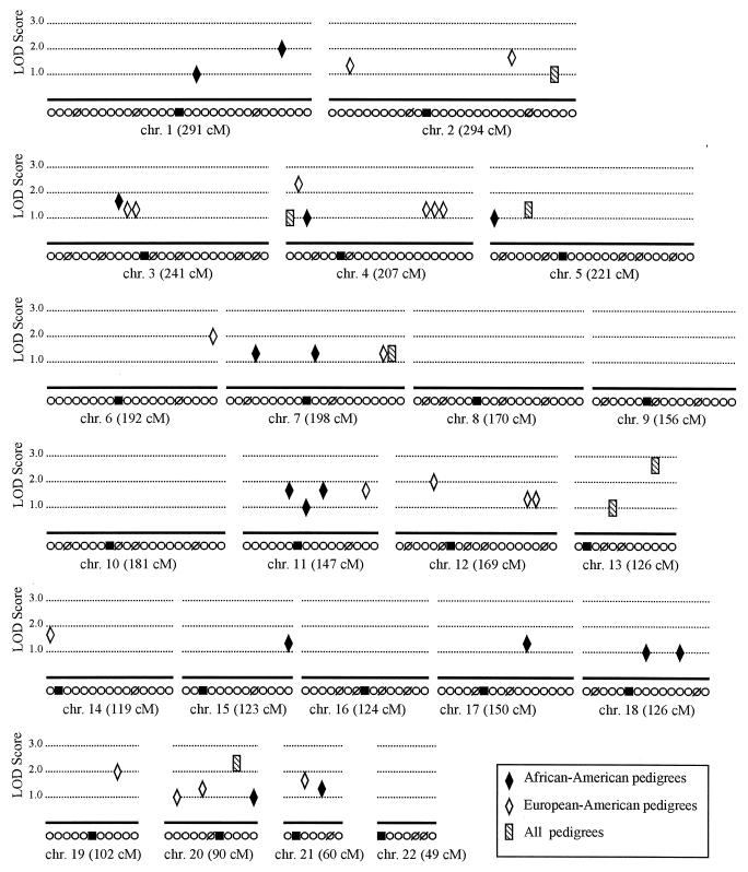
Throughout the somatic chromosomes, 42 markers generated screening lod scores >1 in one of the two racial groups or in the entire pedigree collection (Fig. 1). No evidence for linkage (lod >1) was found for any marker on chromosomes 8, 9, 10, 16, or 22. Maximized lod scores for 16 loci with a screening lod score >1.5 reveal multiple possible genetic effects in lupus (Table 1). Of these, markers FcγRIIA (lod = 3.45), D13s779 (lod = 2.50), and D20s481 (lod = 2.49) had the greatest effect in the combined collection of 94 pedigrees. Seven loci achieved a maximum lod score above 2.0 for the European-American pedigrees. Evidence for linkage in the African-American pedigrees was found on chromosomes 1, 3, and 11.
Figure 1.
Summary of genome-wide scan. Maximum two-point lod scores ≥1.0 obtained by using six screening models for each marker are indicated. Each ○ along the abscissa represents a genotyped marker from the version 8 Weber Screening Set. Markers from this set that have not been evaluated are indicated (Ø). Approximate locations of centromeres are represented by ■.
Table 1.
Maximized parametric analysis of potential human SLE susceptibility loci
| Lod (θ, α) | Chromosomal location | Marker | Ped Grp | Model (penetrance) | GH | ASP |
|---|---|---|---|---|---|---|
| 3.50 (0.15, 1.00) | 1q41 | D1s3462 | AA | D (100, 96) | ||
| 3.45 (0.00, 0.55)* | 1q23 | FcγRIIA | All | R (18, 1) | X | X |
| 3.37 (0.00, 1.00)* | 1q23 | FcγRIIA | AA | R (85, 60) | X | |
| 2.50 (0.00, 0.25) | 13q32 | D13s779 | All | R (100) | ||
| 2.49 (0.00, 0.23) | 20q13 | D20s481 | All | R (100) | X | X |
| 2.21 (0.10, 1.00) | 14q11 | D14s742 | EA | D (81, 1) | ||
| 2.18 (0.07, 1.00) | 4p15 | D4s403 | EA | D (37) | X | X |
| 2.15 (0.00, 0.25) | 11q25 | D11s912 | EA | R (100, 1) | X | |
| 2.10 (0.00, 0.35) | 11q14–23 | D11s2002 | AA | D (100, 68) | X | |
| 2.09 (0.00, 1.00) | 2q32 | D2s1391 | EA | D (21) | X | X |
| 2.05 (0.00, 0.19) | 19q13 | D19s246 | EA | R (100) | ||
| 2.04 (0.00, 0.30) | 6q26–27 | D6s1027 | EA | D (98) | ||
| 2.04 (0.25, 1.00) | 1q31 | lamc1 | All | D (95, 75) | X | |
| 2.01 (0.00, 0.30) | 12p12–11 | D12s1042 | EA | R (65) | X | X |
| 1.88 (0.00, 0.62) | 21q21 | D21s1437 | EA | D (61) | X | |
| 1.87 (0.20, 1.00) | 11p13 | D11s1392 | AA | D (98, 68) | ||
| 1.68 (0.10, 0.59) | 3p21 | D3s1766 | AA | R (100) |
Penetrances of the dominant (D) or recessive (R) screening models were maximized for loci with lod > 1.5 and are given in parentheses as one value for both females and males or two values (female, male penetrance). The pedigree group (Ped Grp) is listed for African-American (AA), European-American (EA), or the entire pedigree collection (All). The maximum lod score is followed by θ, the recombination fraction and α, the proportion of linked pedigrees. Markers where nonparametric multipoint linkage (NPL statistic) from genehunter-plus (GH) or where affected sib-pair (ASP) analysis supported linkage at P < 0.05 are indicated (X).
Maximized models for both All and AA are presented for FcγRIIA.
Evidence for Linkage to Chromosome 1q.
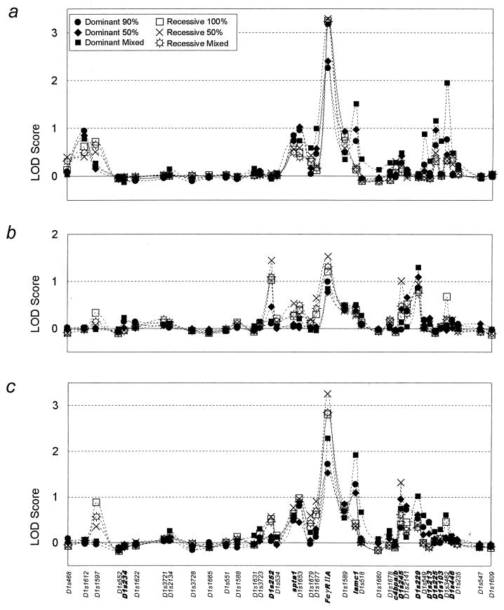
We considered the Ig Fc receptor genes at chromosome 1q23 to be the best candidates for linkage based on data in both humans and murine lupus models (4–6, 13, 22, 23). Also, linkage at 1q41 had been reported (12). The maximum two-point lod scores obtained by using the six screening models shows a major effect at FcγRIIA in the African-American pedigrees (Fig. 2a). Indeed, all of the recessive models tested and the dominant model with mixed penetrance (92% female and 49% male) obtained lod >3.0. Further attempts to maximize the best recessive screening model in the African-Americans yielded a final lod score of 3.37 with 85% penetrance in females and 60% penetrance in males at no recombination (θ = 0) with 100% homogeneity (α = 1.0) (Table 1).
Figure 2.
Screening lod scores for chromosome 1. Six inheritance models were evaluated for linkage at 39 loci from chromosome 1. The maximum lod score for each model is plotted as indicated against each marker positioned according to genetic map distances in cM (total of 291 cM). Additional genotyped markers not included in the version 8 Weber Screening Set are shown in bold. Analysis of (a) 31 African-American pedigrees containing 72 affecteds of 166 subjects, (b) 55 European-American pedigrees containing 129 affecteds of 320 subjects, and (c) the entire collection of 94 pedigrees containing 533 individuals, including eight pedigrees of predominantly Hispanic, Asian, Middle Eastern, or Native American origin.
When the parametric algorithm from genehunter-plus was applied to these data at FcγRIIA, a maximum two-point lod score of 3.93 was obtained in the African-American pedigrees for a dominant model with 99% female and 11% male penetrance. The difference in the maximum lod scores obtained in the African-American pedigrees by the different analytic methods is explained by the elimination of certain unaffected family members of a single large pedigree because of processing limitations of genehunter-plus. An affecteds-only analysis of the African-American pedigrees in linkage produced a lod score of 3.22 under the recessive model, suggesting that the effect observed at FcγRIIA for this mode of inheritance is largely from the African-American SLE affecteds.
Affected sib-pair analyses also support linkage at 1q23 (Table 2). Support for a genetic effect in this region covers nearly 40 cM with the most significant sharing at FcγRIIA (P = 0.0003 for all 78 sib-pairs) where the African-American sib-pairs make the major contribution. These results are also consistent with the possibility that multiple loci are linked with lupus between 1q21 and 1q31 (Fig. 2, Tables 1 and 2).
Table 2.
Affected sib-pair analysis near 1q23 (1q21–1q31)
| Marker | Inter-marker distance, cM | African-American, n = 23 | European-American, n = 48 | All pedigrees, n = 78 |
|---|---|---|---|---|
| SPTA1 | 0.28 | 0.50 | 0.041 | |
| 1 | ||||
| D1s1653 | 0.015 | 0.42 | 0.047 | |
| 8 | ||||
| D1s1679 | 0.0007 | 0.21 | 0.0042 | |
| 3 | ||||
| D1s1677 | 0.0036 | 0.30 | 0.035 | |
| 7 | ||||
| FcγRIIA | 0.0005 | 0.016 | 0.0003 | |
| 13 | ||||
| D1s1589 | 0.029 | 0.038 | 0.008 | |
| 7 | ||||
| lamc1 | 0.036 | 0.056 | 0.016 |
All neighboring loci of FcgRIIA with affected sib-pair sharing P values <0.05 are shown. Distances between the markers spanning this region are given in cM and were estimated from the sex average map distances supplied by Research Genetics for the version 8 Weber Screening Set and the Genome Database (http://gdbwww.gdb.org).
Evidence for linkage at the FcγRIIA locus in the European-American pedigrees was more modest, with a maximum two-point lod score of 1.52 by using a recessive model with 50% penetrance (θ = 0 and α = 0.44). All pedigrees combined yielded a screening lod score of 3.26 under the recessive model with 50% penetrance (θ = 0 and α = 0.48). The maximized lod score for all pedigrees was 3.45; however, the decreased penetrance (18% female and 1% male) and homogeneity (α = 0.55) of this recessive model suggest the effect is mostly related to the contribution of the African-American pedigrees.
A second chromosome 1 effect was found at D1s3462 mapped to 1q41, where the African-American pedigrees obtained a screening lod of 1.95 (Fig. 2a). When maximized, this effect arguably became the highest lod score of the entire study at 3.50 by using a dominant model (Table 1). This marker is 16 cM telomeric to the effect previously reported for D1S229 (12), where the highest parametric screening lod score in our study occurred in the European-Americans at 1.29 (Fig. 2b).
DISCUSSION
Results presented herein support the genetics of lupus being complex and differing among racial groups. Of the 16 potential SLE loci with lod > 1.5 we describe, only three result from the combined pedigrees rather than European-American or African-American pedigrees separately (Table 1). Indeed, the similarities and differences between this genome scan and the one simultaneously reported by Gaffney et al. (24) offer important insights. Their pedigree collection is almost completely composed of European-Americans. Nevertheless, our results are largely supported by those obtained by Gaffney et al. (24). Of the 16 possible effects presented in Table 1, eight have nonparametric linkage scores (Zlr) above 1.0 in one of the nearest neighboring markers in their study, and include D1s3462, D20s481, D14s742, D4s403, D11s2002, D2s1391, lamc1, and D3s1766.
Identification of potential SLE susceptibility loci in humans offers the opportunity for comparison with data from murine models of SLE. The majority of murine SLE susceptibility loci have been mapped in New Zealand hybrid models, with at least 12 located outside the interval containing the murine major histocompatibility complex, H-2. Three regions commonly identified by linkage in the New Zealand models are mapped to murine chromosomes 1, 4, and 7 (13, 25, 26). The murine chromosome 1 loci, Sbw1, Sle1, and Lbw7, are of particular interest. These loci map to a region syntenic with human chromosome 1q23–1q31 that contains FcγRIIA, the locus with the most convincing effect for linkage in our data. The murine chromosome 4 loci, Sle2, nba1, and Lbw2, map to human syntenic regions that include chromosomes 1p32–36 and 9p21, neither of which show evidence for linkage in our data. Linkage of SLE-associated phenotypes to murine chromosome 7 loci, Sle5, Sle3, and Lbw5, span a region with homology to human chromosomes 19q13, 11p15, and 15q11 and includes the D19s246 marker that gave a lod score of 2.05 in our data. Genetic susceptibility to SLE in both murine models and humans obviously is complicated, and linkage studies have yet to identify any particular susceptibility genes for lupus in either species. Whether or not both species share any of the same susceptibility genes for lupus can be known only after the genes are identified.
The linkage at FcγRIIA has potential to be the actual polymorphism responsible for the genetic effect. This biallelic marker encodes for an arginine to histidine change at position 131 of the FcγRIIA that alters receptor capacity to bind human IgG2 and, perhaps, influences clearance of immune complexes (4). Significant evidence for association was not present in African-Americans when unaffected family members or siblings were used as controls (χ2 = 0.5 and 1.7, respectively, P > 0.05). However, this result has not been adjusted for decreased penetrance and heterogeneity. Other candidates nearby that have been implicated in lupus include FcγRIIIB, FcγRIIIA, and the ζ chain of the T cell receptor (22, 23, 27).
In conclusion, results from this genome scan demonstrate evidence for genetic linkage with SLE in African-Americans at 1q23 in a region syntenic to genetic effects also found in murine models of lupus (13, 25, 26) and at 1q41. Multiple loci throughout the genome are virtually certain to be involved in the genetics of SLE and racial origin is likely to influence the specific nature of these effects. Understanding the complex genetics of lupus and, eventually, explaining the biology of lupus undoubtedly will provide important opportunities for improving diagnostic methods, treatment and, perhaps, even strategies to prevent this genetically complicated autoimmune disorder.
Acknowledgments
We thank the patients and their families for their invaluable contribution and the innumerable friends who have referred pedigrees to us. The contributions of Ida Adams, Diana Bozalis, Kim Blythe, Tamara Filer, Teresa Hall, Sharon Johnson, Joyce Mauldin, Allen Molloy, Kevin Schoenhals, Stephanie Susedik, Ross Thanscheidt, and members of the Management Information Systems department at the Oklahoma Medical Research Foundation are appreciated. We thank Dr. Tim Behrens for sharing data before publication (24) and Dr. Jane Olson for critical review of the manuscript. This work was supported by the National Institutes of Health (AR42460, AI24717, AR-5-2221, AR42474, AI31584, AR01981, and AR38889), the Glenn W. Peel Foundation, the Leta McFarlin Chapman Trust, and the Oklahoma Center for the Advancement of Science and Technology (contract no. 5211). Equipment funds were provided by the Oklahoma Chapter of the Lupus Foundation of America and the Oklahoma City Junior Hospitality Club. The majority of this work was conducted at the Robert H. and Lynnie Spahn Center for Genotyping at the Oklahoma Medical Research Foundation. Some genotyping was performed at the National Heart, Lung, and Blood Institute Mammalian Genotyping Service in Marshfield, WI, which is supported by N01-HV-48141. Thirty pedigrees (Cohort A) were obtained from the Lupus Multiplex Registry and Repository (AR-5-2221) (see http://omrf.ouhsc.edu/lupus).
ABBREVIATIONS
- SLE
systemic lupus erythematosus, FcγR, Fc γ receptor
- lod
log10 of the odds for linkage
References
- 1.Hochberg M C. In: Dubois’ Systemic Lupus Erythematosus. 5th Ed. Wallace D J, Hahn B J, editors. Baltimore: Williams & Wilkins; 1997. pp. 49–65. [Google Scholar]
- 2.James J A, Kaufman K M, Farris A D, Taylor-Albert E, Lehman T J, Harley J B. J Clin Invest. 1997;100:3019–3026. doi: 10.1172/JCI119856. [DOI] [PMC free article] [PubMed] [Google Scholar]
- 3.Arnett F C. In: Dubois’ Systemic Lupus Erythematosus. 5th Ed. Wallace D J, Hahn B J, editors. Baltimore: Williams & Wilkins; 1997. pp. 77–117. [Google Scholar]
- 4.Salmon J E, Millard S, Schachter L A, Arnett F C, Ginzler E M, Gourley M F, Ramsey-Goldman R, Peterson M G, Kimberly R P. J Clin Invest. 1996;97:1348–1354. doi: 10.1172/JCI118552. [DOI] [PMC free article] [PubMed] [Google Scholar]
- 5.Duits A J, Bootsma H, Derksen R H, Spronk P E, Kater L, Kallenberg C G, Capel P J, Westerdaal N A, Spierenburg G T, Gmelig-Meyling F H, van de Winkel J G. Arthritis Rheum. 1995;38:1832–1836. doi: 10.1002/art.1780381217. [DOI] [PubMed] [Google Scholar]
- 6.Wu J, Edberg J C, Redecha P B, Bansal V, Guyre P M, Coleman K, Salmon J E, Kimberly R P. J Clin Invest. 1997;100:1059–1070. doi: 10.1172/JCI119616. [DOI] [PMC free article] [PubMed] [Google Scholar]
- 7.Schur P H. In: Dubois’ Systemic Lupus Erythematosus. 5th Ed. Wallace D J, Hahn B J, editors. Baltimore: Williams & Wilkins; 1997. pp. 245–261. [Google Scholar]
- 8.Arnett F C, Shulman L E. Medicine. 1976;55:313–322. doi: 10.1097/00005792-197607000-00003. [DOI] [PubMed] [Google Scholar]
- 9.Buckman K J, Moore S K, Ebbin M, Cox M B, Dubois E L. Arch Intern Med. 1978;138:1674–1676. [PubMed] [Google Scholar]
- 10.Deapen D, Escalante A, Weinrib L, Horwitz D, Bachman B, Roy-Burman P, Walker A, Mack T M. Arthritis Rheum. 1992;35:311–318. doi: 10.1002/art.1780350310. [DOI] [PubMed] [Google Scholar]
- 11.Risch N, Merikangas K. Science. 1996;273:1516–1517. doi: 10.1126/science.273.5281.1516. [DOI] [PubMed] [Google Scholar]
- 12.Tsao B P, Cantor R M, Kalunian K C, Chen C J, Badsha H, Singh R, Wallace D J, Kitridou R C, Chen S L, Shen N, et al. J Clin Invest. 1997;99:725–731. doi: 10.1172/JCI119217. [DOI] [PMC free article] [PubMed] [Google Scholar]
- 13.Drake C G, Rozzo S J, Vyse T J, Palmer E, Kotzin B L. Immunol Rev. 1995;144:51–74. doi: 10.1111/j.1600-065x.1995.tb00065.x. [DOI] [PubMed] [Google Scholar]
- 14.Bias W B, Reveille J D, Beaty T H, Meyers D A, Arnett F C. Am J Hum Genet. 1986;39:584–602. [PMC free article] [PubMed] [Google Scholar]
- 15.Tan E M, Cohen A S, Fries J F, Masi A T, McShane D J, Rothfield N F, Schaller J G, Talal N, Winchester R J. Arthritis Rheum. 1982;25:1271–1277. doi: 10.1002/art.1780251101. [DOI] [PubMed] [Google Scholar]
- 16.Cottingham R W, Jr, Idury R M, Shaffer A A. Am J Hum Genet. 1993;53:252–263. [PMC free article] [PubMed] [Google Scholar]
- 17.Shaffer A A, Gupta S K, Shriram K, Cottingham R W., Jr Hum Hered. 1994;44:225–237. doi: 10.1159/000154222. [DOI] [PubMed] [Google Scholar]
- 18.Terwilliger J D. Am J Hum Genet. 1995;56:777–787. [PMC free article] [PubMed] [Google Scholar]
- 19.Terwilliger J D, Ott J. Handbook of Human Genetic Linkage. Baltimore: Johns Hopkins Univ. Press; 1994. pp. 224–226. [Google Scholar]
- 20.Kruglyak L, Daly M J, Reeve Daly M P, Lander E S. Am J Hum Genet. 1996;58:1347–1363. [PMC free article] [PubMed] [Google Scholar]
- 21.Kong A, Cox N J. Am J Hum Genet. 1997;61:1179–1188. doi: 10.1086/301592. [DOI] [PMC free article] [PubMed] [Google Scholar]
- 22.Clark M R, Liu L, Clarkson S B, Ory P A, Goldstein I M. J Clin Invest. 1990;86:341–346. doi: 10.1172/JCI114706. [DOI] [PMC free article] [PubMed] [Google Scholar]
- 23.Enkel B, Jung D, Frey I. Eur J Immunol. 1991;21:659–663. doi: 10.1002/eji.1830210318. [DOI] [PubMed] [Google Scholar]
- 24.Gaffney P M, Kearns G M, Shark K B, Ortmann W A, Selby S A, Malmgren M L, Rohlf K E, Ockenden T C, Messner R P, King R A, et al. Proc Natl Acad Sci USA. 1998;95:14875–14879. doi: 10.1073/pnas.95.25.14875. [DOI] [PMC free article] [PubMed] [Google Scholar]
- 25.Morel L, Rudofsky U H, Longmate J A, Schiffenbauer J, Wakeland E K. Immunity. 1994;1:219–229. [PubMed] [Google Scholar]
- 26.Kono D H, Burlingame R W, Owens D G, Kuramochi A, Balderas R S, Balomenos D, Theofilopoulos A N. Proc Natl Acad Sci USA. 1994;91:10168–10172. doi: 10.1073/pnas.91.21.10168. [DOI] [PMC free article] [PubMed] [Google Scholar]
- 27.Liossis S N, Ding X Z, Dennis G J, Tsokos G C. J Clin Invest. 1998;101:1448–1457. doi: 10.1172/JCI1457. [DOI] [PMC free article] [PubMed] [Google Scholar]


