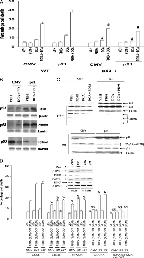
FIGURE 2.
CDK inhibitors enhance p53 expression and promote apoptosis in part via elevated BH3 domain protein expression. Primary hepatocytes were isolated as described under “Experimental Procedures.” Cells were treated with vehicle (VEH) control (DMSO), DCA (50 μm; 150 μm), MEK1/2 inhibitor (PD98059, PD98, 50 μm; PD184352, PD184, 2 μm), or both agents combined, as indicated in each panel. A, in triplicate wild type (WT) and p53–/– mouse hepatocytes were infected 4 h after plating to express nothing (vector, CMV) or p21. Cells were treated 24 h after plating with vehicle, PD184352, DCA, or the agents in combination, and 6 h later cells were isolated and spun onto glass slides for determination of apoptosis as described under “Experimental Procedures.” (n = 3 studies, ±S.E.). #, p < 0.05 apoptosis value less than corresponding value in wild type cells. B, primary rat hepatocytes were infected to express nothing (vector, CMV) or p21 or p27, as indicated. Cells were treated 24 h after plating with vehicle or with PD184352 and DCA and isolated 2 h after treatment. Cells lysed and then were separated into cytosolic and nuclear fractions as noted under “Experimental Procedures.” B, levels of p53 in each fraction were determined in parallel with a protein loading control (n = 2). C, upper section, primary mouse hepatocytes (p21–/–) were infected to express nothing (vector, CMV) or p21, as indicated, and incubated for 24 h. Twenty four hours after infection cells were treated with vehicle (VEH), DCA, PD98059, or both agents combined. Six hours after the addition of DCA/MEK1/2 inhibitor, cells were lysed in SDS-PAGE running buffer, and total cell lysates were subjected to SDS-PAGE and immunoblotting. Data are from a representative experiment (n = 5). Overexpression of p21 for 24 h enhanced total p53 expression in untreated wild type and p21–/– hepatocytes by 3.5 ± 0.4- and 4.6 ± 0.7-fold (S.E.; p < 0.05; n = 5), respectively. Overexpression of p21 reduced total “p90” MDM2 expression in untreated p21–/– hepatocytes by 37 ± 6% (S.E.; p < 0.05; n = 5). Lower section, primary mouse hepatocytes in triplicate were infected to express nothing (vector, CMV) or p21, as indicated and incubated for 24 h. Cells were isolated, lysed, and p53 immunoprecipitated from the lysates. Immunoblotting was performed to determine the amount of ubiquitinated p53 (n = 2). D, primary rat hepatocytes 4 h after plating were transfected with scrambled siRNA (siSCR) or siRNA molecules to knock down PUMA, BAX, or NOXA (or a combination of these siRNA molecules, as indicated). Thirty six hours after transfection, cells were treated with vehicle, PD184352, DCA, or the agents in combination, and 6 h later cells were isolated and spun onto glass slides for determination of apoptosis as described under “Experimental Procedures” (n = 3 studies, ±S.E.). % p < 0.05 apoptosis value less than corresponding value in siSCR cells; %, p < 0.01 apoptosis value less than corresponding value in siSCR cells; &, p < 0.05 apoptosis value greater than corresponding value in siBAX or siPUMA cells. GAPDH, glyceraldehyde-3-phosphate dehydrogenase.

