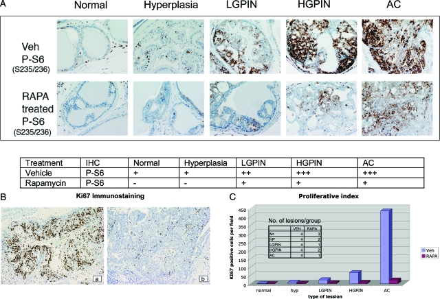
Figure 8.
Rapamycin treatment of prostate lesions. Prostate sections from Pten +/− mice were stained with antibodies to phosphorylated S6 (P-S6: S235/236) (A) and Ki-67 (B). Representative staining pattern in mice treated with vehicle versus rapamycin (RAPA) for phospho-S6 (S235/236) (A) and Ki-67 (B) in vehicle (a) versus rapamycin (b) treated mice. Phospho-S6 positivity (A) was quantified by the following criteria: −, no staining; +, weak staining; ++, moderate staining; +++, strong staining. Sections stained with Ki-67 (C) were quantified as number of positive cells per field (magnification, ×10; n = 2–5 fields examined).

