Abstract
Objective
To determine the pattern of stability changes as a reflection of early healing around single-stage roughened-surface implants in humans utilizing resonance frequency analysis (RFA).
Materials and Methods
Hundred twenty-five patients who demanded dental implants were treated with two different implant (Nobel Biocare Replace™ and Strumman™ ITI) systems. Bone type was classified into four groups. RFA was used for direct measurement of implant stability on the day of implant placement and consecutively at 14, 30 and 60 days after placement. The data were analyzed with Student t test and regression analysis.
Results
Three-hundred four roughened surface implants placed in the maxilla and mandible were evaluated. In Replace™ implants the lowest mean stability measurement was at 30 days for all bone types and the stability did not change significantly in any of the bone types (p>0.05). ITI™ implants demonstrated the lowest stability at 60 days for type 1 and 30 days and baseline for type 2, 3 and 4 bones. In addition, there was significant differences in implant stability between bone types 1 and 4 (P<0.001), 2 and 3 (p<0.05), and bone types 3 and 4 (P=0.07) at all aforementioned times in ITI™ implants. In Replace™ implants, regarding the implant diameter, contrary to ITI implants, no significant stability changes were detected (p>0.05). No significant difference was observed regarding gender, age and lengths in both systems.
Conclusion
In comparison to ITI™ implants, Replace™ implants revealed no significant difference in the pattern of stability changes among different bone types.
Keywords: Dental Implants, Data Interpretation, Dental Prosthesis, Dental Prosthesis, Implant-Supported, Bone Density
INTRODUCTION
Endosseous implants are increasingly being used in maxillofacial, dental and orthopedic surgery [1]. Implant failure and loss may have a number of causes, including factors related to the design of the implant system, a poor surgical technique, excessive loading or unfavorable host reaction. It has been clearly demonstrated that implant-retained prosthesis may be placed successfully and linger functional for many years [7]. There is adequate data suggesting excessive mechanical stresses and poor initial stability at placement as the causes of early failure of implants [2].
Proper primary stability and postponing loading of the implant to about 3–6 months after the surgery have long been considered as the “conditio sine qua non” to provide the required situations for implant osseointegration. However, the necessity to wait that long before loading an implant has been based upon clinical experience and thoughts rather than being evidence based [3,4]. Adequate stability of an implant in the bone is an essential matter for favorable repair process, bone formation and also distribution of mastication forces. Primary stability is critical and believed to be influenced by length, geometry, bone-to-implant contact area, cortical to trabecular bone ratio and the placement technique [3]. Secondary stability is a consequence of secondary woven and lamellar bone formation [1,2,5–9]. Advances in implant dentistry towards improved osseointegration and accelerated loading protocols are based on enhanced implant designs and surface features along with a better understanding of the restorative options for such approaches [9]. High success rates in implant patients following conventional loading protocols can similarly be achieved with the early and immediate loading protocols in appropriately selected cases [10–15]. Accordingly, application of a simple, clinically feasible, noninvasive test to assess implant stability and osseointegration is believed to be highly desirable [16]. The most widely used clinical technique in this matter is the radiographic method which is criticized for being two dimensional and difficult to standardize. It seems a quantitative reproducible method for evaluating the stability of solid dental implants in clinic and it may be helpful. Manual percussion is the simplest form of transient vibration analysis [17]. The Periotest (NIVA, Charlette, NC) is another transient excitation tool that could not be a trustable device due to lack of sensitivity in implant stability measurement [9].
Around the mid-90s, Meredith reported the use of a transducer directly attached to an implant body or to the abutment to achieve a clinical, noninvasive measure of stability to presume osseointegration of implants expressed as Implant Stability Quotient (ISQ) units, which were scaled from 1 to 100 [18–20]. It has been demonstrated that this device is efficacious in assessing changes in interfacial stiffness during osseous healing, osseointegration and the supracrestal dimensions of bone-implant interface[1,3,19–20]. Histomorphometric studies suggest that RF values correlate well with levels of bone-implant interface [21–22]. These findings support the use of RFA in assessing changes in the bone healing and osseointegration processes following implant placement. Although ISQ values cannot be directly linked to actual cellular activities, they provide a reproducible assessment of the condition of the bone-implant interface [18–21].
Therefore, they can be used to monitor and control the biologic conditions of bone-implant interface. More recently, this commercial instrument was modified. This device is now wireless and have an aluminum peg (smart peg) that attached on to the implant, utilizing aluminum peg (smart peg) attached to the implant or the abutment, utilizing electromagnetic pulses across a frequency range and then analyzing the response of the smart peg. The result is two-dimensional through a planar measurement instead of the linear one used with the previous device. This improved technology presents more reproducible and representative results around the implant (360°) via a mathematical algorithm. The aim of this clinical study was to determine the primary stability and to assess changes in implant stability during the early phase of healing, applying the noninvasive RFA technique with the use of a new device Osstell™ Mentor (Osstell AB, Gamlestadsvägen, Göteborg, Sweden) in an attempt to determine the best time for loading of roughened-surface Nobelbiocare Replace Select tapered Tiunite® implants (Nobel Biocare, Guttenberg, Sweden) and ITI SLA® (sandblasted, large-grit, and acid-etched) solid-screw implants (Straumann, BASEL, Switzerland) with different lengths and diameters, placed in different quality types of bone through single-stage surgery.
MATERIALS AND METHODS
This clinical trial was designed to assess implant stability changes with an RF analyzer (Osstell™ Mentor; Integration Diagnostics AB, Sweden) in two different implant systems with different designs in the critical early healing phase (first 60 days after implant placement).
The samples consisted of sixty-eight 18 to 70-year-old patients for each implant system (30 males, 38 females), treated in the Department of Implantology in Tehran University of Medical Sciences during the past two years. Eligible subjects were selected based on inclusion/exclusion criteria (Table 1).
Table 1.
Patient Inclusion/Exclusion Criteria
|
Nobelbiocare Replace Select Tiunite® Tapered implants were 10 mm (n=60) and 13 mm (n=91) long with different diameters of 3.5 mm (n=26), 4.3 mm (n=89) and 5.0 mm (n=38); ITI SLA solid-screw implants were 10 mm (n=75) and 12 mm (n=76) long with different diameters of 3.3 mm narrow body (NB) (n=17), 4.1 mm regular body (RB) (n=95) and 4.8 mm wide body (WB) (n=39). The effect of implant length, diameter, location, bone type, patient age and gender was evaluated on implant stability expressed as ISQ units.
Data were analyzed by descriptive statistics, Student’s t-test and ANOVA using SPSS software.
Clinical protocols
After informed consent forms were signed by the patients, all the implants [61 replace implants (39.89%) in the maxilla and 92 (60.11%) in the mandible and 70 ITI implants (46%) in the maxilla and 81 (54%) in the mandible] were placed using a non-submerged technique, following the manufacturer’s instructions (Table 2).
Table 2.
Distribution of Implants According to Insertion Sites
| Site | Canine | Premolar | Molar |
|---|---|---|---|
| Implant Type | |||
| Replace | 11 (7.20%) | 57 (37.25%) | 85 (55.55%) |
| ITI | 48 (31.78 %) | 77 (51%) | 26 (17.22%) |
Bone density was categorized as type I, II, III or IV at the time of surgery according to Lekholm and Zarb index [23] in 1985 that was approved by the judgment of the tactile sense of the surgeon (Table 3).
Table 3.
Distribution and Number of Placed Implants According to Bone Density
| Bone Type | Number of Replace Implants | Number of ITI Implants |
|---|---|---|
| I | 16 (10.46%) | 5 (3.32%) |
| II | 76 (49.67%) | 110 (72.84%) |
| III | 53 (34.65%) | 27 (17.88%) |
| IV | 8 (5.22%) | 9 (5.96%) |
Immediately after implant placement and at 14-, 30-, and 60-day intervals post-operatively the proper smart peg for each implant (ITI; Types 4 & 17, Nobelbiocare Replace; type 13) was screwed onto the fixture and the implant stability was measured by the RF analyzer and expressed in ISQ units.
An increased ISQ value indicated greater stability than before, whereas decreased values indicated a decrease in implant stability. Readings were performed three times for each implant; one from the top, one from the buccal and one from the lingual side of the smart peg; then the mean was calculated. To reduce observer bias, the previous recordings on the implant were made inaccessible prior to RFA measurement.
RESULTS
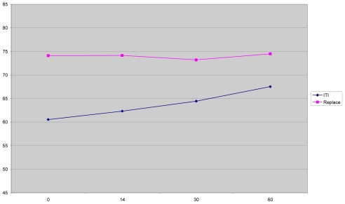
None of the inserted implants failed. ISQ values showed a high level of reproducibility, with an accuracy of ± 2 units. According to the ISQ values, the following results were obtained. In general, ITI implants showed an increase in ISQ values with time, but replace implants remained rather constant.
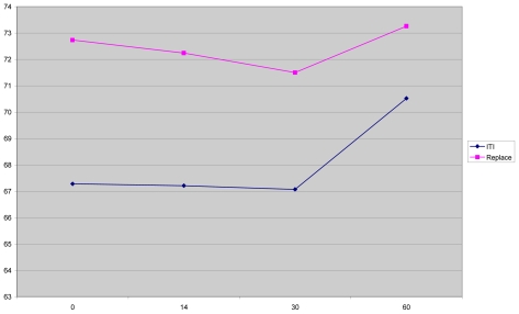
The mean ISQ values for replace implants were higher than those for ITI implants at all times, the difference being significant at all the measurement intervals (p<0.05) (Diagram 1).
Diagram 1.
Primary stability and pattern of stability changes according to mean ISQ values in two different implant systems. It is obvious that tapered implants showed higher stability than parallel ones.
It was observed that in the replace system, more implants with higher ISQ values were present at baseline and at 14- and 30-day intervals (p<0.001). However, no significant differences were observed between the two groups at the 60-day interval (p>0.05). The 10- and 13-mm-long replace implants showed relatively the same ISQ values at all the four measurement intervals (p>0.05), whilst in the ITI implants differences were observed at the 14-day interval and 12-mm-long implants, demonstrating higher stability than 10-mm-long ones; the difference was not significant either (p>0.05). The results showed that ISQ values for 10- and 13-mm-long replace implants were higher than those for 10- and 14-mm-long ITI implants and the values remained rather constant for 10- and 13-mm-long replace implants. Regarding different diameters in ITI implants, the greatest difference was seen at the 30-day interval between 4.8 and 4.1-mm (p>0.05) and also between 4.1- (RB) and 3.3-mm-diameter implants (NB) (p> 0.05). Although 4.8-mm-diameter implants had higher stability compared to 4.1- and 3.3-mm-diameter ones, there was no significant difference in ISQ values with regard to different implant diameters (p>0.05).
Concerning replace tapered implants at all the measurement t intervals, it was noted that the greater the implant diameter, the greater the ISQ value and consequently, the greater the stability (p<0.05); however, as previously discussed, such an increase was not observed in ITI implants.
Although ISQ values for replace implants were found to be more than those for ITI implants with the same diameter, ITI NB and replace NP implants showed relatively equal stability. Regarding type I bone, patterns of stability changes were different for the two implant systems.
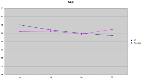
At first, both ITI and replace tapered implants demonstrated high primary stability, with ITI being a little more stable, but as time went by, at the 30-day interval, replace implants showed a non-significant increase (p>0.05) (Diagram 2).
Diagram 2.
Primary stability and pattern of stability changes in two different implant designs in type I bone according to mean ISQ values. Stability levels in this bone type are in the higher limits in both systems but the pattern of stability changes is different.
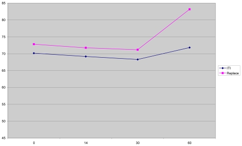
Regarding type II bone, the patterns were rather the same with no significant differences; however, replace tapered implants were slightly more stable (p>0.05) (Diagram 3). Regarding ITI implants, no significant changes in stability from baseline readings were observed in type II bone (p>0.05); however, these changes in other bone types were significant (p<0.05). Contrary to what was seen in ITI implants, these changes were not significant in all bone types in replace implants (p>0.05) and these implants proved more stable compared to ITI implants, particularly when placed in type III and type IV bone (Diagrams 2–5).
Diagram 3.
Primary stability and pattern of stability changes in two different implant designs in type II bone according to mean ISQ values. It is interesting that pattern of stability changes is fairly the same in both implant designs in this bone type.
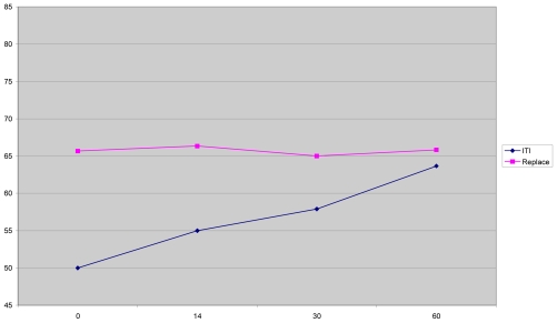
Diagram 5.
Primary stability and pattern of stability changes in two different implant designs in type IV bone according to mean ISQ values. It is interesting that contrary to ITI implants, pattern of stability changes, despite poor bone quality, is fairly similar to other bone types in Replace tapered implants.
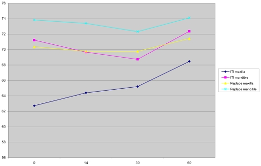
Generally, in both systems, implants placed in the lower jaw were more stable than those in the upper jaw and contrary to what was seen in the maxilla, the pattern of stability changes in the mandible were similar in both systems (p<0.05) (Diagram 6).
Diagram 6.
Implant stability changes according to jaw position. In both systems, ISQ values showed higher values in the mandible compared to the maxilla in all the measurement intervalss during this study.
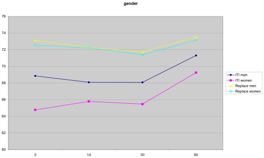
The implant stability was somewhat higher in men, but generally it appeared that gender and age did not have a significant effect on the results (p>0.05) (Diagram 7).
Diagram 7.
Implant stability changes according to gender. In general ISQ values showed higher values in men compare to women.
DISCUSSION
RFA offers a noninvasive stability measurement in the periphery (360º) of implants with Osstell ™ Mentor device. As the smart peg and implant structure are constant, any changes in RFA reveals changes in implant-bone interface, either in quality or quantity. In this study, implant stability was measured at four intervals for each implant; namely, immediately after placement as the primary stability, day 14 as the time for the newly formed woven bone around the implant, day 30 as the time when the woven bone lines most parts of the implant surface and the start of the remodeling phase, and finally day 60 as the time at which the implant surface is lined with lamellar bone as accepted in the literature for loading [24–26]. The present study seems to have allowed a proper evaluation of stability changes in both implant systems during the early stages of healing, leading to valuable and interesting biological and clinical insights. It was noted that the number of implants and mean ISQ values were higher in replace system compared to ITI system at all the measurement intervals. Such a difference might be attributed to different designs and geometry. ITI implants are parallel-sided, differing from replace implants, which are tapered. O’Sullivan [27] reported in 2000 that parallel-sided implants can reach their maximum stability if the coronal part is placed in cortical bone; however, tapered implants apply a lateral compression force to the surrounding walls while being placed, making them more stable at the time. Their higher installation torque value (ITV) compared to the parallel-sided implants might be attributed to the same factor emphasized by Molly in 2006 [28].
Accordingly, the distance between the threads in ITI implants is about twice that in replace implants; thus, incorporating more threads per surface area can make them more stable, especially at the time of installation.
Tiunite coating on replace implants tends to increase the surface area by 37%, while SLA coating on ITI implants accomplishes the same task by 33%; this little difference does not seem to contribute significantly to the dissimilar implant stability during the early healing phase[29–30].
As a rule, although implant surface condition is important during the healing phase, the implant design is the major feature of an implant body during the loading period [31].
Mean implant stability levels were rather equal at baseline, 14- and 30-day intervals and higher at the 60-day interval in each system individually, with no significant differences at the 60-day interval in either of the implant systems, which might be explained by remodeling process at the bone-implant interface and the increase in bone-implant contact area as time goes by [32–34]. The recent finding can also be explained with regard to Carlos Aparicio’s theory in 2005, [34] stating that stability of implants gets closer to each other with time due to bone density homogeneity.
Implant length was not found to be a significantly effective factor influencing stability in both implant systems, which is consistent with the results of other studies [35–38]. Many previous studies have also reported that the success rate and/or the resorption rate of bone do not undergo changes when different implant lengths are used. [34,39,40]. It is probable that once the bone-implant contact is established at the marginal level and the implant is firm, a 2- or 3-mm difference in length in the apical region, which is classically composed of cancellous bone, does not result in a significant increase in the overall implant stability [41]. Accordingly, it is likely that placing a great deal of emphasis on the use of the longest implant applicable is not always the best decision.
In the present study, implant diameter had a positive influence on ISQ values in both systems, which might be attributed to greater implant-bone contact area as the diameter increases. However, in ITI implants this effect was not significant compared to the replace system, which was attributed to the conical design of replace implants, applying more lateral compression force to the surrounding bone with diameter increase; therefore, providing more lateral stiffness and ISQ values. In previous studies, the relationship between implant diameter and ISQ values has been emphasized. [37,42]. Concerning bone type, in ITI implants in the present study, stability patterns in different bone types were noticeably different. Nevertheless, in replace tapered implants, the patterns in all the four bone types were fairly the same. As for bone type I in ITI implants, a rather high primary stability was noted (mean ISQ=75). The reason might be the thick cortical bone layer with a small amount of trabecular core. It might also be attributed to the press-fit of the slightly larger diameter of the implant against cut bone surface [7]. Interestingly, as time went by, the stability demonstrated a slight decline up to day 60, with mainly two possible reasons: 1. Overheating during drilling; the phenomenon is more likely to happen in type I bone than other bone types and might result in marginal bone loss and an increase in effective implant length [28–43] 2. This bone type is almost completely cortical and the capacity of regeneration is impaired because of poor blood supply [44,45]. In both systems in type II bone, a slight non-significant decline was observed in ISQ values during the first 30 days, which began to increase until day 60 afterwards. This finding confirmed the ones in Roberts’ report that bone density/quality is indeed dynamic, changing in relation to implant surface [46]. It appears that type II bone is a proper bone type for both tapered and parallel wall implants from implant stability viewpoint because of the thick cortical layer with a dense trabecular core and good blood supply. However, this non-significant decline during the first month and the subsequent increase reflect a discrepancy with the results of studies by Friberg, [47,48] which might be attributed to the effect of the rough surface coating and the subsequent reaction at the interface[9]. ITI implants in bone type III and IV exhibited considerably lower primary stabilities at the baseline compared to that in type I and type II bones, probably due to less cortical bone and the larger trabecular core with lower density. The subsequent rise in ISQ values after the baseline is consistent with the improved bone formation around the roughened implant surface [9,49]. On the other hand, it has already been shown that implants with lower ISQ values will exhibit greater increase in ISQ values with time [49,9,50,51-53-26].
In replace tapered implants, the primary stability and the pattern of stability changes in all the four bone types were fairly the same. ISQ values in bone types I, II and III were rather high (more than 70); however, in type IV bone, although ISQ values followed the same pattern, they were lower compared to other bone types due to the thinner cortical bone and the larger trabeculae.
These findings might be attributed to the geometry and tapered design of replace implants, which provide more lateral compression and stiffness, [27] compensating lower bone density. A comparison of stability patterns of mandibular and maxillary implants in both systems showed that the overall stability level was higher in the mandible. In replace implants, the similar pattern of stability changes and non-significant values between the jaws in contrast to ITI implants were attributed to the tapered geometry of these implants, which can create higher lateral compression and secure the high primary stability. These results are consistent with reported higher survival rates of implants in the mandible compared to the maxilla, [54,55] as a result of differences in bone density [48–50]. Denser bone exists in the mandible with 25–50% greater integrative success in the anterior mandible compared to the maxillary posterior region [56,43].
In general, it was noted that the denser the bone, the higher the primary stability in both systems; however, replace implants could secure the initial stability and prevail over the bone remodeling stages during the critical first two months of the osseointegration process due to their tapered design and more lateral bone compression during installation, resulting in more lateral stiffness.
As a result, when using replace implants in bone types I, II and III, bone type had no effect on ISQ values in the present study, which is an interesting finding attributable to the implant design. In vivo and histomorphometric studies have confirmed that ISQ values associate well associate well with levels of bone-implant contact area [19,20,22,23,47,48,57]. In a recent study, it was shown that the values measured by the magnetic device used in the present study correlate well with those of the electronic one; the amount measured by the former equals 8–12 units less than that measured by the latter [40]. On the other hand, studies have suggested that implants with ISQ values of more than 60 (measured by the electronic device) are eligible to undergo immediate loading as if a stable fixation exists between the bone and the implant; even minute inter-fragmentary movements can be avoided and dynamic load bearing can be withstood. Therefore, in implants with high primary stability and no significant changes with time, an immediate loading protocol can be indicated [9,11,12,57].
As a result, given the values measured for replace tapered implants in bone types I, II and III, which indicate no significant changes during the study period, it is possible to consider immediate loading for these implants; however, in type IV bone, just to be on the safe side, it would be better to consider early loading protocol because of the poor bone quality and lack of fully acceptable mean primary ISQ values (under 68). In ITI implants, it is difficult to make a firm clinical decision about the immediate loading protocol in type I bone because ISQ values slightly decreased over time. Nonetheless, ISQ values were in the higher limits (more than 65) at all intervals during the study with only a 2% change in mean ISQ value after 30 days. On the other hand, it is suggested that for implants with high primary ISQ values, decrease in implant stability during the first 3 months of healing should be supposed as a common occurrence that does not require modifications in routine follow-up procedures [48]. Due to the ISQ values in type I bone, which were over 65 with little decrease in high primary stability after two months in the present study, immediate loading in ITI SLA implants in type I bone is tempting.
However, continuous decreases in ISQ levels in type I bone and the least mean ISQ values after 60 days compared to the other three earlier measurements favor the early loading protocols. It appears that in ITI implants, proper conditions for immediate loading protocol were only seen in type II bone.
As histological bone analysis has been established as the gold standard to determine the bone type in the literature, [28] perhaps it was better for us to determine the bone type in this manner. Anyway, the large number of samples and the highly professional surgeons who were involved in this study made our results more accurate. On the other hand, a large number of previous studies have utilized the surgeon’s professional common sense to determine the bone type [9,23,38,41,42,58]. In addition, Trisi [59] showed that the surgeon’s sense can determine the bone type appropriately.
In general, this study appears to have provided valuable insights into implant stability changes in the two systems throughout the important early stages of healing. As there is a recent interest in immediate loading of single-unit restorations and none of the implants were immediately loaded in this study, a study involving monitoring of the stability patterns of single-unit, immediately loaded, roughened-surface implants would offer more results to validate our results.
The effect of splinting versus non-splinting will possibly be compared in an RFA study on immediate hybrids and immediate single-unit restorations. It would also be valuable in these studies to examine occlusal factors as potential variables in the healing process.
CONCLUSION
This study demonstrated that in parallel wall implants the primary stability and pattern of stability changes are different between different bone types, but tapered implants can inhibit decreases in primary stability in all bone types. Bone type and geometry of the implants are the most important factors for implant stability during the first 60 days of healing. In parallel and tapered wall implants, with regard to primary stability and pattern of stability changes, maybe immediate and early loading protocols are appropriate alternatives in type II and types I, II, III bone, respectively. Implant diameter was found to be ineffective in ITI parallel wall system, but in replace tapered system, wider implants were more stable. Patient sex, age and implant length were not significantly effective in implant stability according to ISQ values in either of the two systems.
Maybe future studies, examining occlusal factors as possible variables in the healing process and also evaluating the effects of splinting versus non-splinting procedures can be beneficial to a better understanding of the results of the present study.
Diagram 4.
Primary stability and pattern of stability changes in two different implant designs in type III bone according to mean ISQ values.
ACKNOWLEDGMENTS
This study was partly supported by the Dental Research Center, Tehran University of Medical Sciences. The authors would like to thank Dr. M. Khrazifard for statistical testing and his contributions to the study.
REFERENCES
- 1.Meredith N. Assessment of implant stability as a prognostic determinant. Int J Prosthodont. 1998 Sep-Oct;11(5):491–501. [PubMed] [Google Scholar]
- 2.Albrektsson T. On Long-term maintenance of the osseointegrated. Aust Prosthet J. 1993;7:15–24. [PubMed] [Google Scholar]
- 3.Schroeder A, Pohler O, Sutter F. Tissue reaction to an implant of a titanium hollow cylinder with a titanium surface spray layer. SSO Schweitz Monatsschr Zahnheilhad. 1976 Jul;86(7):713–27. [PubMed] [Google Scholar]
- 4.Branemark P-I, Hansson BO, Adell R, Breine U, Lindström J, Hallén O, et al. Osseointegrated implants in the treatment of the edentulous jaw. Experience from a 10-year period. Scand J Plast Reconstr Surg. 1977;16:1–132. [PubMed] [Google Scholar]
- 5.Friberg B, Jemt T, Lekholm U. Early failures in 4641 consecutively placed Branemark dental implants. A study from stage 1 surgery to the connection of completed prostheses. Int J Oral Maxillofac Implants. 1991 Summer;6(2):142–6. [PubMed] [Google Scholar]
- 6.Albrektsson T. Dental implants: A review of clinical approaches. Aust Prosthodon Soc Bull. 1985 Dec;15:7–25. [PubMed] [Google Scholar]
- 7.Brunski JB. Biomechanical factors affecting the bone dental implant interface. Clin Mater. 1992;10(3):153–201. doi: 10.1016/0267-6605(92)90049-y. [DOI] [PubMed] [Google Scholar]
- 8.Zarb GA, Albrektsson T. Osseointegration: A requiem for the periodontal ligament? Int J Periodont Rest Dent. 1991;11:58–91. [Google Scholar]
- 9.Barewal RM, Oates TW, Meredith N, Cochran DL. Resonance frequency measurement of implant stability in vivo on implant with a sand blasted and acid etched surface. In J Oral Maxillofac Implants. 2003 Sep-Oct;18(5):641–51. [PubMed] [Google Scholar]
- 10.Tarnow DP, Emtiaz S, Classi A. Immediate loading of threaded implants at stage 1 surgery in edentulous arches: ten consecutive case reports with 1-to 5-year data. Int J Oral Maxillofac Implants. 1997 May-Jun;12(3):319–24. [PubMed] [Google Scholar]
- 11.Chiapasco M, Gatti C. Implant-retained mandibular over dentures with immediate loading: A 3- to 8-year prospective study of 328 implants. Clin Implant Dent Relat Res. 2003;5(1):29–38. doi: 10.1111/j.1708-8208.2003.tb00179.x. [DOI] [PubMed] [Google Scholar]
- 12.Chiapasco M, Abati S, Romeo E, Vogel G. Implant-retained mandibular overdentures with Branemark System MKII implants: a prospective comparative study between delayed and immediate loading. Int J Oral Maxillofac Implants. 2001 Jul-Aug;16(4):537–46. [PubMed] [Google Scholar]
- 13.Schnitman PA, Wohrle PS, Rubenstein JE. Immediate fixed interim prostheses supported by two-stage threaded implants: methodology and results. J Oral Implantol. 1990;16(2):96–105. [PubMed] [Google Scholar]
- 14.Schnitman PA. Branemark implants loaded with fixed provisional prostheses at fixture placement: nine-year-follow-up. J Oral Implantol. 1995;21:235–45. [Google Scholar]
- 15.Schnitman PA, Wohrle PS, Rubinstein JE, DaSilva JD, Wang NH. Ten-year results for Branemark implants immediately loaded with fixed prostheses at implant placement. Int J Oral Maxillofac Implants. 1997 Jul-Aug;12(4):495–503. [PubMed] [Google Scholar]
- 16.Cochran DL, Buser D, ten Bruggenkate CM, Weingart D, Taylor TM, Bernard JP, et al. The use of reduced healing times on ITI implants with a sandblasted and acid-etched (SLA) surface: early results from clinical trials on ITI SLA implants. Clin Oral Implants Res. 2002 Apr;13(2):144–53. doi: 10.1034/j.1600-0501.2002.130204.x. [DOI] [PubMed] [Google Scholar]
- 17.Huang HM, Pan L, Lee SY, Chiu CL, Fan KH, Ho KN. Assessing the implant/bone interface by using natural frequency analysis. Oral Surg Oral Med Oral Pathol Oral Radiol Endod. 2000 Sep;90(3):285–91. doi: 10.1067/moe.2000.108918. [DOI] [PubMed] [Google Scholar]
- 18.Meredith N, Alleyne D, Cawley P. Quantitative determination of the stability of the implant-tissue interface using resonance frequency analysis. Clin Oral Implants Res. 1996 Sep;7(3):261–7. doi: 10.1034/j.1600-0501.1996.070308.x. [DOI] [PubMed] [Google Scholar]
- 19.Meredith N, Book K, Friberg B, Jemt T, Sennerby L. Resonance frequency measurements of implant stability in vivo. Clin Oral implants Res. 1997 Jun;8(3):226–33. doi: 10.1034/j.1600-0501.1997.080309.x. [DOI] [PubMed] [Google Scholar]
- 20.Meredith N, Sennerby F, Alleyne D, Sennerby L, Cawley P. The application of resonance frequency measurements to study the stability of titanium implants during healing in the rabbit tibia. Clin Oral Impl Res. 1997 Jun;8(3):234–43. doi: 10.1034/j.1600-0501.1997.080310.x. [DOI] [PubMed] [Google Scholar]
- 21.Piattelli A, Ruggieri A, Franchi M, Romasco N, Trisi P. A histologic and histomorphometric study of bone reactions to unloaded and loaded non-submerged single implants in monkeys: A pilot study. J Oral Implantol. 1993;19(4):314–20. [PubMed] [Google Scholar]
- 22.Rasmusson L, Meredith N, Cho IH, Sennerby L. The influence of simultaneous versus delayed placement on the stability of titanium implants in only bone grafts. Int J Oral Maxillofac Implants. 1999 Jun;28(3):224–31. [PubMed] [Google Scholar]
- 23.Lekholm U, Zarb GA. Patient selection and preparation. In: Branemark PI, Zarb GA, Albrektsson T, editors. Tissue integrated prosthesis: Osseointegration in clinical dentistry. Chicago: Quintessence, Publishing Co; 1985. pp. 199–208. [Google Scholar]
- 24.Berglundh T, Abrahamsson I, Lang NP, Lindhe J. De novo alveolar bone formation adjacent to endosseous implants.a model study in the dog. Clin Oral Implants Res. 2003 Jun;14(3):251–62. doi: 10.1034/j.1600-0501.2003.00972.x. [DOI] [PubMed] [Google Scholar]
- 25.Lindhe j, Berglundh T, Niklaus P. Clinical periodontology and implant dentistry. 5th ed. Blackwell; 2008. pp. 99–107. [Google Scholar]
- 26.Albrektsson T, Eriksson AR, Friberg B, Lekholm U, Lindahl I, Nevins M, et al. Histologic investigations on 33 retrieved Nobelpharma implants. Clin Mat. 1993;12(1):1–9. doi: 10.1016/0267-6605(93)90021-x. [DOI] [PubMed] [Google Scholar]
- 27.O’Sullivan D, Sennerby L, Meredith N. Measurements comparing the initial stability of five designs of dental implants: a human cadaver study. Clin Implant Dent Relat Res. 2000;2(2):85–92. doi: 10.1111/j.1708-8208.2000.tb00110.x. [DOI] [PubMed] [Google Scholar]
- 28.Molly L. Bone density and primary stability in implant therapy. Clin Oral Implants Res. 2006 Oct;17( Suppl 2):124–35. doi: 10.1111/j.1600-0501.2006.01356.x. [DOI] [PubMed] [Google Scholar]
- 29.Wennerberg A, Albrektsson T, Andersson B. An animal study of c.p. titanium screws with different surface topographies. J Materials Sci Materials in Med. 1995;6:302–9. [Google Scholar]
- 30.Wennerberg A, Ektessabi A, Albrektsson T, Johansson C, Andersson B. A 1-year follow-up of implants of differing surface roughness placed in rabbit bone. Int J Oral Maxillofac Implants. 1997 Jul-Aug;12(4):486–94. [PubMed] [Google Scholar]
- 31.Piattelli Adriano, Misch Carl E, Pontes Ana Emilia Farias, et al. Contemporary implant dentistry. 3th ed. Mosby: Elsevier; 2008. pp. 599–620. [Google Scholar]
- 32.Huwiler MA, Pjetursson BE, Bosshardt DD, Salvi GE, Lang NP. Resonance frequency analysis in relation to jawbone characteristics and during early healing of implant installation. Clin Oral Implants Res. 2007 Junb;18(3):275–80. doi: 10.1111/j.1600-0501.2007.01336.x. [DOI] [PubMed] [Google Scholar]
- 33.Abrahmsson I, Berglundh T, Linder E, Lang NP, Lindhe J. Early bone formation adjacent to rough and turned endosseous implant surfaces. An experimental study in the dog. Clin Oral Implants Res. 2004 Aug;15(4):381–92. doi: 10.1111/j.1600-0501.2004.01082.x. [DOI] [PubMed] [Google Scholar]
- 34.Aparicio C, Lang NP, Rangert B. Validity and clinical significance of biomechanical testing of implant/bone interface. Clin Oral Imp Res. 2006 Oct;17( Suppl 2):2–7. doi: 10.1111/j.1600-0501.2006.01365.x. [DOI] [PubMed] [Google Scholar]
- 35.Farzad P, Andersson L, Gunnarsson S, Sharma P. Implant stability, tissue conditions, and patients self-evaluation after treatment with osseointegrated implants in the posterior mandible. Clin Implant Dent Relat Res. 2004;6(1):24–32. doi: 10.1111/j.1708-8208.2004.tb00024.x. [DOI] [PubMed] [Google Scholar]
- 36.Bischof M, Nedir R, Szmukler-Moncler S, Bernard JP, Samson J. Implant stability measurement of delayed and immediately loaded implants during healing. Clin Oral Implants Res. 2004 Oct;15(5):529–39. doi: 10.1111/j.1600-0501.2004.01042.x. [DOI] [PubMed] [Google Scholar]
- 37.Horwitz J, Zuabi O, Peled M. Resonance frequency analysis in immediate loading of dental implants. Refuat Hapeh Vehashinayim. 2003 Jul;20(3):80–8. 104. [PubMed] [Google Scholar]
- 38.Balleri P, Cozzolino A, Ghelli L, Momicchioli G, Varriale A. Stability measurements of osseointegrated implants using Osstell in partially edentulous jaws after 1 year of loading: a pilot study. Clin Implant Dent Relat Res. 2002;4(3):128–32. doi: 10.1111/j.1708-8208.2002.tb00162.x. [DOI] [PubMed] [Google Scholar]
- 39.Hobkirk JA, Wiskott HW Working group 1. Biochemical aspects of oral implants. Consensus report of working Group. Clin Oral Imp Res. 2006 Oct;17( Suppl 2):52–4. doi: 10.1111/j.1600-0501.2006.01358.x. [DOI] [PubMed] [Google Scholar]
- 40.Misch CE, Steigenga J, Barboza E, Misch-Dietsh F, Cianciola LJ, Kazor C. Short dental implants in posterior partial edentulism: a multicenter retrospective 6-year case series study. J Periodontol. 2006 Aug;77(8):1340–7. doi: 10.1902/jop.2006.050402. [DOI] [PubMed] [Google Scholar]
- 41.Valderrama P, Oates TW, Jones AA, Simpson J, Schoolfield JD, Cochran DL. Evaluation of two different resonance frequency devices to detect implant stability: a clinical trial. J Periodontol. 2007 Feb;78(2):262–72. doi: 10.1902/jop.2007.060143. [DOI] [PubMed] [Google Scholar]
- 42.Ostman PO, Hellman M, Wendelhag I, Sennerby L. Resonance frequency analysis measurements of implants at placement surgery. Int J Prosthodont. 2006 Jan-Feb;19(1):77–83. discussion 84. [PubMed] [Google Scholar]
- 43.Misch CE. Density of bone: effect on treatment plans, surgical approach, healing, and progressive bone loading. Int J Oral Implantol. 1990;6(2):23–31. [PubMed] [Google Scholar]
- 44.Rhinelander FW. The normal circulation of bone and its response to surgical intervention. J Biomed Mater Res. 1974 Jan;8(1):87–90. doi: 10.1002/jbm.820080111. [DOI] [PubMed] [Google Scholar]
- 45.Chanavaz M. Anatomy and histophysiology of the periosteum: classification of the periosteal blood supply to the adjacent bone with 85Sr and gamma spectrometry. J Oral Implantol. 1995;21(3):214–9. [PubMed] [Google Scholar]
- 46.Roberts WE. Bone tissue interface. J Dent Educ. 1988 Dec;52(12):804–9. [PubMed] [Google Scholar]
- 47.Friberg B, Sennerby L, Meredith N, Lekholm U. A comparison between cutting torque and resonance frequency measurements of maxillary implants. A 20-month clinical study. Int J Oral Maxillofac Surg. 1999 Aug;28(4):297–303. [PubMed] [Google Scholar]
- 48.Friberg B, Sennerby L, Linden B, Grondahl K, Lekholm U. Stability measurements of one-stage Branemark implants during healing in mandibles: A clinical resonance frequency analysis study. Int J Oral Maxillofac Surg. 1999 Aug;28(4):266–72. [PubMed] [Google Scholar]
- 49.Nedir R, Bischof M, Szmukler-Moncler S, Bernard JP, Samson J. Predicting osseointegration by means of implant primary stability. Clin Oral Implants Res. 2004 Oct;15(5):520–8. doi: 10.1111/j.1600-0501.2004.01059.x. [DOI] [PubMed] [Google Scholar]
- 50.Friberg B, Sennerby L, Roos J, Lekholm U. Identification of bone quality in conjunction with insertion of titanium implants. Clin Oral Implants Res. 1995 Dec;6(4):213–9. doi: 10.1034/j.1600-0501.1995.060403.x. [DOI] [PubMed] [Google Scholar]
- 51.Friberg B, Sennerby L, Roos J, Johansson P, Strid CG, Lekholm U. Evaluation of bone density using cutting resistance measurements and microradiography: An in vitro study in pig ribs. Clin Oral Implants Res. 1995 Sep;6(3):164–71. doi: 10.1034/j.1600-0501.1995.060305.x. [DOI] [PubMed] [Google Scholar]
- 52.Becker W, Sennerby L, Bedrossian E, Becker BE, Lucchini JP. Implant stability measurements for implants placed at the time of extraction: a cohort, prospective clinical trial. J Periodontol. 2005 Mar;76(3):391–7. doi: 10.1902/jop.2005.76.3.391. [DOI] [PubMed] [Google Scholar]
- 53.Sjostrom M, Lundgren S, Nilson H, Sennerby L. Monitoring of implant stability in grafted bone using resonance frequency analysis. A clinical study from implant placement to 6 months of loading. Int J Oral Maxillofac Surg. 2005 Jan;34(1):45–51. doi: 10.1016/j.ijom.2004.03.007. [DOI] [PubMed] [Google Scholar]
- 54.Cochran DL. A comparison of endosseous dental implant surfaces. J Periodontol. 1999 Dec;70(12):1523–39. doi: 10.1902/jop.1999.70.12.1523. [DOI] [PubMed] [Google Scholar]
- 55.Esposito M, Hirsch J, Lekholm U, Thomsen P. Biological factors contributing to failures of osseointegrated oral implants. Eur J Oral Sci. 1998 Jun;106(3):721–64. doi: 10.1046/j.0909-8836..t01-6-.x. [DOI] [PubMed] [Google Scholar]
- 56.Misch C. Divisions of available bone in implant dentistry. Int J Oral Implantol. 1990;7(1):9–17. [PubMed] [Google Scholar]
- 57.Schenk RK. Cytodynamics and histodynamics of primary bone repair. In: Lane JM, editor. Fracture Healing Bristol – Myers/Zimmer Orthopaedic Symposium. New York: Churchill-Livingstone; 1987. pp. 23–32. [Google Scholar]
- 58.Rasouli Ghahroudi A, Talaeepour A, Mesgarzadeh A, Rokn A, Khorsand A, Mesgarzadeh N, Kharazi Fard M. Radiographic vertical bone loss evaluation around dental implants following one year of functional loading. J Dent (Tehran) 2010 Spring;7(2):89–97. [PMC free article] [PubMed] [Google Scholar]
- 59.Trisi P, Rao W. Bone classification: clinical-histomorphometric comparison. Clin Oral Implants Res. 1999 Feb;10(1):1–7. doi: 10.1034/j.1600-0501.1999.100101.x. [DOI] [PubMed] [Google Scholar]


