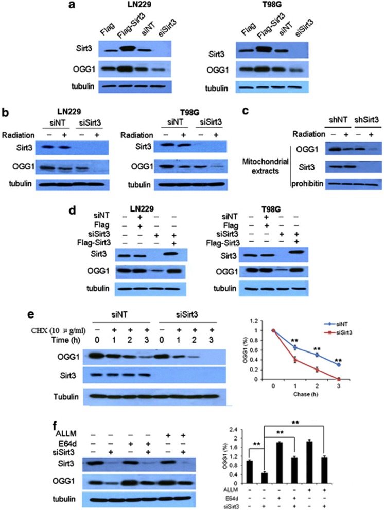
Figure 3.
Silencing of Sirt3 expression promotes the degradation of OGG1 by calpain. (a) LN229 or T98G cells were transfected with a Sirt3 siRNA or a Flag-Sirt3 plasmid. The levels of OGG1 and Sirt3 were examined by western blot. Tubulin was used as a loading control. (b) LN229 or T98G cells were transfected with a non-_targeting RNA or a Sirt3-_targeted siRNA and then were treated or untreated with irradiation. The levels of Sirt3 and OGG1 were examined by western blot. (c) LN229 cells expressing a Sirt3 shRNA or non-_targeting RNA were treated or untreated with irradiation. At the end of the treatment, the mitochondria were isolated using a mitochondrial isolation kit (Pierce). The protein levels of Sirt3 and OGG1 were examined by western blot. Prohibitin was used as a loading control. (d) LN229 or T98G cells were transfected with a Sirt3 siRNA that _targets only the noncoding sequences of the Sirt3 mRNA, followed by transfection with a Flag-Sirt3 expression plasmid. The protein levels of OGG1 and Sirt3 were examined by western blot. (e) LN229 cells were transfected with a non-_targeting RNA or an siRNA _targeting Sirt3, followed by treatment with 10 μg/ml CHX. At the indicated time points, OGG1 protein was examined by western blot. Each point represents the mean±S.E. of three experiments. **P<0.01. (f) LN229 cells with or without silencing of Sirt3 expression were either untreated or treated with ALLM or E64d. Twenty-four hours later, the protein levels of Sirt3 and OGG1 were examined by western blot. Each bar represents the mean±S.E. of three experiments **P<0.01

