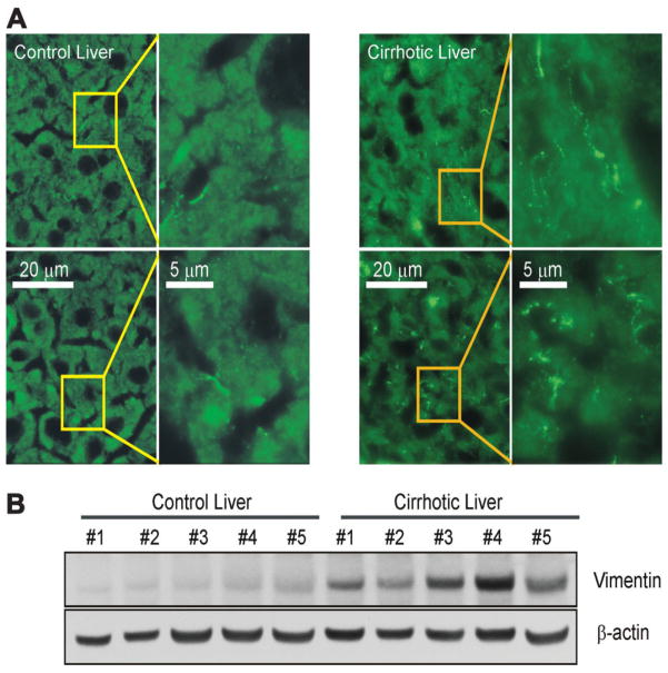
Fig. 1.
Vimentin in cirrhotic livers. (A) Vimentin in normal and cirrhotic mouse livers was visualized by immunofluorescence staining. Each panel represents a separate experiment. Note the characteristic fibrous staining of vimentin in hepatocytes in cirrhotic liver. In contrast, little staining was observed in hepatocytes in control liver. (B) Western blot analysis of liver lysates from control (n = 5) and cirrhotic mice (n = 5). All lysates are from five separate experiments and exhibit overexpression of vimentin in cirrhotic livers.

