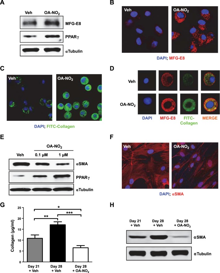
Figure 5.
NFAs reverse fibrotic changes in vitro and in vivo. A–D) Murine alveolar macrophages were treated with OA-NO2 (1 μM) for 24 h. A) MFG-E8 and PPARγ in whole-cell lysates were assessed by Western blotting. B) MFG-E8 expression (red) was evaluated by confocal microscopy. C) Cells were incubated for 30 min with FITC-conjugated type 1 collagen, and uptake was evaluated by confocal microscopy. D) Images demonstrating collagen uptake and MFG-E8 expression were merged. E, F) Fibroblasts derived from lungs of patients with IPF (n=6) were incubated with OA-NO2 (1 μM for confocal) for 24 h, after which α-SMA and PPARγ in whole-cell lysates were assessed by Western blotting (E), and α-SMA expression (red) was assessed by confocal microscopy (F); representative images from a single patient with IPF are shown. G, H) Pulmonary fibrosis was induced in mice by a single intratracheal injection of bleomycin (0.025 U) in saline (50 μl). Beginning 21 d later and continuing daily for 7 d, mice received OA-NO2 (25 μg) intratracheally. Before initiation of OA-NO2 treatment and 24 h after the final OA-NO2 administration lung collagen content (G) was measured, and α-SMA (H) was assessed by Western blotting. Data are representative of 2–3 independent experiments; n = 6 mice/group (G, H); AMs from 1–3 mice for each treatment group (A–D). *P < 0.05, **P < 0.01, ***P < 0.001.

