Figure 5.
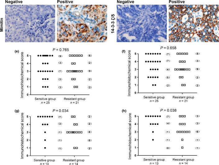
Immunohistochemistry was used to detect the expression of mimitin and 14-3-3 ζ/δ in the first cohort of 46 ovarian cancer patients. In specimens not expressing mimitin and 14-3-3 ζ/δ (negative control), there was no brown staining in the cytoplasm (a,c). Mimitin and 14-3-3 ζ/δ were stained brown in the cytoplasm (b,d). Different immunohistochemical staining scores were recorded and the plots represented the numbers of patients (e–h). When patients with different regimens of chemotherapy enrolled, no significant correlation of immunohistochemical staining and chemotherapy response was observed (Mann–Whitney U-test: mimitin, P = 0.765; 14-3-3 ζ/δ, P = 0.658, Fig.5e,f). While 27 patients with only regimens containing paclitaxel enrolled, a significant difference was observed in the expression levels of mimitin and 14-3-3 ζ/δ between paclitaxel-sensitive and paclitaxel-resistant patients (Mann-Whitney U-test: mimitin, P = 0.034; 14-3-3 ζ/δ, P = 0.038, Fig.5g,h).

