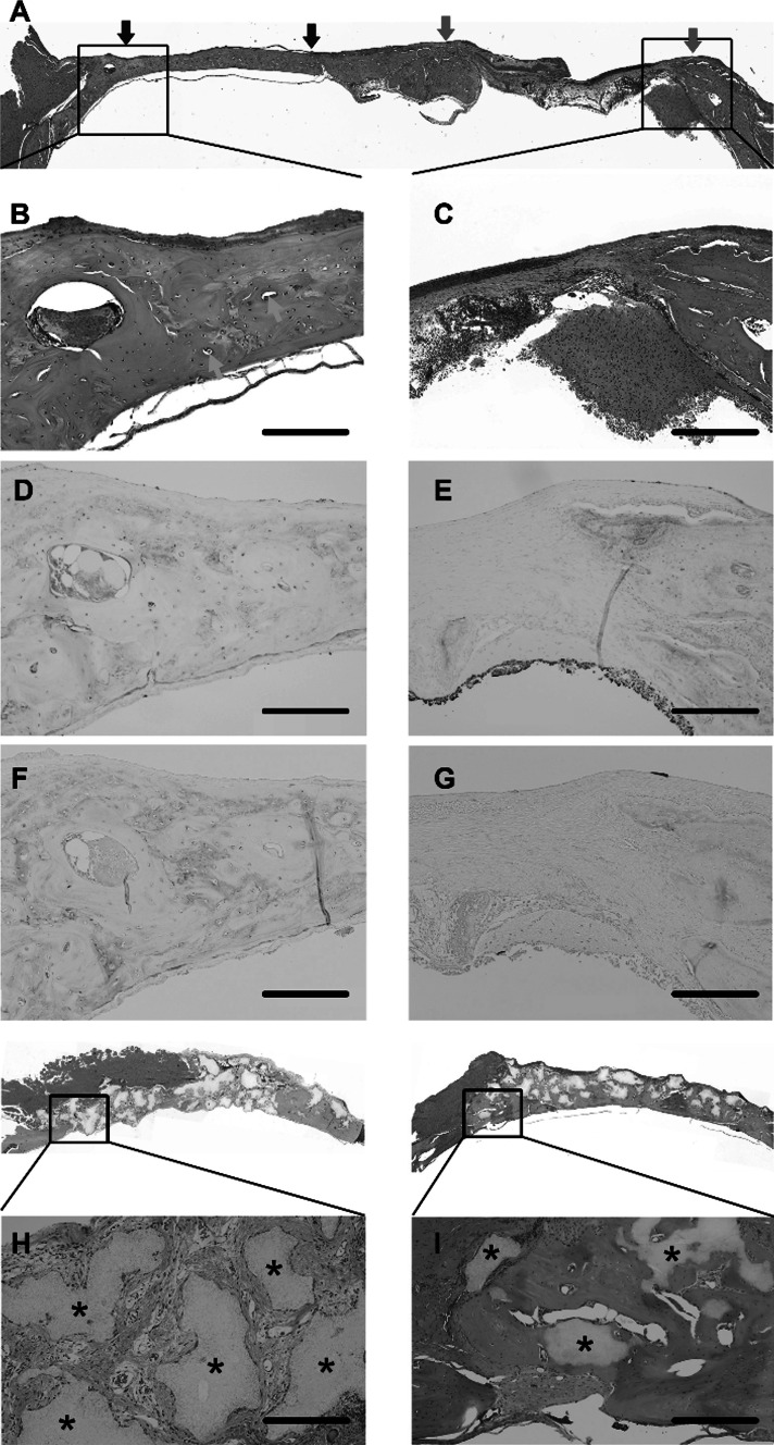
Fig. 4.
Histological assessment of bone regeneration at 8 weeks post-implantation: Bone regeneration was examined in calvarial bone defects at 8 weeks after implantation of mesenchymal stem cell (MSC) spheroids, β-TCP granules alone, and MSC spheroids + β-TCP, and was compared with the untreated defect sites. a Hematoxylin and eosin (HE) staining of the whole bone section showing the implanted area (left side) and the untreated control area (right side). b, c Magnified view of the HE-stained, MSC spheroid-implanted site (b) and control site (c). The control site showed only a thin band of fibrous connective tissue in the defect area along with minimal new bone formation (c). In contrast, at the MSC spheroid-implanted site, new vascularization was apparent, along with a significant amount of new bone and bone proteins throughout the defect area (b). d, e Immunohistochemical staining to visualize distribution of osteocalcin, MSC-implanted site (d) and untreated defect site (e). f, g Immunohistochemical staining to visualize distribution of osteopontin in the MSC-implanted site (f) and untreated defect site (g). h, i Defect site implanted with β-TCP granules alone (h) and β-TCP + MSC spheroids (i). The β-TCP implant site showed disintegrating tissue, fibrous tissue, and blood vessels between β-TCP granules (shown by asterisks). The site implanted with spheroids + β-TCP showed formation of new bone with fewer interspersed β-TCP granules (shown by asterisks). Scale, 500 μm

