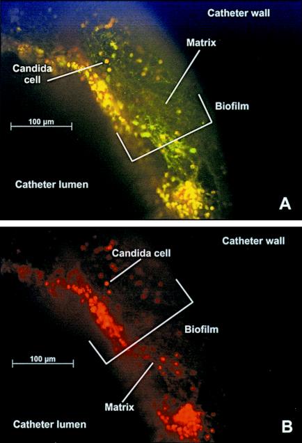
FIG. 4.
Fluorescence images of in vivo C. albicans biofilm with both FUN-1 and ConA stains after 24 h of development. A view of the catheter wall and intraluminal biofilm in an end-on orientation is shown. Magnification, ×20. (A) Image capture was set for simultaneous visualization of both green and red fluorescence. (B) Image capture was set for visualization of red fluorescence. Cells fluorescing red are metabolically active.

