Abstract
Molecular _targeted therapy has the potential to dramatically improve cancer patient survival. However, complete and durable responses to _targeted therapy are rare in advanced-stage solid cancer patients. Even the most effective _targeted therapies generally do not induce a complete tumor response, resulting in residual disease and tumor progression that limits patient survival. We discuss the emerging need to more fully understand the molecular basis of residual disease as a prelude to designing principled therapeutic strategies to minimize or eliminate it so that we can move from temporary to chronic control or cure in advanced-stage solid cancer patients. Ultimately, we propose a shift from the current reactive paradigm of analyzing and treating acquired drug resistance to a pre-emptive paradigm of defining the mechanisms of residual disease in order to _target and limit this disease reservoir.
Introduction
We live in an unprecedented era of precision medicine in which many advanced-stage solid cancers respond profoundly to _targeted therapy against a specific molecular alteration driving tumor growth1–5,6. Prominent examples of _targeted therapies include kinase inhibitors against EGFR (epidermal growth factor receptor), ALK (anaplastic lymphoma kinase), ROS1 (ROS proto-oncogene 1), and RET (ret proto-oncogene) in advanced-stage lung cancer and against BRAF (B-RAF proto-oncogene) and MEK (Mitogen-Activated Protein Kinase Kinase 1) in advanced melanoma. The unprecedented clinical efficacy and improved safety of _targeted therapies arises largely because these agents precisely and selectively suppress molecular events essential for tumor-cell survival with relative sparing of normal cells, in contrast to conventional cytotoxic chemotherapy _targeting general cell proliferative processes.5,7
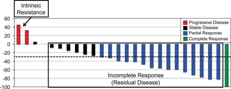
However, _targeted therapy typically induces an incomplete tumor response that is followed by therapy-resistant tumor progression in advanced-stage solid cancer patients (Figure 1)7. The incomplete anti-tumor activity of many _targeted therapies across different cancers has increasingly illuminated the widespread problem of residual disease. Here, we define residual disease as a population of tumor cells within a largely therapy-sensitive tumor that survives initial treatment resulting in a drug-resistant population that, in turn, enables eventual therapy failure and tumor progression during continuous treatment. Consistent with the concept that this reservoir of residual disease cells can ultimately drive therapy failure and tumor progression, there is a correlation in clinical trials between tumor therapy response rates and progression free survival in certain cancers8. This residual disease may either be detected early during therapy by conventional radiographic imaging showing an incomplete tumor response (Figure 1) or occult, wherein an initial complete response to the therapy is followed by eventual therapy failure and tumor progression during continuous treatment3. The biological mechanisms underlying residual disease in patients simultaneously with maximal initial therapy response remain poorly understood, largely due to the lack of direct analysis of patient-derived residual disease samples and the lack of cancer models that faithfully recapitulate human tumor responses (with a few notable exceptions).
Figure 1. The clinical problem of residual disease during _targeted cancer therapy.

A prototypical waterfall plot of best response to therapy highlighting the difference between intrinsic drug resistance and the residual tumor burden observed even amongst patients deemed to have an objective tumor response by RECIST criteria.
Understanding the biological links between incomplete response, residual disease, and therapy-resistant tumor progression and identifying and therapeutically _targeting residual disease cells is essential to enhance response and prevent or minimize therapy failure. The incomplete therapy response as observed in patients has been observed in select few mouse solid cancer models. Though, some examples do exist such as those for mouse mammary tumors caused by induced expression of oncogenes including MYC that fail to regress completely upon de-induction of oncogene expression in vivo for unclear reasons, resulting in residual tumor cells and recurrence9. These data suggest more potent, next generation oncogene-_targeted inhibitors will not be sufficient to overcome the problem of residual disease, as we discuss below.
In this Perspective, we create a framework for understanding and _targeting residual disease in oncogene-driven solid cancer. We highlight the current knowledge of the etiology of clinical residual disease, which is poorly understood compared to innate or acquired resistance (reviewed elsewhere3,7). We propose principled strategies to fill the knowledge gaps of residual disease and accelerate clinical progress through rational therapeutic strategies that directly combat the residual disease state in oncogene-driven solid cancers, with the goal of either chronically managing or potentially eradicating disease persistence and progression. In some cases, the goal of therapy _targeting residual disease will be to achieve chronic disease management, whereas in others it will be disease cure.
The presence and etiology of residual disease
Principles of residual disease
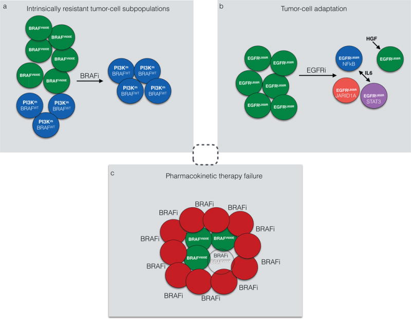
In principle, three primary types of residual disease may be linked to the incomplete response and residual disease that ultimately drives therapy-resistant tumor progression in advanced-stage solid cancers (Figure 2): intrinsic resistance of tumor-cell subpopulations within a generally sensitive tumor, therapy-induced adaptation of tumor-cell subpopulations enabling tumor-cell survival, and pharmacokinetic therapy failure resulting in incomplete drug impact. Multiple mechanisms can operate within an individual metastatic tumor or between different metastatic tumors in an individual patient to promote residual disease and therapy failure (Figures 2–3)10–14.
Figure 2. Modes of residual disease and therapy failure.

a, Intrinsic resistance describes the survival of subpopulations of drug-resistant tumor cells within a generally sensitive tumor during initial therapy. Shown is a pre-treatment melanoma harboring different clones of cells, some with BRAFV600E and some with BRAFWT that instead have mutant PI3K. These resistant BRAFWT cells form a drug-resistant niche during initial BRAFi treatment that results in incomplete response and eventual therapy failure and tumor progression. b, Tumor-cell adaptation can occur via therapy-induced changes in the tumor cells that enable adaptive survival and/or drug-tolerance, fueling a drug-resistant residual disease niche at maximal response. Here, the NF-kB-STAT3 axis, a JARID1A-mediated epigenetic program, and/or secreted factors including IL6 and HGF promote a drug-tolerant state that enables lung cancer cell persistence during initial EGFR inhibitor (EGFRi) treatment. c, Pharmacokinetic therapy failure can result from either pharmacologic limitations or dose-limiting toxicities or tumor intrinsic barriers to drug penetration into the tumor-cell compartment. Shown is poor penetration of a BRAF inhibitor (BRAFi) into a melanoma with dense stromal cell infiltration (red), resulting in low efficiency kill of BRAFV600E-mutant tumor cells (green). The centered dotted circle (grey) indicates the functional overlap and continuum across these modes of residual disease driving therapy failure.
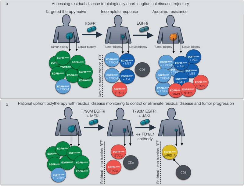
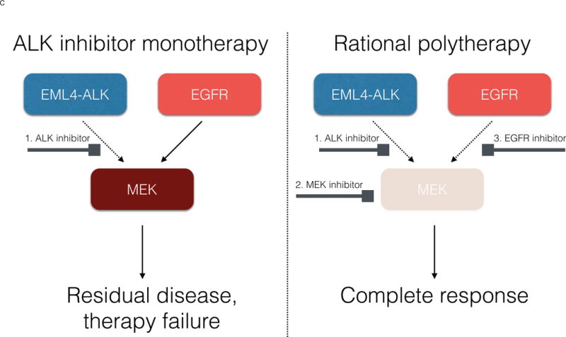
Figure 3. Multimodal characterization of residual disease and rational therapeutic _targeting via upfront, iterative polytherapy.


a, Multimodal longitudinal characterization of the molecular and cellular features of the tumor before therapy and at incomplete maximal response to reveal co-_targets is necessary to understand the biologic underpinnings of residual disease (highlighted in the shaded box; measured and characterized as the residual tumor fraction, RTF). This analysis could be accomplished via serial sampling of tumor and liquid biopsies, which offer key complementary information regarding genetic clones, adaptive signaling events, and cellular constituents within the tumor microenvironment during the evolution of residual disease and therapy failure. Emerging single-cell genetic and proteomic analytic methods could be incorporated73,74. Here, multiple modes of residual disease including, (1) therapy-induced selection of resistant clones and (2) induction of tumor-cell adaptive programs, contribute to the drug-resistant residual disease niche that enables eventual therapy-resistant tumor progression. b, Shown is a conceptual approach to rational upfront polytherapy that addresses the relevant tumor-cell clones present prior to therapy by co-inhibiting a critical downstream effector MEK together with a key on-_target EGFR inhibitor resistance mutation (EGFRT790M). Serial analysis of the residual state, smaller upon (b) polytherapy vs. (a) monotherapy, following initial polytherapy then identifies persistent tumor cells with adaptive STAT3 activation that is, in turn, treated with combined JAK inhibition together with an immune-cell activating therapy (here, anti-PD1). This results in further tumor-cell adaptation that is then controlled by the therapy-mediated activation of the host immune system. This strategy highlights the potential of rational upfront polytherapy, here using simultaneous and sequential combination regimens, to control and limit residual disease as well as toxicity, preventing overt tumor progression via continuous monitoring that guides iterative rational polytherapy. Arrows indicate tumor or liquid biopsy, as in (a). c, Shown are monotherapy versus rational polytherapy for ALK+ lung cancers, where either EGFR or MEK inhibition is paired with ALK inhibition to limit EGFR and MEK signaling that blunts ALK inhibitor response. _targeting upstream EGFR in ALK+ cancer cells may block MAPK and also PI3K/mTOR signaling among others, but may not be effective in patients, for example, with RAS gene amplification or mutation. Inhibiting MEK may more broadly _target escape pathways in ALK+ cancer regardless of the upstream initiator of MEK activation (for example, EGFR or RAS), but may allow other escape pathways (for example, PI3K or NF-kB).
Intrinsic resistance
In tumor-cell subpopulations within a generally sensitive, tumor intrinsic resistance can promote incomplete response to therapy and residual disease. One mechanism underlying tumor-cell intrinsic resistance is incomplete suppression of the pathway that is _targeted by the signal transduction inhibitor. For example, the _targeted agent can induce rapid pathway reactivation to immediately compensate for pathway inhibition, as observed with some BRAF inhibitors that paradoxically activate the RAF-MEK-ERK pathway that they are intended to block, via several mechanisms in certain cells (Figure 2)15,16,17,18,19.
Tumor genetic heterogeneity is present in the treatment-naïve state. This manifests as both intra-tumoral heterogeneity in which multiple different tumor-cell clones with distinct mutational profiles exist in individual tumors and as inter-tumoral heterogeneity in which there are genetic differences between different metastatic lesions. Furthermore, differences in the stromal microenvironment may be also substantial (e.g. lung vs. liver vs. brain), within an individual patient and also promote residual disease20. Different clonal populations of cells within a metastatic tumor may harbor the relevant _target of a particular drug while others do not, and some may contain a drug-resistant mutant form of the _target (e.g. the Thr790Met mutation in EGFR that causes resistance to first generation EGFR inhibitors) or a genetic alteration in a different _target than the one impacted by the drug, thereby enabling survival during therapy (Figure 2)13,21–26,27,28. The presence of different lineage-specific gene expression programs within tumor cell subpopulations is another form of heterogeneity that may enable residual disease during initial therapy29.
Tumor-cell adaptation
Tumor-cell adaptation during initial treatment can promote incomplete response and residual disease (Figure 2). This residual tumor-cell survival can be caused by signaling or metabolic adaptations occurring either within tumor cells or within the tumor microenvironment early during initial treatment, permitting a drug-tolerant state in a subpopulation of tumor cells30–34. For example, EGFR-_targeted therapies may elicit an epigenetically-regulated ‘persister’ cell phenotype and adaptive activation of the transcription factor NF-kappaB which then promotes IL6-JAK-STAT signaling cascade activation to enable survival and drug-tolerance during initial treatment in certain oncogene-driven lung cancer cells (Figure 2)31,32. Secreted growth factors emanating either from tumor cells or tumor-resident stromal cells may also enable tumor-cell survival during initial treatment, as well as intrinsic resistance (Figure 2)30,31,35,36. Hence, biological events limiting _targeted drug efficacy may operate across different modes of residual disease, depending upon the strength and kinetics of activation of the rescue signal.
Pharmacokinetic therapy failure
Pharmacokinetic therapy failure can result in residual disease, as pharmacokinetic issues that prevent the _targeted drug from sufficiently accessing all tumor cells can lead to incomplete anti-tumor effects37–39. To distinguish it from the types of residual disease discussed above in which the _targeted agent reaches the intended _target in cells, this type of failure can arise via the presence of drug efflux pumps in tumor cells and stromal and physical barriers that restrict drug delivery, resulting in insufficient drug concentrations to impact the intended tumor therapeutic _target (Figure 2)40,41. Pharmacokinetic failure due to pharmacologic limitations in drug solubility, distribution, concentration, and dose-limiting toxicity can also result in incomplete suppression of the _targeted pathway, resulting in insufficient pathway blockade and residual disease in vivo39,42. An example is the lack of CNS-penetration and activity of many _targeted agents in clinical use such as certain inhibitors of EGFR (such as erlotinib) and ALK (such as crizotinib), resulting in CNS disease persistence that is strikingly different from extra-cranial tumor response (albeit typically incomplete) in patients 43. The multifactorial basis of pharmacokinetic failure makes it a substantial challenge to the ultimate success of _targeted therapy.
_targeting residual disease
Several potential therapeutic strategies may combat residual disease (Figure 3). These strategies include next-generation _targeted therapies with activity against mutant forms of a particular oncoprotein, such as those resistant to a first-generation _targeted drug, agents with favorable chemical properties and that are not substrates for CNS barrier efflux pumps to enhance CNS penetration, and rational polytherapy to intercept multiple survival (or anti-apoptotic) signals enabling residual disease (Figure 3). The diversity and heterogeneity in strategies that tumors use to evade _targeted therapy emphasizes the need to understand and therapeutically address this complexity to improve clinical responses. Yet, within this complexity a potential commonality across the types of failure is the apoptotic evasion of certain tumor cells in the population during _targeted therapy10–14. If confirmed in residual disease patient samples, this observation suggests the general strategy of using agents (such as BCL-2 protein family inhibitors26,44) to prime or lower the apoptotic threshold necessary for _targeted therapy-induced tumor death, perhaps as part of a polytherapy that also includes an oncoprotein-_targeted agent.
Rational polytherapy
An emergent theme in cancer therapy is the potential for rational _targeted polytherapy to induce a more complete and durable tumor responses than monotherapy because of the ability of polytherapy to address the diverse and multifactorial basis of therapy failure. Clinical responses to treatment with next-generation _targeted agents that, unlike earlier generation drugs, can block mutant, drug-resistant forms of the intended _target remain incomplete, and acquired resistance to such newer agents often occurs earlier than with the initial oncogene-_targeted therapy45,46,47. One such example is the third-generation EGFR inhibitor osimertinib that blocks the mutant protein EGFRT790M that causes resistance to the first generation EGFR inhibitor erlotinib in lung cancer47. Clinical responses to osimertinib are often profound but remain incomplete, resulting in residual disease and eventual tumor progression47. These observations indicate diminishing returns for the use of sequential monotherapy strategies, even with improved individual _targeted agents. It is also not clear that upfront use of such next generation inhibitors will significantly delay resistance as use of these drugs may select for different resistance mechanisms at a similar pace. For example, use of the third-generation EGFR inhibitors may select for the EGFRC797S resistance mutation equally fast as a first-generation EGFR inhibitor selects for EGFRT790M 48. Further enhancing clinical response will likely require rational _targeted polytherapy with an agent against the primary tumor driver plus a drug against a biological event driving residual disease.
Clinical context
Another general theme in cancer therapy is the importance of the clinical context in which therapies designed to enhance patient survival are deployed. Therapies to overcome _targeted therapy resistance are typically tested in the second-line treatment setting, in patients who have failed first-line treatment (Table 1)46,45, 47. However, in many cases second-line polytherapy against the primary oncogene _target plus a critical bypass track component has shown muted clinical efficacy, as exemplified by results from clinical trials testing EGFR plus PI3K (phosphoinositide 3-kinase) inhibitor treatment in lung cancer (Table 1)49,50,42,51. An example of the potential importance of utilizing rational polytherapy as a first-line therapeutic strategy is the use of BRAF inhibitor plus MEK inhibitor polytherapy in BRAFV600E-mutant melanoma patients to overcome the re-activation of RAF-MEK-ERK signaling that can occur during BRAF inhibitor monotherapy and cause resistance to BRAF inhibition alone52,14. Whereas combined MEK inhibitor and BRAF inhibitor treatment was ineffective at overcoming acquired BRAF inhibitor resistance49, upfront BRAF-MEK inhibitor polytherapy improved survival compared to BRAF inhibitor monotherapy and elicited an improved tumor response rate that reflects the activity of this upfront polytherapy against residual disease, consistent with the hypothesis that residual tumor cells drive disease relapse on monotherapy (here, BRAF inhibitor)53,54. Upfront polytherapy may better neutralize or eliminate residual disease cells that fuel eventual tumor progression. Thus, the timing of the polytherapy is critical in battling residual disease and drug resistance. However, clinical responses to effective upfront polytherapy (e.g. BRAF + MEK inhibition in melanoma) are still typically incomplete and not curative53,54. Combining agents which induce non-overlapping mechanisms of resistance in tumors is warranted55,56, but such efforts should be guided by understanding the residual disease state to define what agents should be combined and measure the efficacy of the polytherapy against residual disease.
Table 1. Rational co-_targets and select polytherapy strategies in specific solid cancers.
Shown are select tumor types with the primary _target and _targeted therapy, resistance event, polytherapy and treatment line of clinical testing, outcome of polytherapy, and reference to the primary data (where available) or the relevant clinical trial.gov assignment.
| Tumor Type | Primary _target | Primary _targeted therapy | Resistance event/co-_target | Polytherapy Therapy | line | Outcome | Reference |
|---|---|---|---|---|---|---|---|
| Melanoma | BRAF V600E | Vemurafenib, dabrafenib | RAF-MEK pathway re-activation | Dabrafenib + trametinib | 1 | Improved OR, PFS; FDA approval | 54 |
| Melanoma | PD1 | Nivolumab | CTLA-4 | Nivolumab + ipilumumab | 2 | Improved OR, PFS | 75 |
| Lung adenocarcinoma | EGFR L858R, exon19 del | Erlotinib, gefitinib, afatinib | MET upregulation | METMab or Tivantinib | 2 | Trials completed, low efficacy in EGFR mutant patients |
76, J Clin Oncol 29: 2011 (suppl; abstr 7505) |
| Lung adenocarcinoma | EGFR L858R, exon19 del | Erlotinib, gefitinib, afatinib | PI3K activation | Erlotinib + everolimus | 2 | Trial completed, low efficacy | 77 |
| Lung adenocarcinoma | EGFR L858R, exon19 del | Erlotinib | Epigenetic-induced drug-tolerance or EMT | Entinostat | 2 | Trial completed, lack of efficacy | 78 |
| Lung adenocarcinoma | EGFR L858R, exon19 del | Erlotinib | Bypass kinase activation (multiple HSP90 clients) | Erlotinib + AUY922 | 2 | Trial completed, lack of efficacy and high toxicity | 79 |
| Lung adenocarcinoma | BRAF V600E | Dabrafenib | RAF-MEK pathway reactivation | Dabrafenib + trametinib | 2 | Improved OR, ongoing | J Clin Oncol 33, 2015 suppl; abstr 8006 |
| Colon cancer | BRAF V600E | Vemurafenib | EGFR | Vemurafenib plus cetuximab or panitumumab | 1 | Improved OR, ongoing | 14,80 |
| Breast cancer | HER2 | Trastuzumab | PI3K/mTOR activation | Trastuzumab + everolimus | 1 | Improved PFS | 81 |
| Breast cancer | HER2 | Trastuzumab | HER2 | Trastuzumab +pertuzumab | 1 | Improved PFS | 82 |
| Prostate cancer | AR | Abiraterone | AR activation | Abiraterone + enzalutamide | 2 | Ongoing | NCT01949337 |
| Glioblastoma | EGFR VIII | Lapatinib | PTEN loss (PI3K signaling) | Gefitinib + everolimus | 2 | Completed, results pending | NCT00085566 |
Redefining the _target and therapeutic landscape
We advocate caution and careful re-prioritization of strategies to enhance _targeted therapy clinical response, given the generally incremental rather than transformative clinical progress to date toward the goal of turning most advanced-stage solid cancers into chronic or curable conditions. Developing better ways to prioritize therapy regimens in appropriate patient subsets that are defined by molecular criteria is necessary to accelerate progress. Indeed, current strategies to prioritize specific therapies (or polytherapies) for clinical testing remain inadequate to support systematic _targeted polytherapy clinical development. The number of potential combination regimens vastly exceeds the number of clinical trials possible, particularly in molecular subclass-specific trials where a minority fraction of patients are selected for enrollment57. Currently, standard preclinical models including cell lines and models are used to define and prioritize most therapy strategies. Yet, while these models can be useful, they lack sufficient predictive power for systematic accurate and effective translation into clinical application58. These conventional tumor models often do not accurately represent the salient features of human cancer, such as genetic heterogeneity and micro-environmental influences, and may distort tumor-cell signaling properties present in humans59.
A new strategy
We propose a distinct strategy to develop and prioritize treatment regimens based upon the residual state of disease at the point of maximal therapy response in patients. This residual state would be accessed via tumor and “liquid” biopsies60 and would allow quantification and molecular-definition of residual disease cells for ex vivo study (potentially leveraging single-cell analyses). This would create the opportunity for more precise and accurate understanding of residual disease and the generation of more appropriate patient-derived models, such as xenografts and organoids, for investigation of the functional properties of residual disease (Figure 3)61,62,58. Multimodal characterization of primary patient (and patient-derived) tumors obtained before and at maximal response by genetic, epigenetic, proteomic, and cellular phenotypic analyses to define resident tumor, stromal, and immune cell types present in the residual tumor may provide a cohesive and complete view of the ecological properties of the residual disease state. An open and important question to address in research studies is the feasibility of such multimodal characterization in the clinical setting.
Such an approach would enable simultaneous exploration of key open questions: Is incomplete response a consequence of genetic heterogeneity that is apparent through phylogenetic tree analysis, and if so to what extent21,63? Are residual cancers characterized by reversion to certain phylogenetic truncal genomic events that often represent early, clonal drivers of oncogenesis21? To what extent does _targeted therapy induce a drug-tolerant epigenetic or apoptotically anergic state in residual disease cells in patients?
The relevance of immune strategies
Furthermore, the above strategy could define the contribution and biologic character of stromal and infiltrating immune cells in the residual disease state64. Determining whether certain immune cell subsets are present and active in the tumor at the maximal therapeutic response of residual cancers could offer a strong biologic rationale to investigate combined inhibition of certain immunosuppressive checkpoint proteins64 such as PD-1 (programmed cell death protein 1) expressed on T and B cells and PD-L1 (programmed death ligand 1) expressed on tumor cells and oncogene-inhibitor treatment. This approach could be pursued either as simultaneous initial polytherapy or as sequential therapy with each agent. A strategy where a tumor response is initially induced by an oncoprotein inhibitor followed by augmentation or consolidation of the initial (yet incomplete) tumor response with an immunotherapy is attractive to achieve durable clinical remissions with less toxicity, similar to certain leukemias treated with induction chemotherapy followed by a distinct consolidation chemotherapy regimen65,66 – but characterizing the residual disease state is essential for the necessary biologic foundation of such an induction-consolidation strategy.
Indefinite versus discontinuation therapy
Finally, longitudinal biological phenotyping of residual disease during treatment would allow the determination of whether indefinite continuous therapy is necessary for durable disease control, or if _targeted therapy discontinuation can be safely recommended in select advanced-stage solid cancer patients. Residual disease monitoring could be used to indicate sustained disease elimination or control, or the need for therapy re-initiation in such patients.
Clinical endpoints
Understanding and _targeting residual disease will require additional clinical metrics and endpoints. Current clinical endpoints such as conventional radiographic response, progression free survival, and overall survival are inadequate measures of residual disease. We propose the use of the ‘residual tumor fraction (RTF)’ (Figure 3) to quantify the residual tumor cell population that remains at the maximal treatment response. This would be quantified via positron emission tomography (PET)-based, molecular imaging probes, and/or molecular phenotyping by tumor and liquid biopsies that may accelerate studies of the residual disease state and polytherapy clinical testing67. This approach would enable a more precise and early readout of residual disease burden and characteristics such as tumor metabolic activity or pathway signaling in the upfront setting. For instance, measurement of nuclear NF-kB or STAT3 levels in tumor cells in a lung adenocarcinoma biopsy obtained at maximal response to EGFR inhibitor treatment could offer both molecular biomarkers and therapeutic _targets to quantify and eliminate residual disease tumor cells in individual patients. Similarly, measuring the level of intratumor heterogeneity in solid cancers, via established metrics22, before and during initial treatment could serve as a biomarker of response magnitude and duration, and pinpoint discrete clonal mutations for co-_targeting to serially manage or eliminate residual disease. Such clinical measures and endpoints might allow rapid triage of upfront polytherapies in trials to determine, for example, whether blockade of upstream signaling components such as EGFR or downstream nodes such as MEK are most effective at blocking escape pathways in ALK+ tumors treated with ALK inhibitors11,68 (Figure 3).
Clinical trials
Clinical trials incorporating residual disease endpoints and metrics are needed to address these questions for each disease context and therapy. Beyond trials in metastatic patients, neo-adjuvant (i.e. pre-operative) trials using _targeted therapy for limited time periods prior to surgical tumor resection would allow treatment to (near) maximal response followed by tumor removal. The resected tumor sample may enable extensive and multimodal characterization of residual disease following _targeted therapy and allow establishment of ex vivo models, potentially circumventing the challenges of isolating adequate tumor samples from metastatic tumor biopsies in advanced-stage patients69,70. As oncogene-_targeted therapies have not yet moved from the metastatic setting to incorporation into curative-intent regimens for early-stage disease, the time is ripe to establish clinical trial protocols that will achieve these dual goals. The incorporation of “liquid biopsies” that measure circulating tumor DNA in the blood of individuals20,71,48,72, and potentially of advanced liquid biopsy methods that reliably capture intracellular signaling events as well as genetic mutations, into the metastatic or neo-adjuvant clinical trials to identify and monitor the key cellular and genetic features of the disease longitudinally during treatment, including at incomplete maximal response, would inform the biology of residual disease and its functional relationship to the treatment-naïve and acquired resistance states in solid cancer patients (Figure 3).
Efforts to understand residual disease will potentially provide the strongest possible rationale for testing polytherapies that can have challenging toxicity. For instance, the data may offer the biological rationale for combining agents with non-overlapping toxicities or using specific sequential or alternating drug regimens to maximize efficacy and minimize toxicity. Understanding clinical residual disease will strengthen the rationale for testing drug combinations using agents from different pharmaceutical company portfolios. This remains a challenge, as each individual company has different, often competing, development priorities for individual agents that should be combined as rational polytherapy. Data from residual disease samples would potentially further encourage inter-company collaboration.
Capturing and studying patient-derived residual disease samples faces challenges, including subjecting patients to repeat biopsies and blood tests at maximal therapeutic response, obtaining material of sufficient quantity and quality for analysis, and covering the cost of the repeat biopsies. However, with the use of modern, safe tumor sampling methods (such as image-guided biopsies) and liquid biopsies, obtaining and studying residual disease from advanced-stage solid cancer patients on _targeted therapy is feasible and can provide important insights31. Until the utility of repeat tumor (and blood) sampling is proven through clinical trials, the cost of such repeat tumor sampling will probably need to be covered by research programs in academic, community, and commercial (diagnostic and pharmaceutical) organizations. We believe this cost is an invaluable investment towards the diagnostic and therapeutic advances that are necessary to substantially improve outcomes for patients and continue to generate therapies that all stakeholders, including government and private insurance payers, will value over the longer term, as is the case now for many costly on-treatment imaging tests such as positron emission tomography – (PET) – scanning. Patient engagement is critical to the success of such an initiative and will empower the individuals who stand to benefit the most.
Future Directions
We call on the research community to harness the available resources to re-focus efforts to better diagnosis, monitor, and therapeutically eliminate residual disease. This new directive could catalyze dramatically improved survival in solid cancer patients through definitive, upfront polytherapy regimens that combat residual disease and induce durable responses and potentially cures.
Acknowledgments
T.G.B. acknowledges funding support from the following sources: NIH Director’s Office and Common Fund (New Innovators Award), NCI (R01), HHMI (Collaborative Innovation Award Program), Searle Scholars Program, Pew Charitable Trust and the Addario Lung Cancer Foundation. R.C.D. acknowledges funding support from the V Foundation for Cancer Research and the University of Colorado Lung Cancer SPORE.
Footnotes
Potential competing interests: T.G.B. has ownership in and is a consultant to Driver Group, and is a consultant to Novartis, Astellas, Natera, Array Biopharma, Ariad, and a recipient of research grants from Servier and Ignyta. R.C.D. has received honoraria from AstraZeneca, Clovis, Pfizer, is a consultant to Array BioPharma, Ariad, has received research grants from Loxo Oncology, Mirati Therapeutics, Abbott Molecular and licensing fees from Chugai, Ariad, Blueprint Medicines, GVKbio, Abbott Molecular.
References
- 1.Collins FS, Varmus H. A new initiative on precision medicine. The New England journal of medicine. 2015;372:793–795. doi: 10.1056/NEJMp1500523. [DOI] [PMC free article] [PubMed] [Google Scholar]
- 2.Varmus H. Ten years on–the human genome and medicine. The New England journal of medicine. 2010;362:2028–2029. doi: 10.1056/NEJMe0911933. [DOI] [PubMed] [Google Scholar]
- 3.Sawyers CL. The 2011 Gordon Wilson Lecture: overcoming resistance to _targeted cancer drugs. Transactions of the American Clinical and Climatological Association. 2012;123:114–123. discussion 123–115. [PMC free article] [PubMed] [Google Scholar]
- 4.Sawyers CL. Lessons learned from the development of kinase inhibitors. Clinical advances in hematology & oncology: H&O. 2009;7:588–589. [PubMed] [Google Scholar]
- 5.Sawyers CL. Shifting paradigms: the seeds of oncogene addiction. Nature medicine. 2009;15:1158–1161. doi: 10.1038/nm1009-1158. [DOI] [PubMed] [Google Scholar]
- 6.de Bono JS, Ashworth A. Translating cancer research into _targeted therapeutics. Nature. 2010;467:543–549. doi: 10.1038/nature09339. [DOI] [PubMed] [Google Scholar]
- 7.Garraway LA, Janne PA. Circumventing cancer drug resistance in the era of personalized medicine. Cancer discovery. 2012;2:214–226. doi: 10.1158/2159-8290.CD-12-0012. [DOI] [PubMed] [Google Scholar]
- 8.Blumenthal GM, et al. Overall response rate, progression-free survival, and overall survival with _targeted and standard therapies in advanced non-small-cell lung cancer: US Food and Drug Administration trial-level and patient-level analyses. Journal of clinical oncology: official journal of the American Society of Clinical Oncology. 2015;33:1008–1014. doi: 10.1200/JCO.2014.59.0489. [DOI] [PMC free article] [PubMed] [Google Scholar]
- 9.Boxer RB, Jang JW, Sintasath L, Chodosh LA. Lack of sustained regression of c-MYC-induced mammary adenocarcinomas following brief or prolonged MYC inactivation. Cancer cell. 2004;6:577–586. doi: 10.1016/j.ccr.2004.10.013. [DOI] [PubMed] [Google Scholar]
- 10.Zhang Z, et al. Activation of the AXL kinase causes resistance to EGFR-_targeted therapy in lung cancer. Nature genetics. 2012;44:852–860. doi: 10.1038/ng.2330. [DOI] [PMC free article] [PubMed] [Google Scholar]
- 11.Hrustanovic G, et al. RAS-MAPK dependence underlies a rational polytherapy strategy in EML4-ALK-positive lung cancer. Nature medicine. 2015 doi: 10.1038/nm.3930. [DOI] [PMC free article] [PubMed] [Google Scholar]
- 12.Yu HA, et al. Analysis of tumor specimens at the time of acquired resistance to EGFR-TKI therapy in 155 patients with EGFR-mutant lung cancers. Clinical cancer research: an official journal of the American Association for Cancer Research. 2013;19:2240–2247. doi: 10.1158/1078-0432.CCR-12-2246. [DOI] [PMC free article] [PubMed] [Google Scholar]
- 13.Van Allen EM, et al. The genetic landscape of clinical resistance to RAF inhibition in metastatic melanoma. Cancer discovery. 2014;4:94–109. doi: 10.1158/2159-8290.CD-13-0617. [DOI] [PMC free article] [PubMed] [Google Scholar]
- 14.Ahronian LG, et al. Clinical Acquired Resistance to RAF Inhibitor Combinations in BRAF-Mutant Colorectal Cancer through MAPK Pathway Alterations. Cancer discovery. 2015;5:358–367. doi: 10.1158/2159-8290.CD-14-1518. [DOI] [PMC free article] [PubMed] [Google Scholar]
- 15.Poulikakos PI, Zhang C, Bollag G, Shokat KM, Rosen N. RAF inhibitors transactivate RAF dimers and ERK signalling in cells with wild-type BRAF. Nature. 2010;464:427–430. doi: 10.1038/nature08902. [DOI] [PMC free article] [PubMed] [Google Scholar]
- 16.Lito P, Rosen N, Solit DB. Tumor adaptation and resistance to RAF inhibitors. Nature medicine. 2013;19:1401–1409. doi: 10.1038/nm.3392. [DOI] [PubMed] [Google Scholar]
- 17.Poulikakos PI, et al. RAF inhibitor resistance is mediated by dimerization of aberrantly spliced BRAF(V600E) Nature. 2011;480:387–390. doi: 10.1038/nature10662. [DOI] [PMC free article] [PubMed] [Google Scholar]
- 18.Lito P, et al. Relief of profound feedback inhibition of mitogenic signaling by RAF inhibitors attenuates their activity in BRAFV600E melanomas. Cancer cell. 2012;22:668–682. doi: 10.1016/j.ccr.2012.10.009. [DOI] [PMC free article] [PubMed] [Google Scholar]
- 19.Prahallad A, et al. Unresponsiveness of colon cancer to BRAF(V600E) inhibition through feedback activation of EGFR. Nature. 2012;483:100–103. doi: 10.1038/nature10868. [DOI] [PubMed] [Google Scholar]
- 20.Alizadeh AA, et al. Toward understanding and exploiting tumor heterogeneity. Nature medicine. 2015;21:846–853. doi: 10.1038/nm.3915. [DOI] [PMC free article] [PubMed] [Google Scholar]
- 21.de Bruin EC, et al. Spatial and temporal diversity in genomic instability processes defines lung cancer evolution. Science. 2014;346:251–256. doi: 10.1126/science.1253462. [DOI] [PMC free article] [PubMed] [Google Scholar]
- 22.Hiley C, de Bruin EC, McGranahan N, Swanton C. Deciphering intratumor heterogeneity and temporal acquisition of driver events to refine precision medicine. Genome biology. 2014;15:453. doi: 10.1186/s13059-014-0453-8. [DOI] [PMC free article] [PubMed] [Google Scholar]
- 23.Burrell RA, Swanton C. Tumour heterogeneity and the evolution of polyclonal drug resistance. Molecular oncology. 2014;8:1095–1111. doi: 10.1016/j.molonc.2014.06.005. [DOI] [PMC free article] [PubMed] [Google Scholar]
- 24.Turke AB, et al. Preexistence and clonal selection of MET amplification in EGFR mutant NSCLC. Cancer cell. 2010;17:77–88. doi: 10.1016/j.ccr.2009.11.022. [DOI] [PMC free article] [PubMed] [Google Scholar]
- 25.Rosell R, et al. Pretreatment EGFR T790M mutation and BRCA1 mRNA expression in erlotinib-treated advanced non-small-cell lung cancer patients with EGFR mutations. Clinical cancer research: an official journal of the American Association for Cancer Research. 2011;17:1160–1168. doi: 10.1158/1078-0432.CCR-10-2158. [DOI] [PubMed] [Google Scholar]
- 26.Lin L, et al. The Hippo effector YAP promotes resistance to RAF- and MEK-_targeted cancer therapies. Nature genetics. 2015;47:250–256. doi: 10.1038/ng.3218. [DOI] [PMC free article] [PubMed] [Google Scholar]
- 27.Janku F, et al. PIK3CA mutations frequently coexist with RAS and BRAF mutations in patients with advanced cancers. PloS one. 2011;6:e22769. doi: 10.1371/journal.pone.0022769. [DOI] [PMC free article] [PubMed] [Google Scholar]
- 28.Hata AN, et al. Tumor cells can follow distinct evolutionary paths to become resistant to epidermal growth factor receptor inhibition. Nature medicine. 2016 doi: 10.1038/nm.4040. [DOI] [PMC free article] [PubMed] [Google Scholar]
- 29.Muller J, et al. Low MITF/AXL ratio predicts early resistance to multiple _targeted drugs in melanoma. Nature communications. 2014;5:5712. doi: 10.1038/ncomms6712. [DOI] [PMC free article] [PubMed] [Google Scholar]
- 30.Straussman R, et al. Tumour micro-environment elicits innate resistance to RAF inhibitors through HGF secretion. Nature. 2012;487:500–504. doi: 10.1038/nature11183. [DOI] [PMC free article] [PubMed] [Google Scholar]
- 31.Blakely CM, et al. NF-kappaB-Activating Complex Engaged in Response to EGFR Oncogene Inhibition Drives Tumor Cell Survival and Residual Disease in Lung Cancer. Cell reports. 2015;11:98–110. doi: 10.1016/j.celrep.2015.03.012. [DOI] [PMC free article] [PubMed] [Google Scholar]
- 32.Sharma SV, et al. A chromatin-mediated reversible drug-tolerant state in cancer cell subpopulations. Cell. 2010;141:69–80. doi: 10.1016/j.cell.2010.02.027. [DOI] [PMC free article] [PubMed] [Google Scholar]
- 33.Lee HJ, et al. Drug resistance via feedback activation of Stat3 in oncogene-addicted cancer cells. Cancer cell. 2014;26:207–221. doi: 10.1016/j.ccr.2014.05.019. [DOI] [PubMed] [Google Scholar]
- 34.Haq R, et al. Oncogenic BRAF regulates oxidative metabolism via PGC1alpha and MITF. Cancer cell. 2013;23:302–315. doi: 10.1016/j.ccr.2013.02.003. [DOI] [PMC free article] [PubMed] [Google Scholar]
- 35.Wilson TR, et al. Widespread potential for growth-factor-driven resistance to anticancer kinase inhibitors. Nature. 2012;487:505–509. doi: 10.1038/nature11249. [DOI] [PMC free article] [PubMed] [Google Scholar]
- 36.Obenauf AC, et al. Therapy-induced tumour secretomes promote resistance and tumour progression. Nature. 2015;520:368–372. doi: 10.1038/nature14336. [DOI] [PMC free article] [PubMed] [Google Scholar]
- 37.Bollag G, et al. Clinical efficacy of a RAF inhibitor needs broad _target blockade in BRAF-mutant melanoma. Nature. 2010;467:596–599. doi: 10.1038/nature09454. [DOI] [PMC free article] [PubMed] [Google Scholar]
- 38.Szmulewitz RZ, Ratain MJ. Vemurafenib oral bioavailability: an insoluble problem. Journal of clinical pharmacology. 2014;54:375–377. doi: 10.1002/jcph.277. [DOI] [PubMed] [Google Scholar]
- 39.Undevia SD, Gomez-Abuin G, Ratain MJ. Pharmacokinetic variability of anticancer agents. Nature reviews. Cancer. 2005;5:447–458. doi: 10.1038/nrc1629. [DOI] [PubMed] [Google Scholar]
- 40.Neesse A, Algul H, Tuveson DA, Gress TM. Stromal biology and therapy in pancreatic cancer: a changing paradigm. Gut. 2015;64:1476–1484. doi: 10.1136/gutjnl-2015-309304. [DOI] [PubMed] [Google Scholar]
- 41.Jain RK. Antiangiogenesis strategies revisited: from starving tumors to alleviating hypoxia. Cancer cell. 2014;26:605–622. doi: 10.1016/j.ccell.2014.10.006. [DOI] [PMC free article] [PubMed] [Google Scholar]
- 42.Janne PA, et al. Phase I safety and pharmacokinetic study of the PI3K/mTOR inhibitor SAR245409 (XL765) in combination with erlotinib in patients with advanced solid tumors. J Thorac Oncol. 2014;9:316–323. doi: 10.1097/JTO.0000000000000088. [DOI] [PubMed] [Google Scholar]
- 43.Lin NU. _targeted therapies in brain metastases. Current treatment options in neurology. 2014;16:276. doi: 10.1007/s11940-013-0276-z. [DOI] [PMC free article] [PubMed] [Google Scholar]
- 44.Thompson CB. _targeting the anti-apoptotic signaling pathway. Clinical advances in hematology & oncology: H&O. 2009;7:819–822. [PubMed] [Google Scholar]
- 45.Shaw AT, et al. Ceritinib in ALK-rearranged non-small-cell lung cancer. The New England journal of medicine. 2014;370:1189–1197. doi: 10.1056/NEJMoa1311107. [DOI] [PMC free article] [PubMed] [Google Scholar]
- 46.Sequist LV, et al. Rociletinib in EGFR-mutated non-small-cell lung cancer. The New England journal of medicine. 2015;372:1700–1709. doi: 10.1056/NEJMoa1413654. [DOI] [PubMed] [Google Scholar]
- 47.Janne PA, et al. AZD9291 in EGFR inhibitor-resistant non-small-cell lung cancer. The New England journal of medicine. 2015;372:1689–1699. doi: 10.1056/NEJMoa1411817. [DOI] [PubMed] [Google Scholar]
- 48.Thress KS, et al. Acquired EGFR C797S mutation mediates resistance to AZD9291 in non-small cell lung cancer harboring EGFR T790M. Nature medicine. 2015 doi: 10.1038/nm.3854. [DOI] [PMC free article] [PubMed] [Google Scholar]
- 49.Johnson DB, et al. Combined BRAF (Dabrafenib) and MEK inhibition (Trametinib) in patients with BRAFV600-mutant melanoma experiencing progression with single-agent BRAF inhibitor. Journal of clinical oncology: official journal of the American Society of Clinical Oncology. 2014;32:3697–3704. doi: 10.1200/JCO.2014.57.3535. [DOI] [PMC free article] [PubMed] [Google Scholar]
- 50.Charakidis M, Boyer M. _targeting MET and EGFR in NSCLC-what can we learn from the recently reported phase III trial of onartuzumab in combination with erlotinib in advanced non-small cell lung cancer? Translational lung cancer research. 2014;3:395–396. doi: 10.3978/j.issn.2218-6751.2014.09.03. [DOI] [PMC free article] [PubMed] [Google Scholar]
- 51.Riely GJ, et al. Prospective assessment of discontinuation and reinitiation of erlotinib or gefitinib in patients with acquired resistance to erlotinib or gefitinib followed by the addition of everolimus. Clinical cancer research: an official journal of the American Association for Cancer Research. 2007;13:5150–5155. doi: 10.1158/1078-0432.CCR-07-0560. [DOI] [PubMed] [Google Scholar]
- 52.Wagle N, et al. MAP kinase pathway alterations in BRAF-mutant melanoma patients with acquired resistance to combined RAF/MEK inhibition. Cancer discovery. 2014;4:61–68. doi: 10.1158/2159-8290.CD-13-0631. [DOI] [PMC free article] [PubMed] [Google Scholar]
- 53.Flaherty KT, et al. Combined BRAF and MEK inhibition in melanoma with BRAF V600 mutations. The New England journal of medicine. 2012;367:1694–1703. doi: 10.1056/NEJMoa1210093. [DOI] [PMC free article] [PubMed] [Google Scholar]
- 54.Long GV, et al. Combined BRAF and MEK inhibition versus BRAF inhibition alone in melanoma. The New England journal of medicine. 2014;371:1877–1888. doi: 10.1056/NEJMoa1406037. [DOI] [PubMed] [Google Scholar]
- 55.Bhang HE, et al. Studying clonal dynamics in response to cancer therapy using high-complexity barcoding. Nature medicine. 2015;21:440–448. doi: 10.1038/nm.3841. [DOI] [PubMed] [Google Scholar]
- 56.Bozic I, et al. Evolutionary dynamics of cancer in response to _targeted combination therapy. eLife. 2013;2:e00747. doi: 10.7554/eLife.00747. [DOI] [PMC free article] [PubMed] [Google Scholar]
- 57.Schwaederle M, et al. On the Road to Precision Cancer Medicine: Analysis of Genomic Biomarker Actionability in 439 Patients. Molecular cancer therapeutics. 2015;14:1488–1494. doi: 10.1158/1535-7163.MCT-14-1061. [DOI] [PubMed] [Google Scholar]
- 58.Crystal AS, et al. Patient-derived models of acquired resistance can identify effective drug combinations for cancer. Science. 2014;346:1480–1486. doi: 10.1126/science.1254721. [DOI] [PMC free article] [PubMed] [Google Scholar]
- 59.Gao D, et al. Organoid cultures derived from patients with advanced prostate cancer. Cell. 2014;159:176–187. doi: 10.1016/j.cell.2014.08.016. [DOI] [PMC free article] [PubMed] [Google Scholar]
- 60.Gingras I, Salgado R, Ignatiadis M. Liquid biopsy: will it be the ‘magic tool’ for monitoring response of solid tumors to anticancer therapies? Current opinion in oncology. 2015 doi: 10.1097/CCO.0000000000000223. [DOI] [PubMed] [Google Scholar]
- 61.Li X, et al. Oncogenic transformation of diverse gastrointestinal tissues in primary organoid culture. Nature medicine. 2014;20:769–777. doi: 10.1038/nm.3585. [DOI] [PMC free article] [PubMed] [Google Scholar]
- 62.Karthaus WR, et al. Identification of multipotent luminal progenitor cells in human prostate organoid cultures. Cell. 2014;159:163–175. doi: 10.1016/j.cell.2014.08.017. [DOI] [PMC free article] [PubMed] [Google Scholar]
- 63.Piotrowska Z, et al. Heterogeneity Underlies the Emergence of EGFR T790 Wild-Type Clones Following Treatment of T790M-Positive Cancers with a Third Generation EGFR Inhibitor. Cancer discovery. 2015 doi: 10.1158/2159-8290.CD-15-0399. [DOI] [PMC free article] [PubMed] [Google Scholar]
- 64.Nakasone ES, et al. Imaging tumor-stroma interactions during chemotherapy reveals contributions of the microenvironment to resistance. Cancer cell. 2012;21:488–503. doi: 10.1016/j.ccr.2012.02.017. [DOI] [PMC free article] [PubMed] [Google Scholar]
- 65.Mayer RJ, et al. Intensive postremission chemotherapy in adults with acute myeloid leukemia. Cancer and Leukemia Group B. The New England journal of medicine. 1994;331:896–903. doi: 10.1056/NEJM199410063311402. [DOI] [PubMed] [Google Scholar]
- 66.Coombs CC, Tallman MS, Levine RL. Molecular therapy for acute myeloid leukaemia. Nature reviews Clinical oncology. 2015 doi: 10.1038/nrclinonc.2015.210. [DOI] [PMC free article] [PubMed] [Google Scholar]
- 67.Condeelis J, Weissleder R. In vivo imaging in cancer. Cold Spring Harbor perspectives in biology. 2010;2:a003848. doi: 10.1101/cshperspect.a003848. [DOI] [PMC free article] [PubMed] [Google Scholar]
- 68.Doebele RC, et al. Mechanisms of resistance to crizotinib in patients with ALK gene rearranged non-small cell lung cancer. Clinical cancer research: an official journal of the American Association for Cancer Research. 2012;18:1472–1482. doi: 10.1158/1078-0432.CCR-11-2906. [DOI] [PMC free article] [PubMed] [Google Scholar]
- 69.Van Allen EM, et al. Whole-exome sequencing and clinical interpretation of formalin-fixed, paraffin-embedded tumor samples to guide precision cancer medicine. Nature medicine. 2014;20:682–688. doi: 10.1038/nm.3559. [DOI] [PMC free article] [PubMed] [Google Scholar]
- 70.De Silva N, et al. Molecular effects of Lapatinib in the treatment of HER2 overexpressing oesophago-gastric adenocarcinoma. British journal of cancer. 2015;113:1305–1312. doi: 10.1038/bjc.2015.342. [DOI] [PMC free article] [PubMed] [Google Scholar]
- 71.Siravegna G, et al. Clonal evolution and resistance to EGFR blockade in the blood of colorectal cancer patients. Nature medicine. 2015;21:795–801. doi: 10.1038/nm.3870. [DOI] [PMC free article] [PubMed] [Google Scholar]
- 72.Murtaza M, et al. Non-invasive analysis of acquired resistance to cancer therapy by sequencing of plasma DNA. Nature. 2013;497:108–112. doi: 10.1038/nature12065. [DOI] [PubMed] [Google Scholar]
- 73.Macosko EZ, et al. Highly Parallel Genome-wide Expression Profiling of Individual Cells Using Nanoliter Droplets. Cell. 2015;161:1202–1214. doi: 10.1016/j.cell.2015.05.002. [DOI] [PMC free article] [PubMed] [Google Scholar]
- 74.Behbehani GK, et al. Mass Cytometric Functional Profiling of Acute Myeloid Leukemia Defines Cell-Cycle and Immunophenotypic Properties That Correlate with Known Responses to Therapy. Cancer discovery. 2015;5:988–1003. doi: 10.1158/2159-8290.CD-15-0298. [DOI] [PMC free article] [PubMed] [Google Scholar]
- 75.Larkin J, et al. Combined Nivolumab and Ipilimumab or Monotherapy in Untreated Melanoma. The New England journal of medicine. 2015;373:23–34. doi: 10.1056/NEJMoa1504030. [DOI] [PMC free article] [PubMed] [Google Scholar]
- 76.Sequist LV, et al. Randomized phase II study of erlotinib plus tivantinib versus erlotinib plus placebo in previously treated non-small-cell lung cancer. Journal of clinical oncology: official journal of the American Society of Clinical Oncology. 2011;29:3307–3315. doi: 10.1200/JCO.2010.34.0570. [DOI] [PubMed] [Google Scholar]
- 77.Price KA, et al. Phase II trial of gefitinib and everolimus in advanced non-small cell lung cancer. J Thorac Oncol. 2010;5:1623–1629. doi: 10.1097/JTO.0b013e3181ec1531. [DOI] [PMC free article] [PubMed] [Google Scholar]
- 78.Witta SE, et al. Randomized phase II trial of erlotinib with and without entinostat in patients with advanced non-small-cell lung cancer who progressed on prior chemotherapy. Journal of clinical oncology: official journal of the American Society of Clinical Oncology. 2012;30:2248–2255. doi: 10.1200/JCO.2011.38.9411. [DOI] [PMC free article] [PubMed] [Google Scholar]
- 79.Johnson ML, et al. Phase I/II Study of HSP90 Inhibitor AUY922 and Erlotinib for EGFR-Mutant Lung Cancer With Acquired Resistance to Epidermal Growth Factor Receptor Tyrosine Kinase Inhibitors. Journal of clinical oncology: official journal of the American Society of Clinical Oncology. 2015;33:1666–1673. doi: 10.1200/JCO.2014.59.7328. [DOI] [PMC free article] [PubMed] [Google Scholar]
- 80.Yaeger R, et al. Pilot trial of combined BRAF and EGFR inhibition in BRAF-mutant metastatic colorectal cancer patients. Clinical cancer research: an official journal of the American Association for Cancer Research. 2015;21:1313–1320. doi: 10.1158/1078-0432.CCR-14-2779. [DOI] [PMC free article] [PubMed] [Google Scholar]
- 81.Hurvitz SA, et al. Combination of everolimus with trastuzumab plus paclitaxel as first-line treatment for patients with HER2-positive advanced breast cancer (BOLERO-1): a phase 3, randomised, double-blind, multicentre trial. The Lancet Oncology. 2015;16:816–829. doi: 10.1016/S1470-2045(15)00051-0. [DOI] [PubMed] [Google Scholar]
- 82.Swain SM, et al. Pertuzumab, trastuzumab, and docetaxel in HER2-positive metastatic breast cancer. The New England journal of medicine. 2015;372:724–734. doi: 10.1056/NEJMoa1413513. [DOI] [PMC free article] [PubMed] [Google Scholar]


