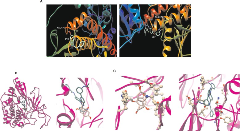
Fig. 8.
Molecular Docking. a PH shows 3 BS of protein with 3H-bond, Sor shows binding near to active site (with 2-H bond). b N-Sh2, C-Sh2, and PTP domains of Shp1 protein (2B3O) structure. The four binding sites (BS) identified from Swiss dock models are marked in the imaged along with the predicted binding affinities for Sor (SFB) and PH (PHL). c Docking of PH in ATP binding site of AKT1 protein (PDB id: 4EJN). PH and 0R4 are in rosy brown and sea green respectively (Left)

