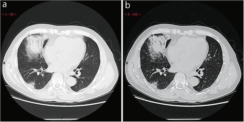
Fig. 9.

CT imaging of consolidation stage. A 65 years old male with fever (maximum temperature of 39 ℃). Laboratory test: hypoproteinemia (decreased total protein (62.20 g/L), decreased albumin (35.70 g/L)), abnormal liver function (increased alanine aminotransferase (79 U/L), increased aspartate aminotransferase (72 U/L)), increased procalcitonin (0.10 ng/ml), increased C-reaction protein (53 mg/L), decreased white blood cells (3.72 × 10⁹/L), decreased lymphocytes (0.9 × 10⁹/ L), mildanemia (decreased red blood cells (4.10 × 10¹²/L), decreased hemoglobin (131.10 g/L), decreased hematocrit (39.0%). Imaging examination: a (thin layer CT) and b (high-resolution CT) showedmultiple patchyand large consolidation in right middle lobe, posterior and basal segment of right lower lobe and outer and basal segment of left lower lobe, with air-bronchogram inside
